Full Text-1 - African Index Medicus
advertisement
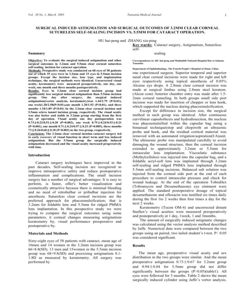
Vol. 20 No. 1, March 2005 Tanzania Medical Journal 1 SURGICAL INDUCED ASTIGMATISM AND SURGICAL OUTCOMES OF 3.2MM CLEAR CORNEAL SUTURELESS SELF-SEALING INCISION VS. 5.5MM FOR CATARACT OPERATION. Summary HU hai-peng and ZHANG xiu-ping Key wards: Cataract surgery, Astigmatism, Sutureless selfsealing Objective: To evaluate the surgical induced astigmatism and other surgical outcomes in 3.2mm and 5.5mm clear corneal sutureless self-sealing incision for cataract operation. Methods: Prospective study was conducted on 68 eyes of 58 patients out of which 35 eyes were in 3.2mm and 33 eyes in 5.5mm incision groups. Except the incision size, lens type, and implantation technique, the surgical methods were identical. Uncorrected visual acuity, keratometry were measured preoperatively, one day, one week, one month and three months postoperatively. Results: Eyes in 3.2mm clear corneal incision group had significantly less surgical induced astigmatism than 5.5mm incision group. One day after surgery, the surgical induced astigmatism(vector analysis, keratometry)was 1.44/2.79 (P<0.01), one week1.28/2.58(P<0.01),one month 1.20/1.92 (P<0.01), and three months 1.10/1.89 (P<0.01) In the 3.2mm clear corneal incision and 5.5mm sclera tunnel incision group, respectively. The visual acuity was also better and stable in 3.2mm group starting from the first day of operation. Visual acuity one day postoperation was 0.73±0.22/0.51±0.20 (P=0.002), one week 0.75±0.24/0.53±0.21 (P=0.001), one month 0.71±0.24/0.57±0.23 (P=0.005), three months 73±0.26/0.60±0.18 (P=0.003) in the two group, respectively. Conclusion: The 3.2mm clear corneal incision cataract surgery led to early recovery of visual function in short term and less induced astigmatism. But the 5.5mm group the surgically induced astigmatism decreased and the visual acuity increased progressively with time. Introduction Cataract surgery techniques have improved in the past decades. Self-sealing incision are recognized to improve intraoperative safety and reduce postoperative inflammation and complications. The small incision surgery has a number of surgical advantages: It is easy to perform, is faster, offer's better visualization and cosmetically attractive because there is minimal bleeding and no need of retrobulbar or pribulbar injection for anesthesia. Sutureless clear corneal incisions are the preferred approach for phacoemulsification; that is 3.2mm for foldable lens and 5.5mm for ridged PMMA lens implantation. In this prospective study we were trying to compare the surgical outcomes using some parameters, it corneal changes measuring astigmatism keratometry by, visual performance preoperative and postoperative by. Materials and Methods Sixty-eight eyes of 58 patients with cataract, mean age of 16men and 14 women in the 3.2mm incision group was 66+8.8(SD), 13 men and 15women in the 5.5mm incision group was 68+9.8(SD) and preexising astigmatism 0.11.8D as measured by keratometry. All surgery was performed by Correspondence to: HU hai-peng and Muhimbili National Hospital Dar es Salaam, Tanzania. Department of Ophthalmology, The Fourth People’s Hospital of Jinan, China one experienced surgeon. Superior temporal and superior nasal clear corneal incisions were made for right and left eyes respectively using topical anesthesia of 0.05% Alcaine eye drops. A 2.8mm clear corneal incision was made at surgical limbus using 2.8mm steel keratom. (Alcon com) Anterior chamber entry was made after 1.52mm corneal tunneling. In both groups small side port incision was made for insertion of chopper or lens hook, which supported the nucleus during phacoemulsification. Except for difference in incision size, the surgical method in each group was identical. After continuous curvilinear capsulorhexis and hydrodissection, the nucleus was phacoemulsified within the capsular bag using a bimanual technique(stop and chop)with an ultrasonic probe and hook, and the residual cortical material was removed with an automated irrigation/aspiration(I/A)unit. The ultrasonic probe was manipulated carefully to avoid damaging the wound structure, then the corneal incision extended to approximately 3.2mm or 5.5mm for intraocular lens implantation. Viscoelatic substance (Methylcellulos) was injected into the capsular bag, and a foldable acryl-soft lens was implanted through 3.2mm self-sealing and ridged PMMA lens implanted through 5.5mm self-sealing incisions. Balanced salt solution was injected from the corneal side port at the end of each procedure to control intraocular pressure and check for wound leakage. At the end of the operation TobraDex (Tobramycin and Dexamethasone) eye ointment were applied. The standard postoperative dosage of topical dexamethasone and ofloxacin was instilled six times daily during the first 1to 2 weeks then four times a day for the next 2 weeks. Keratometry (Tocon OM-4) and uncorrected distant Snellen’s visual acuities were measured preoperatively and postoperatively at 1 day, 1week, 1 and 3months. The amount of surgically induced astigmatic changes was calculated using the vector analysis method described by Jaffe. Numerical data were compared between the two groups using an paired, two tailed student’s t-test. P< 0.05 was considered significant. Results The mean age, preoperative visual acuity and sex distribution in the two groups were similar. And the mean preoperative astigmatism 0.73±0.67 for 3.2mm group and 0.94±0.84 for 5.5mm group did not differ significantly between the groups (P>0.05)(table1). All eyes were followed for 3 months. Table 2 shows the mean surgically induced cylinder using Jaffe’s vertor analysis. Vol. 20 No. 1, March 2005 Tanzania Medical Journal At all postoperative period, the mean induced astigmatism was large in the CCI-5.5mm group (P<0.01). Mean preoperative uncorrected visual acuity (UCVA) were 0.15±0.12/0.13±0.12 (P>0.05) for 3.2mm and 5.5mm groups respectively. The postoperative uncorrected visual acuity was much better in CCI-3.2mm group from the first day of operation till the third month follow up time (P<0.01). In the CCI-3.2mm group the mean UCVA at day one and three months postoperative was almost the same, but in CCI-5.5mm group, there was progressive increment from postoperative day one till the third month of follow up (table-3). No intraoperative complications were observed in either group. No suture was needed because the wounds were self-sealing. Other complications such as fibrin formation, prolonged inflammation, secondary glaucoma, IOL dislocation were not seen. Table 1. 1-age, preoperative vusual acuity (VA) and astigmatisms (AS) Age Preop VA Preop AS CCI-3.2.mm 66±8.8(SD) 0.13±0.14 0.93±0.67 CCi-5.5.mm 68± 9.8(SD 0.12±0.17 094±0.84 P-value P>0.05 P>0.05 P>0.05 *CCI=velar corneal incision Table 2. Mean surgical induced astigmatism (vertor analysis) through time from keratometry data 1 –day 1 – week 1 – mont 3 - months CCI-3.2.mm 1.68±1.30 1.44±1.20 1.35±0.96 1.16±0.84 CCi-5.5.mm 2.86±1.6 2.68±1.5 2.36±1.2 1.76±1.6 P-value P =0.0067 P = 0.00078 P = 0.0007 P = 0.004 Table 3. Mean Pre and postoperative UCVA Pre 1 day 1 week 1 mont 3 months CCI-3.2mm 0.15±0.12 0.73±0.22 0.75±0.24 0.71±0.24 0.73±0.26 CCi-5.5.mm 0.13±012 0.15±0.20 0.53±0.21 0.57±0.23 0.60±0.18 P-value 0.21 0.02 0.001 0.005 0.003 corneal astigmatism could be minimized, as K-Muller and B.Barlin corneal tunnel incision at 12o’clock only in eyes with preoperative WTR astigmatism of more than 1.0D, in eyes with preoperative WTR astigmatism between 0.5and 1.0D to use corneoscleral incision which induces considerably less astigmatism, in eyes WTR astigmatism of 0.5D or less, ATR astigmatism and spherical cornea lateral corneal tunnel incision. However the superior 5.5mm clear corneal incision for ridged PMMA lens implantation compare to other incision type such as sclera tunnel or corneoscleral incision of classic ECCE, it has obvious advantages: it could be done using topical anesthesia with minimal or no bleeding, less discomfort, short operation time, fast recovery of wound and visual performance. Xie lixin’s study showed that surgical induced astigmatism was very small after a corneal incision in phacoemultification without a suture. If the incision was placed on the steepest meridian, the corneal astigmatism was significantly reduced postoperatively. From our study, though of small sample size and short duration of followup we can conclude that 3.2mm is superior in every aspect the 5.5mm incision group except it is expensive. But 5.5mm clear corneal incision is also a good alternative for normal PMMA lens based on its many advantages. Furthermore it is better to consider preoperative astigmatism the dioptre and direction from keratometry readings to determine the incision site on the cornea and its size (3.3mm and 5.5mm). So, the patient can choose which incision (3.2mm or 5.5mm), he or she prefer depending on their financial capability because foldable lens were expensive than ridged lens. Finally we suggest long term comparative study between different types of clear corneal incision and sclerrocorneal tunnel or scleral incision to see which incision type has minimal induced astigmatism and less complications. Reference 1. Discussion Our results showed a significantly higher amount of surgically induced astigmatism with 5.5mm sclera tunnel incision than 3.2mm corneal incision based on vector analysis of keratometric data. The uncorrected visual acuity was also significantly better in the 3.2mm group than in 5.5mm group throughout the follow time. In 3.2mm the mean visual acuity become stable after oneweek postoperatively. Whereas in 5.5mm group the visual acuity progressively improves with time though not statistically significant. Keratometric change induced by a 3.2mm incision was not related to uncorrected visual rehabilitation. The relatively poor uncorrected visual acuity in the 5.5mm incision group might be due to high-induced astigmatism, produced by relatively big wound gap. It is known that different incision types have beneficiary effect if they are in accordance with preexisting astigmatism because the final post operative 2 2. 3. K.Muller-Jensen, B.Borlinn. Long term astigmatic changes after clear corneal cataract surgery. J cataract Refract surg; 1997, 23; 354-357. Aameniads CD, Borwck A,Knolle GE. Effect of incision length location and shape on local corneoscleral deformation during cataract surgery. J cataract Refract surg; 1990;16;83-87 Xie lixin, ZHU Gang, WANG Xu. Clinical observation of astigmatism induced by corneal incision after phacoemultification. Chinese journal of ophthalmology;2001;37;108-110.
