Microsoft Word - Lab 3 Resting and Exercise Electrocardiograph1
advertisement
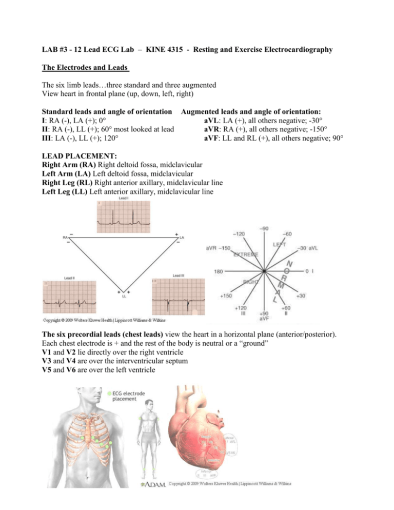
LAB #3 - 12 Lead ECG Lab – KINE 4315 - Resting and Exercise Electrocardiography The Electrodes and Leads The six limb leads…three standard and three augmented View heart in frontal plane (up, down, left, right) Standard leads and angle of orientation Augmented leads and angle of orientation: I: RA (-), LA (+); 0° aVL: LA (+), all others negative; -30° II: RA (-), LL (+); 60° most looked at lead aVR: RA (+), all others negative; -150° III: LA (-), LL (+); 120° aVF: LL and RL (+), all others negative; 90° LEAD PLACEMENT: Right Arm (RA) Right deltoid fossa, midclavicular Left Arm (LA) Left deltoid fossa, midclavicular Right Leg (RL) Right anterior axillary, midclavicular line Left Leg (LL) Left anterior axillary, midclavicular line The six precordial leads (chest leads) view the heart in a horizontal plane (anterior/posterior). Each chest electrode is + and the rest of the body is neutral or a “ground” V1 and V2 lie directly over the right ventricle V3 and V4 are over the interventricular septum V5 and V6 are over the left ventricle LEAD PLACEMENT: Right Arm (RA) Right deltoid fossa, midclavicular Left Arm (LA) Left deltoid fossa, midclavicular Right Leg (RL) Right anterior axillary, midclavicular line, to the right of the umbilicus Left Leg (LL) Left anterior axillary, midclavicular line, to the left of the umbilicus V1 4th interspace, just to the right of the sternum V2 4th interspace, just to the left of the sternum V3 Midway between V2 and V4 V4 5th interspace left midclavicular line V5 Left anterior axillary line on the same level as V4 V6 Left midaxillary line on the same level as V4 and V6 Skin Preparation for ECG – This is an essential step for recording high quality ECGs during exercise. Failure to prepare the skin properly and consistently for an exercise test will result in an ECG signal that cannot be continuously monitored or accurately interpreted because of artifact. 1. Have the patient disrobe to the waist and lie down face up on the prep table. (Women should wear a sports bra for this step). 2. Shaving may be required for men over the sites for the electrode placement. Place the shaver in the biohazard container when you are finished. 3. Identify the general areas to be used for electrode placement (consider marking each electrode location with an ‘X’ using a felt-tip pen). 4. Rub the skin with an abrasive pad such as sandpaper to remove the outer layer of the dead skin. The area should be red, but do not press too hard, as you will cause pain, remove the skin and/or cause bleeding. 5. Use an alcohol swab to remove any excess oils before marking the spot. 6. Electrode Application - The electrode gel at center of electrode should be placed directly over the area abraded. Smooth the adhesive part of the electrode with your finger to insure proper adhesion to the skin. Do NOT press on the center of the electrode. 7. Attach the lead wires from the ECG recorder to the patient electrodes. Caution: be sure to connect the lead wires to the proper electrodes. ECG Quick Reference Guide P wave: atrial depolarization Normal P wave = 0.04 sec. P wave should be upright. QRS Complex: ventricular depolarization Normal QRS = 0.08-0.12 sec = amplitude depends on situation T wave: ventricular repolarization Normal T wave = 1/3 -1/2 amplitude of QRS complex. T wave should be upright and not inverted PR interval: measures the time from the start of atrial depolarization to the start of ventricular depolarization. Normal: 0.12-0.20 seconds or 3-5 boxes. ST segment: records the time from the end of ventricular depolarization to the start of ventricular repolarization. Normal: Should be at the baseline or isoelectric line. QT interval: measures the time from the start of ventricular depolarization to the end of ventricular repolarization Rate, Rhythm, Axis Rate can be determined by counting the number of small boxes between two R waves and dividing them into 1500. Or a standard counting system can be used by labeling the number of large boxes between two R waves. See below: Rhythm refers to whether the strip is normal or has deviations. An abnormal ECG is called an arrhythmia. It may or may not be clinically significant. Several basic things to look for when evaluating rhythm. 1) Does every QRS have a P wave and is the P wave normal. 2) Is the QRS complex normal. Normal duration of the QRS is .08-.12 seconds or 2-3 boxes. 3) Is the rhythm regular or irregular. Meaning is the space between each R wave the same. Deviations can be caused by slightly heavy breathing but most often due to a AV node conduction delay problem. Axis The term axis refers to the direction of the mean electrical vector, representing the average direction of current flow. Normally the axis lies between 0-90 degrees showing the dominance of the left ventricle over the right ventricle. An abnormal or deviated axis is usually applied to left or right ventricular hypertrophy. Three things happen to a wave on the ECG when a chamber hypertrophies 1) The chamber may take longer to depolarize. Therefore the ECG wave will increase in duration. 2) The chamber may generate more current and thus a larger voltage. Therefore the wave may increase in amplitude. 3) The mean electrical vector (or the electrical axis) of the ECG may shift or deviate. Basic determination of axis is based on the QRS complex. If the QRS is predominately positive when the height of the complex above the isoelectric line is greater than that below. The converse is true when a QRS is negative. Lead I (0°) + + Normal Axis aVF (90°) + + Left Axis Deviation Axis Left axis deviation Right axis deviation Extreme right axis deviation!!! Normal Assignment: Prep one other student. Run a 12 lead supine ECG. Depending on the size of the lab you may work in groups of 4. Everyone should to prep another member of the class and evaluate their ECG. THIS IS NOT A GROUP PROJECT. Evaluate the rate, rhythm and the axis. Determine lengths as normal or abnormal when evaluating the P and T waves, QRS complexes, and PR intervals. Determine rate by either hand method. Show your work. Determine if there is any artifact? What do you think the cause of the artifact is? Use the data sheet provided. Turn in your EKG strips with the data sheet. Save this information for the practical. LAB #3: EKG Name:_______________________________ Subject’s name:________________________ Lying: Rate:________________________________ Rhythm:_____________________________ Pwave:________________________ PR interval length:_______________ QRS width:_____________________ T wave:________________________ Axis:________________________________ Artifact:______________________________ Date:___________________________
