DRE & ANOSCOPY - School of Medicine, Queen`s University
advertisement
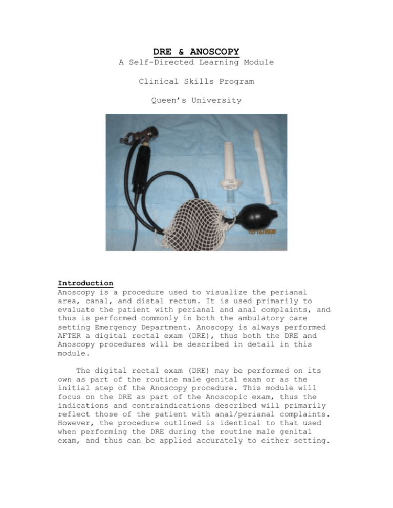
DRE & ANOSCOPY A Self-Directed Learning Module Clinical Skills Program Queen’s University Introduction Anoscopy is a procedure used to visualize the perianal area, canal, and distal rectum. It is used primarily to evaluate the patient with perianal and anal complaints, and thus is performed commonly in both the ambulatory care setting Emergency Department. Anoscopy is always performed AFTER a digital rectal exam (DRE), thus both the DRE and Anoscopy procedures will be described in detail in this module. The digital rectal exam (DRE) may be performed on its own as part of the routine male genital exam or as the initial step of the Anoscopy procedure. This module will focus on the DRE as part of the Anoscopic exam, thus the indications and contraindications described will primarily reflect those of the patient with anal/perianal complaints. However, the procedure outlined is identical to that used when performing the DRE during the routine male genital exam, and thus can be applied accurately to either setting. Objectives Describe the indications for DRE and Anoscopy Describe the contraindications for DRE and Anoscopy Describe the equipment and preparation required for these procedures Describe the stepwise approach to both procedures Describe potential complications of these procedures and the relevant prevention and management strategies Indications Initial evaluation of rectal bleeding (hemorroids, proctitis, neoplasm) Anal/perianal pain (thrombosed hemorrhoids, fissures) Perianal itching Abdominal pain Change in bowel habit Anal discharge/prolapse Palpable mass on DRE Retrieval of foreign body Evaluation of anal trauma Evaluation of fecal impaction Evaluation of intra-anal condyloma Treatment of prolapsing hemorrhoids by rubber band ligation (using a slotted anoscope) Contraindications Absolute: Imperforate Anus Unwilling patient Relative: Severe rectal pain may preclude an awake anoscopy and may require sedation Recent acute myocardial infarction Acute abdomen Patients with valvular heart disease or prosthetic valves require prophylactic antibiotics before the procedure Patients with coagulopathy (or anticoagulation medication) if biopsy is to be considered Major rectal trauma Post-operative status (anal surgery) Equipment The following is a list of the equipment required to perform both the DRE and Anoscopy: Gloves (3) Anoscope (one of 2 models) Light source (flashlight, headlight, or gooseneck lamp if anoscope not equipped with a light source) Lubricant Topical anesthetic if local discomfort is anticipated (lidocaine 2% jelly) Large tipped cotton swabs (these may be too large if using the small anoscope) Biospy forceps (if biopsy to be performed) Anoscope- the anoscope (pictured below) consists of 2 parts: a hollow tapered cylinder and an obturator that fits inside. The anoscope may be made of metal (reusable once sterilized), clear or white opaque plastic (single use, disposable). Clear plastic anoscopes may allow better visualization of mucosal tissues compared to opaque devices. The length of the anoscope ranges from 7 to 10 cm, and the tapered external diameter is approximately 1.9cm. Some anoscopes can be attached to a battery light source while others require an external source (gooseneck lamp, headlight). Another variety of anoscope is the slotted anoscope. This type is made of metal but has an open slot running the length of the device. This type is generally only used by surgeons to band hemorroids. You should be familiar with the equipment available at your institution prior to attempting to perform the procedure. A B C A- anoscope with obturator inserted, B- obturator, Couter cylinder of anoscope Patient Preparation Due to the nature and anatomical location of the procedure, it is natural that the patient will feel anxiety and embarrassment at the prospect of the DRE and Anoscopy. For the evaluation to be successful the patient must be cooperative and relaxed, thus adequate mental preparation for the patient is essential. Frank admission that the procedure will be unpleasant and uncomfortable, but not painful, is helpful. The patient should be warned they may experience a sensation of having to defecate during the exam. If the exam is to be performed to evaluate anal pain (e.g. anal fissure), the patient may be extremely tender and apprehensive. In addition, the anal sphincter contracts, making the examination difficult. In this setting, application of 2% lidocaine 5 minutes prior to the procedure can markedly reduce the patient's discomfort and make the examination easier to perform. The procedure must always be explained in detail to the patient, and consent must be obtained prior to any exam. Procedure In addition to this module, the DRE is described in detail in your Queen's Physical Examination Manual (blue book), and a Bates video is available for your viewing on the Clinical Skills website. It is recommended that all three resources be utilized to prepare for perfoming a DRE. 1. Patient positioning: Place the patient in the left lateral decubitus position with the patient's buttocks close to the near edge of the examining table (pictured below). The patients hips and knees should be flexed. Drape the patient so that only the buttocks is exposed. This position is adequate for most DREs and anoscopies. Note: this picture is meant only to illustrate the position of the patient. In reality, the patient may wear their shirt or a gown, and a drape should be placed over their legs so that only the buttocks is exposed. 2. DRE Having a nurse in the room for the procedure is recommended, they may act both as a chaperone as well as assistant. Don your gloves, placing 2 gloves on your dominant hand and one on your non-dominant hand. Utilize universal precautions as appropriate during exams. Inspection: Inspect the buttocks Use both hands to gently spread the buttocks apart to inspect the perianal and sacrococcygeal regions Note any lesions, lumps, ulcers, inflammation, rashes, or excoriations Ask the patient to bear down and observe for any hemorrhoid or mucosal prolapse Palpation: Apply lubricant to the index finger of your DOUBLE GLOVED hand and to the patient's anus Instruct the patient to relax their anal sphincter Once the anal sphincter is relaxed, insert your finger pointed towards the umbilicus with your palm facing towards the patient's head. Insert your finger slowly and in a step-wise fashion, feeling the anal canal initially and then advancing as far as possible Slowly rotate your hand clockwise to palpate the right side of the patient's rectum, then counterclockwise to palpate the left side of the patient's rectum Have the patient squeeze their anus and then bear down. Note sphincter tone, any tenderness, induration, irregularities, or nodules Palpation of the Prostate in Male Patients: Reassure the patient that when palpating the prostate they may feel the urge to urinate but that they will not do so. Then, turn your body so that you can further rotate your finger counterclockwise to palpate the posterior surface of the prostate gland. Identify the lateral lobes and medial sulcus Note the size, shape, and consistency Identify any nodules or tenderness Palpation of the Cervix in Female Patients: Inform the patient that you will be feeling for the cervix of the uterus. Note any tenderness, irregularities, and mass effect. Removal of the Finger: Ask the patient to strain, once the patient relaxes their sphincter, withdraw your finger from the anus and note the presence of fecal matter (its colour and consistency) and the presence of blood Remove and discard the now dirty glove from your dominant hand (taking care not to contaminate the clean one underneath) 3. Anoscopy Lubricate the anoscope well with the obturator in place Spread the buttocks and gently insert the anoscope (with obturator) into the anal canal o Asking the patient to take a few deep, gentle breaths and to bear down slightly may make the insertion easier Gently advance the instrument towards the umbilicus until the full length is inserted o If the patient complains of pain during insertion, note the location and quality and correlate the pain with clinical symptoms Remove the obturator and visualize the anal mucosa o Any fecal matter can be removed with a large swab o Note the gross appearance of mucous membranes and vasculature as well as the presence of pus, mucous, blood, ulceration, and hemorrhoidal tissue Slowly rotate the anoscope (with the obturator still removed) as it is withdrawn, inspect the anal canal as the device is extracted, looking for mass lesions, hemorrhoids or fissures. A clear plastic anoscope allows the examiner to visualize the mucosa both through the walls of the anoscope as well as at the opening of the device Masses or polyps visible through the anoscope should not be sampled as the instrument is too short to get a good appreciation of the extent of the mass lesion noted. A sigmoidoscope, either rigid or flexible is best used here. Once the device is fully withdrawn from the patient's anal canal, wipe away any excess lubricant with tissue Anal fissure (arrow) seen through the wall of a clear plastic anoscope Complications When performed properly, Anoscopy has few complications: Discomfort post examination Tearing of the perianal skin or mucosa Abrasion or tearing of hemorrhoidal tissue Infection post procedure is possible, but very rarely occurs Most discomfort and abrasion can be prevented by the adequate use of lubricant. If the patient is experiencing severe discomfort with the procedure, the use of local anesthetic can be considered or the procedure may be postponed. Infection is a very rare complication, prophylactic antibiotics can be considered in certain high risk populations (see relative contraindications). Self-Assessment Questions Question 1 Which of the following are indications for Anoscopy? A. Initial evaluation of rectal bleeding B. Peri-anal pain C. Change in bowel habit D. Evaluation of anal discharge E. All of the above Question 2 True or False The DRE is only performed prior to anoscopy in extenuating circumstances. Queston 3 Which of the following is NOT a contraindication to Anoscopy? A. Anal fissures B. Fecal incontinence C. Imperforate anus D. Unwilling patient Question 4 Which of the following statements is FALSE? A. A clear plastic anoscope may provide better visualization of the mucosa than the opaque devices B. Anoscopes consist of a tapered cylinder and a solid obturator that fits inside C. The anoscope should always be inserted without the obturator in the cylinder. The obturator is then placed in the cylinder to better visualize the mucosa D. The patient should be positioned in the left lateral decubitus position for both the DRE and the Anoscopy procedures Credits Congratulations! You have now completed the DRE & Anoscopy Module. Credits This module was written and developed by Nicole Rocca for the Queen's University Faculty of Health Sciences Patient Simulation Lab. Contributors: Dr. Paul Belliveau and Dr. Linda O'Connor The module was created using exe :eLearning XHTML editor with support the Queen's University School of Medicine MedTech Unit. License This module is licensed under the Creative Commons Attribution Non-Commercial No Derivatives license. The module may be redistributed and used provided that credit is given to the author and it is used for noncommercial purposes only. The contents of this presentation cannot be changed or used individually. For more information on the Creative Commons license model and the specific terms of this license, please visit creativecommons.ca. References 1. Muller AF: Proctoscopy, Sigmoidoscopy, and Rectal Biopsy. In Toghill PJ(eds): Essential Medical Procedures. New York, Arnold, 1997, p 48-52. 2. Barnes J: Proctoscopy, Sigmoidoscopy, and the Treatment of Piles. In Brown JS(eds): Procedures in General Practice. London, BMJ, 1997, p 132-142. 3. Jones DJ: Proctoscopy and Sigmoidoscopy. In Scott N(eds): Procedures in Practice. London, BMJ, 1994, p 244-252. 4. Tuggy ML, Garcia J. Procedures Consult. Anoscopy. 2007, Elsevier Inc. www.proceduresconsult.com 5. The content of this module was based, in part, on the Queen's Physical Examination Manual, 5th edition, Queen's University Clinical Skills Program 2007.
