The Lungs and Thorax
advertisement
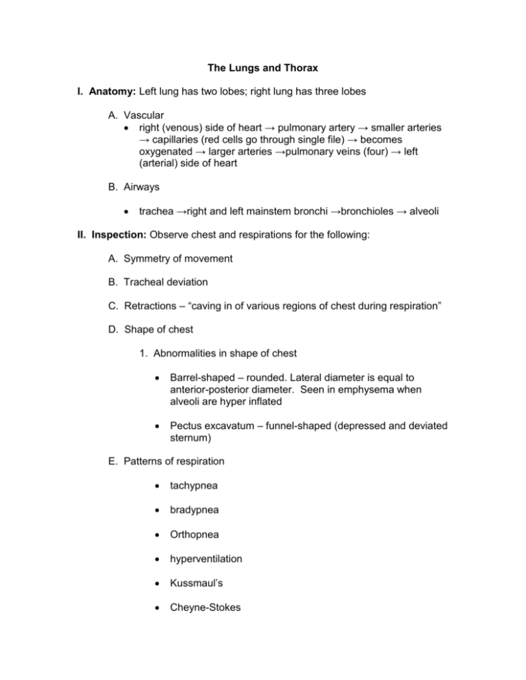
The Lungs and Thorax I. Anatomy: Left lung has two lobes; right lung has three lobes A. Vascular right (venous) side of heart → pulmonary artery → smaller arteries → capillaries (red cells go through single file) → becomes oxygenated → larger arteries →pulmonary veins (four) → left (arterial) side of heart B. Airways trachea →right and left mainstem bronchi →bronchioles → alveoli II. Inspection: Observe chest and respirations for the following: A. Symmetry of movement B. Tracheal deviation C. Retractions – “caving in of various regions of chest during respiration” D. Shape of chest 1. Abnormalities in shape of chest Barrel-shaped – rounded. Lateral diameter is equal to anterior-posterior diameter. Seen in emphysema when alveoli are hyper inflated Pectus excavatum – funnel-shaped (depressed and deviated sternum) E. Patterns of respiration tachypnea bradypnea Orthopnea hyperventilation Kussmaul’s Cheyne-Stokes III. Palpation A. Respiratory expansion 1. Place thumbs along each costal margin at the back 2. Slide hands medially to raise loose skinfolds between thumbs 3. Ask patient to inhale deeply 4. Note range and symmetry B. Tactile fremitus – use bony part of palm at base of fingers to palpate vibrations to chest wall when patient speaks. Ask to repeat “ninety-nine” or “one-one-one.” Vibrations are increased when lung tissue is consolidated (“Made solid” by virtue of being filled with mucus and secretions). This technique is not as valuable as assessing for bronchial breath sounds where they shouldn’t be IV. ercussion A. Used to determine diaphragmatic excursion which is the distance between levels of dullness on full expiration and full inspiration. Normally is 3-6 cm (textbooks vary) B. Assess posteriorly 1. ask patient to take a deep breath and hold it 2. percuss up from lower torso 3. note area where dullness changes to resonance 4. mark a line with skin pencil 5. then ask patient to exhale and hold it 6. percuss up till resonance again noted 7. mark line with skin pencil 8. normal diaphragmatic excursion is 3 to 6 cms (textbooks vary) V. Auscultation-assisted percussion A. Start at apex on the patient’s back (posterior thorax) B. Move to corresponding area on the other side C. Move back and forth between scapulae D. When below scapulae, move back and forth listening to corresponding area and comparing sounds E. Note patient’s anterior chest is not a reliable area for hearing abnormal breath sounds. Best to listen anteriorly and laterally too, but always listen posteriorly F. However, right middle lobe can only be heard anteriorly, so if patient has a pneumonia there, that is where you must listen G. Technique Listen to one respiratory cycle in each area Raise and lower your arm to indicate to the patient to breath in then out Instruct patient to breathe through the mouth Instruct patient to breathe deeper than normal Patient should be sitting if at all possible H. Normal breath sounds 1. Vesicular soft low pitched over peripheral lung fields where the alveoli are located inspiration longer than expiration 2. Bronchial – Listen over trachea anteriorly at the manubrium or posteriorly between the scapulae. Breath sound louder and higher in pitch. Expiratory sounds as long as inspiratory or expiratory may be longer than inspiratory. 3. Bronchovesicular – Heard over junction between bronchi and alveoli. Anteriorly, heard over bronchi in first and second interspaces. Inspiratory and expiratory phases equal in length. I. Abnormal (adventitious) sounds and associated pathophysiology 1. Discontinous sounds a. Crackles (râles in French) caused by airways popping open late, because of fluid or secretion accumulation fine, high-pitched heard at end of inspiration heard most often at bases in CHF, the higher up in the lung fields that crackles extend is an indication of the degree of failure may not indicate pathology in which case they clear with coughing in pneumonia, crackles will be heard over involved lobe 2. Continuous sounds a. Wheezes may be called sibilant wheeze are high-pitched and musical or whistling caused by air passing through narrowed airways such as in asthma most often heard on expiration, but may be heard throughout the respiratory cycle b. Rhonchi may be called sonorous wheeze are low-pitched, rumbling most often associated with air passing through thick secretions c. Bronchial breath sounds If these are heard over the peripheral lung (where vesicular breath sounds should be), that indicates consolidation (alveoli filled with fluid, pus, mucus) In the periphery (over alveoli) you should not hear expiration. If you do, that represents bronchial breath sounds and suggests consolidation d. Pleural friction rubs continuous grating sound like two pieces of leather being rubbed together caused by inflamed visceral and parietal pleura heard over inflamed area of lung may be intermittent e. Stridor crowing sound caused by inflammation/edema of laryngo-tracheal area – “croup” in children often accompanied by barking cough often occurs at night may be relieved by humidity (placing child in bathroom with shower on) or taking outside into cool night air can be heard in adults post-extubation needs to be reported/evaluated because of danger of edema obstructing airway completely f. Other abnormal sounds: If abnormalities are noted in tactile fremitus, percussion or auscultation, check spoken and whispered sounds using stethoscope. i) Bronchophony occurs when lungs are “solid” or filled wirh secretions (consolidation) ask patient to say “99” normally, sound is muffled over consolidated lungs, it is easily audible (bronchophony) ii) Egophony again, heard over consolidated lungs have patient say “e” and it will sound like “a” over consolidated lungs (egophony) it will sound like “e” over normal lungs iii) Whispered pectoriloquy with stethoscope over consolidated lungs, listen while patient whispers normally sound is soft with consolidation, it is easily audible VI. Signs of respiratory distress A. Tachypnea B. Retractions – pulling inward of intercostals cartilages, at the suprasternal notch or at the xiphoid process C. Nasal flaring D. Expiratory grunt VII. Common Abnormalities A. Asthma wheezes can be life-threatening – call healthcare provider unless you know patient history B. Atelectasis decreased breath sounds crackles probably a fever C. Bronchitis Rhonchi wheezes maybe, crackles “smoker’s cough” with chronic bronchitis D. Pleural effusion muted, distant, or decreased breath sounds E. Pneumonia crackles fever F. Influenza maybe as mild as common cold OR a deadly pneumonia crackles wheezes fever G. Pneumothorax decreased or absent breath sounds tympany on percussion H. Hemothorax muted, distant, or decreased breath sounds I. Chronic Obstructive Pulmonary Disease (COPD) “Pack-year” – one pack a day for 50 years is a 50 pack-year smoking history. Two packs a day for 25 years is a 50 pack-year smoking history, etc. A 50 pack-year history is almost certainly associated with COPD crackles wheezes Rhonchi dyspnea and Orthopnea often, barrel chest – especially with emphysema J. Croup (laryngotracheobronchitis) viral infection of laryngeal (larynx) area stridor over larynx
