Pediatric/Neonatal Intubation
advertisement
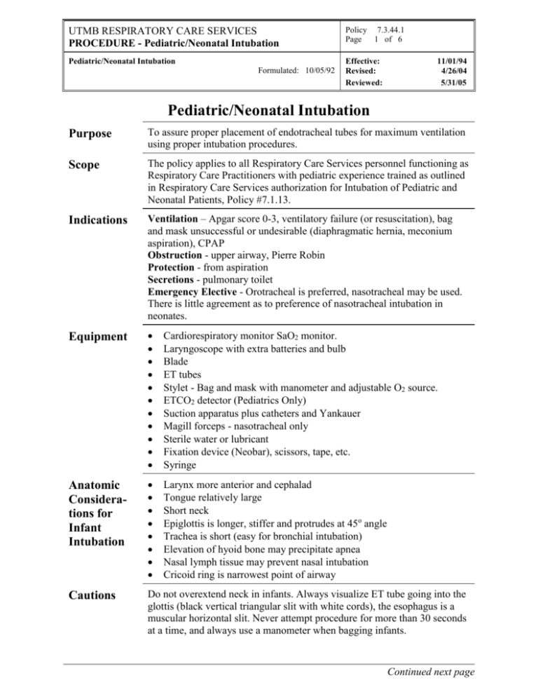
UTMB RESPIRATORY CARE SERVICES PROCEDURE - Pediatric/Neonatal Intubation Policy 7.3.44.1 Page 1 of 6 Pediatric/Neonatal Intubation Effective: Revised: Reviewed: Formulated: 10/05/92 11/01/94 4/26/04 5/31/05 Pediatric/Neonatal Intubation Purpose To assure proper placement of endotracheal tubes for maximum ventilation using proper intubation procedures. Scope The policy applies to all Respiratory Care Services personnel functioning as Respiratory Care Practitioners with pediatric experience trained as outlined in Respiratory Care Services authorization for Intubation of Pediatric and Neonatal Patients, Policy #7.1.13. Indications Ventilation – Apgar score 0-3, ventilatory failure (or resuscitation), bag and mask unsuccessful or undesirable (diaphragmatic hernia, meconium aspiration), CPAP Obstruction - upper airway, Pierre Robin Protection - from aspiration Secretions - pulmonary toilet Emergency Elective - Orotracheal is preferred, nasotracheal may be used. There is little agreement as to preference of nasotracheal intubation in neonates. Equipment Cardiorespiratory monitor SaO2 monitor. Laryngoscope with extra batteries and bulb Blade ET tubes Stylet - Bag and mask with manometer and adjustable O2 source. ETCO2 detector (Pediatrics Only) Suction apparatus plus catheters and Yankauer Magill forceps - nasotracheal only Sterile water or lubricant Fixation device (Neobar), scissors, tape, etc. Syringe Anatomic Considerations for Infant Intubation Larynx more anterior and cephalad Tongue relatively large Short neck Epiglottis is longer, stiffer and protrudes at 45o angle Trachea is short (easy for bronchial intubation) Elevation of hyoid bone may precipitate apnea Nasal lymph tissue may prevent nasal intubation Cricoid ring is narrowest point of airway Cautions Do not overextend neck in infants. Always visualize ET tube going into the glottis (black vertical triangular slit with white cords), the esophagus is a muscular horizontal slit. Never attempt procedure for more than 30 seconds at a time, and always use a manometer when bagging infants. Continued next page UTMB RESPIRATORY CARE SERVICES PROCEDURE - Pediatric/Neonatal Intubation Policy 7.3.44.1 Page 2 of 6 Pediatric/Neonatal Intubation Effective: Revised: Reviewed: Formulated: 10/05/92 Complications 11/01/94 4/26/04 5/31/05 Insertion Hypoxia (maximum 15 seconds) Trauma (poor technique), hemorrhage, broken teeth, spinal cord damage Aspiration (gag reflex), vomitus, blood, arrhythmias/ hypotension vagal stimulation, apnea, bronchospasm/laryngospasm Improper Position In esophagus In pharynx In right mainstream Beveling at carina. Obstruction Secretions Kinking Cuff herniation Cuff Leak - not enough air, tube too small, hole in cuff or balloon, increased tracheal diameter (malacia) Overinflation - necrosis (Tracheomalacia) tracheal stenosis, fistula, vessel rupture), cuff herniation Improper Care Contamination Oral/nasal necrosis Body Response Edema, BPD Granulomas Cord paralysis Atelectasis Barotrauma Tracheal & pharyngeal perforation Eliminates physiological PEEP Procedure Continued Orotracheal Procedure: Step Action 1 Assemble and prepare equipment: Insure scope light, suction and bag & mask works. Select 2-3 tube sizes. Check cuff function. Cardiorespiratory monitoring is a must. Continued next page UTMB RESPIRATORY CARE SERVICES PROCEDURE - Pediatric/Neonatal Intubation Policy 7.3.44.1 Page 3 of 6 Pediatric/Neonatal Intubation Effective: Revised: Reviewed: Formulated: 10/05/92 Procedure Continued 11/01/94 4/26/04 5/31/05 Heat source for infant. Orotracheal Procedure: Step Action 2 Prepare and Identify Patient: Infant- Sniff position (rolled towel under shoulders), supine - do not hyperextend Older child- Hyperextension is preferred Suction oropharynx and nasopharynx if needed Ventilate and oxygenate- For 1 minute if possible Hyperventilate with enriched O2 (100% is most commonly used) Pressure: 20-30 cm H2O Do not bag if: meconium aspiration, diaphragmatic hernia, upper airway obstruction Monitor vital signs and SaO2 3 Intubate - Position self at patient's head, hold scope in left hand, open mouth with fingers (not blade), insert blade into right side of mouth, move blade to center of mouth pushing tongue to the left side, slowly advance blade and lift epiglottis till larynx is visualized. If esophagus is seen first, withdraw blade slightly. Position curved blade under top of epiglottis and lift. 4 Visualization may be improved by: Suctioning of pharynx Gentle "lifting" of the scope Gentle pressure on the hyoid bone 5 Insert ET tube into right side of mouth using right hand and pass alongside of blade (not through the groove). Advance tube 1-2 cm through cords while maintaining visualization. A stylet may be used but ensure it is at least 1 cm back from tip. 5 Hold tube in place (note position) and gently withdraw laryngoscope Inflate cuff (larger tubes) Attach bag with ETCO2 detector and hyperventilate and oxygenate (Pedi only) Continued next page UTMB RESPIRATORY CARE SERVICES PROCEDURE - Pediatric/Neonatal Intubation Policy 7.3.44.1 Page 4 of 6 Pediatric/Neonatal Intubation Effective: Revised: Reviewed: Formulated: 10/05/92 Procedure continued Orotracheal Procedure Continued: Step Procedure 11/01/94 4/26/04 5/31/05 Action 6 Confirm position by: ETCO2 detector(Pedi only) Auscultation of chest and stomach Chest excursion Improved color Improved heart rate Improved SaO2 Stat x-ray All the above should occur within seconds 7 Correct position is: 1-2 cm above carina (children), midway between carina and clavicle in small infants Listen at L mid-axillary, advance tube until BS (in mainstem bronchus), then pull back the appropriate distance. 8 Secure tube: refer to policy 7.3.39. Cut tube 1 cm above tape (after confirming x-ray) A small leak should exist around cuffless tube. (@ 10 cm H2O pressure) Nasotracheal Intubation: Step 1 Action Same as orotracheal with following exceptions: ET tube is lubricated with H2O soluble lubricant ET tube is inserted via nose Keep orotracheal tube in place (if already present): when the nasotracheal tube is just above the glottis, have an assistant pull the oral tube out, apply gentle pressure to hyoid bone and advance the tube through the cords. 2 A Magill forceps is often cumbersome in neonate but may be used to guide the tube. Continued next page UTMB RESPIRATORY CARE SERVICES PROCEDURE - Pediatric/Neonatal Intubation Policy 7.3.44.1 Page 5 of 6 Pediatric/Neonatal Intubation Effective: Revised: Reviewed: Formulated: 10/05/92 Documentation 11/01/94 4/26/04 5/31/05 Document on Respiratory Care Services Flow Sheet and Treatment Card per Respiratory Care Services Policies # 7.1.1and # 7.1.2. Continued next page UTMB RESPIRATORY CARE SERVICES PROCEDURE - Pediatric/Neonatal Intubation Policy 7.3.44.1 Page 6 of 6 Pediatric/Neonatal Intubation Effective: Revised: Reviewed: Formulated: 10/05/92 11/01/94 4/26/04 5/31/05 Infection Control Follow procedures outlined in Healthcare Epidemiology Policies and Procedures #2.24; Respiratory Care Services. http://www.utmb.edu/policy/hcepidem/search/02-24.pdf References AARC Clinical Practice Guidelines, Management of Airway Emergencies Respiratory Care, 1995; 40(7); 749-760 van den Berg AA, Mphanza T. Choice of tracheal tube size for children: finger size or age-related formula? Anaesthesia. 1997; 52:701-703. Shukla HK, Hendricks-Munoz KD, Atakent Y, Rapaport S. Rapid estimation of insertional length of endotracheal intubation in newborn infants. Journal of Pediatrics. 1997; 131:561-564. Khine HH, Corddry DH, Kettrick RG, et al. Comparison of cuffed and uncuffed endotracheal tubes in young children during general anesthesia. Anesthesiology. 1997; 86:627-631. Roth B, Lundberg D. Disposable CO2-detector, a reliable tool for determination of correct tracheal tube position during resuscitation of a neonate. Resuscitation. 1997; 35:149-150 Rivera R, Tibballs J. Complications of endotracheal intubation and mechanical ventilation in infants and children. Critical Care Medicine. 1992; 20:193-1999.
