spine
advertisement
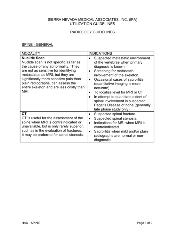
SIERRA NEVADA MEDICAL ASSOCIATES, INC. (IPA) UTILIZATION GUIDELINES RADIOLOGY GUIDELINES SPINE - GENERAL MODALITY Nuclide Scan Nuclide scan is not specific as far as the cause of any abnormality. They are not as sensitive for identifying metastases as MRI, but they are significantly more sensitive pain than plain radiographs, can assess the entire skeleton and are less costly than MRI. CT CT is useful for the assessment of the spine when MRI is contraindicated or unavailable, but is only rarely superior, such as in the evaluation of fractures. It may be preferred for spinal stenosis. RAD - SPINE INDICATIONS Suspected metastatic environment of the vertebrae when primary diagnosis is known. Screening for metastatic involvement of the skeleton. Occasional cases of sacroiilitis (quantitative imaging is more accurate) To localize level for MRI or CT In attempt to quantitate extent of spinal involvement in suspected Paget’s Disease of bone (generally late phase study only). Suspected spinal fracture. Suspected spinal stenosis. Indications for MRI when MRI is contraindicated. Sacroiilitis when mild and/or plain radiographs are normal or nondiagnostic. Page 1 of 2 MRI MRI is best for the evaluation of known or possible tumors, including primary bone tumors, skeletal metastases and intradural masses. It can provide a non-invasive, nonradiating serial evaluation of the response to anti-cancer treatment. It is best to show infection such as spondylitis, osteomyelitis, discitis, and abscesses. It is best to show the demyelination of multiple sclerosis if the spinal cord is involved and MRI of the brain is negative. It is best to diagnose spinal disc degeneration, distinguish it from facet or spur problems, and to diagnose spinal cord diseases. It may be preferred for spinal stenosis. It eliminates the need for myelography to diagnose metastases and may make it unnecessary prior to disc surgery. It is best for all levels of the spine. In general, MRI scans of the spine should be preceded by plain radiographs. RAD - SPINE Suspected vertebral, paraspinal or intraspinal metastases or infections. Suspected primary bone tumors. Suspected multiple sclerosis or spinal cord disease (myelopathy). Suspected disc herniation, based on progressive and persistent symptoms of back and leg pain, four to eight weeks without improvement. Radiculopathy of unknown cause. Suspected spinal stenosis when low back pain is progressive and heaviness after exercise is relieved by rest. Follow-up of treatment for malignancy or infection. Evaluation of recurrent radicular symptoms after spinal surgery (do plain films and/or CT if only back pain). Sacroiilitis (some cases); mainly for sclerosis. MRI after spinal surgery should be performed with/without gadolinium injection. Cord compression. Page 2 of 2
