Management of Head Trauma: - Waterworks Road Vet Surgery
advertisement
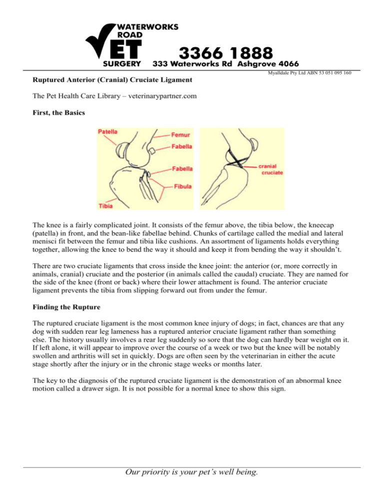
Myalldale Pty Ltd ABN 53 051 095 160 Ruptured Anterior (Cranial) Cruciate Ligament The Pet Health Care Library – veterinarypartner.com First, the Basics The knee is a fairly complicated joint. It consists of the femur above, the tibia below, the kneecap (patella) in front, and the bean-like fabellae behind. Chunks of cartilage called the medial and lateral menisci fit between the femur and tibia like cushions. An assortment of ligaments holds everything together, allowing the knee to bend the way it should and keep it from bending the way it shouldn’t. There are two cruciate ligaments that cross inside the knee joint: the anterior (or, more correctly in animals, cranial) cruciate and the posterior (in animals called the caudal) cruciate. They are named for the side of the knee (front or back) where their lower attachment is found. The anterior cruciate ligament prevents the tibia from slipping forward out from under the femur. Finding the Rupture The ruptured cruciate ligament is the most common knee injury of dogs; in fact, chances are that any dog with sudden rear leg lameness has a ruptured anterior cruciate ligament rather than something else. The history usually involves a rear leg suddenly so sore that the dog can hardly bear weight on it. If left alone, it will appear to improve over the course of a week or two but the knee will be notably swollen and arthritis will set in quickly. Dogs are often seen by the veterinarian in either the acute stage shortly after the injury or in the chronic stage weeks or months later. The key to the diagnosis of the ruptured cruciate ligament is the demonstration of an abnormal knee motion called a drawer sign. It is not possible for a normal knee to show this sign. Our priority is your pet’s well being. Myalldale Pty Ltd ABN 53 051 095 160 The Drawer Sign The veterinarian stabilizes the position of the femur with one hand and manipulates the tibia with the other hand. If the tibia moves forward (like a drawer being opened), the cruciate ligament is ruptured. Another method is the tibial compression test where the veterinarian stabilizes the femur with one hand and flexes the ankle with the other hand. If the ligament is ruptured, again the tibia moves abnormally forward. If the rupture occurred some time ago, there will be swelling on side of the knee joint that faces the other leg. This is called a medial buttress and is a sign that arthritis is well along. It is not unusual for animals to be tense or frightened at the vet’s office. Tense muscles can temporarily stabilize the knee, preventing demonstration of the drawer sign during examination. Often sedation is needed to get a good evaluation of the knee. This is especially true with larger dogs. Eliciting a drawer sign can be difficult if the ligament is only partially ruptured so a second opinion may be a good idea if the initial examination is inconclusive. Since arthritis can set in relatively quickly after a cruciate ligament rupture, radiographs to assess arthritis are helpful. Another reason for radiographs is that occasionally when the cruciate ligament tears, a piece of bone where the ligament attaches to the tibia breaks off as well. This will require surgical repair and the surgeon will need to know about it before beginning surgery. Arthritis present prior to surgery limits the extent of the recovery after surgery though surgery is still needed to slow or even curtail further arthritis development. How Rupture Happens Several clinical pictures are seen with ruptured cruciate ligaments. One is a young athletic dog playing roughly who takes a bad step and injures the knee. This is usually a sudden lameness in a young largebreed dog. A recent study identified the following breeds as being particularly at risk for this phenomenon: Neapolitan mastiff, Newfoundland, Akita, St. Bernard, Rottweiler, Chesapeake Bay retriever, and American Staffordshire terrier. On the other hand, an older large dog, especially if overweight, can have weakened ligaments and slowly stretch or partially tear them. The partial rupture may be detected or the problem may not become apparent until the ligament breaks completely. In this type of patient, stepping down off the bed or a small jump can be all it takes to break the ligament. The lameness may be acute but have features of more chronic joint disease or the lameness may simply be a more gradual/chronic problem. Larger overweight dogs that rupture one cruciate ligament frequently rupture the other one within a year's time. An owner should be prepared for another surgery in this time frame. Our priority is your pet’s well being. Myalldale Pty Ltd ABN 53 051 095 160 What Happens if the Cruciate Rupture is Not Surgically Repaired (almost) normal damaged Without an intact cruciate ligament, the knee is unstable. Wear between the bones and meniscal cartilage becomes abnormal and the joint begins to develop degenerative changes. Bone spurs called osteophytes develop resulting in chronic pain and loss of joint motion. This process can be arrested or slowed by surgery but cannot be reversed. Osteophytes are evident as soon as 1 to 3 weeks after the rupture in some patients. This kind of joint disease is substantially more difficult for a large breed dog to bear, though all dogs will ultimately show degenerative changes. Typically, after several weeks from the time of the acute injury, the dog may appear to get better but is not likely to become permanently normal. In one study, a group of dogs was studied for 6 months after cruciate rupture. At the end of 6 months, 85% of dogs less than 30 pounds of body weight had regained near normal or improved function while only 19% of dogs over 30 pounds had regained near normal function. Both groups of dogs required at least 4 months to show maximum improvement. What Happens in Surgical Repair? There are four different surgical repair techniques commonly used. Extracapsular Repair Our priority is your pet’s well being. Myalldale Pty Ltd ABN 53 051 095 160 This surgery is currently favored as it can be performed in a relatively shorter time than the other procedures and does not require specialized equipment. The knee joint is opened and inspected. The torn or partly torn cruciate ligament is removed. Any bone spurs of significant size are bitten away with an instrument called a rongeur. If the meniscus is torn, the damaged portion is removed. A large, strong suture is passed around the fabella behind the knee and through a hole drilled in the front of the tibia. This tightens the joint to prevent the drawer motion, effectively taking over the job of the cruciate ligament. Typically, the dog may carry the leg up for a good 2 weeks after surgery but will increase knee use over the next 2 months, eventually returning to normal. Typically, the dog will require 8 weeks of exercise restriction after surgery (no running, only outside on a leash, including the backyard). The suture placed will break 2 to 12 months after surgery and the dog's own healed tissue will hold the knee. Tibial Plateau Leveling Osteotomy (TPLO) Lateral orthopedic wire is shown taking the place of the anterior cruciate ligament. Usually thick suture is used rather than wire but for illustrative purposes the wire shows where the suture would This procedure uses a fresh be placed around the approach to the knee. biomechanics of the knee joint and is meant to address the lack of success seen with the above technique long term in larger dogs. Unfortunately, the technique has not yielded clearly superior results and controversy continues. With this surgery, the tibia is cut and rotated in such a way that the natural weight-bearing of the dog actually stabilizes the knee joint. As before, the knee joint still must be opened and damaged meniscus removed. The cruciate ligament remnants may or may not be removed depending on the degree of damage. This surgery is complex and involves specific training in this technique. Many radiographs are necessary to calculate the angle of the osteotomy (the cut in the tibia). This procedure typically costs substantially more than the extracapsular repair as it is more invasive to the joint. Typically, most dogs are touching their toes to the ground by 10 days after surgery, although it can take up to 3 weeks. As with other techniques, 8 to 12 weeks of exercise restriction are needed. Full function is generally achieved 3 to 4 months after surgery and the dog may return to normal activity. Tibial Tuberosity Advancement (TTA) Our priority is your pet’s well being. Myalldale Pty Ltd ABN 53 051 095 160 The TTA represents another take on how to use the biomechanics of the knee to create stabilization. The theory behind this procedure is that when the cruciate ligament is torn, the tibial plateau (the top of the tibia) and the patellar ligament should be repositioned at 90 degrees to one another to combat the shear force generated as the dog walks. To make this happen, the tibial tuberosity (front of the tibia where the patellar ligament attaches) is separated and anchored in its new position by a titanium or steel cage, “fork,” and plate. Bone grafts are used to assist healing. This procedure was developed in 2002 at the University of Zurich and since then over 20,000 patients worldwide have had this surgery. Some experts prefer it to the TPLO while others prefer the TPLO. Both procedures require specific equipment and expertise and, again, no procedure has demonstrated clear superiority; controversy continues. Typically the leg is bandaged for a week after surgery. The patient's activity must be restricted and confinement is a must with gradually increased activity over 3 to 4 months. Most dogs can return to normal activity by 4 months after surgery. Triple Tibial Osteotomy (TTO) A thin wedge of bone on the front of the tibia is cut from its position and pushed forward so that it sits slightly away from the tibia. A second and third cut is made into the tibia, to remove a wedge of bone. When this wedge of bone is removed the bone leans forward, causing the top of the tibia to create a flat surface across the top. The bones are held in this position with a metal bone plate specifically shaped to fit the bone and screwed in place on the inside of the leg. The purpose of these last three steps is to create a flat top to the tibia, stopping the femur and tibia wanting to slide against one another, so minimizing damage in the joint. Again, the dog may not bear weight for a good 2 weeks after surgery and will likely require 2 months to return to normal function. Again, 8 to 12 weeks of exercise restriction will be necessary for healing. General Rehabilitation after Surgery Our priority is your pet’s well being. Myalldale Pty Ltd ABN 53 051 095 160 Rehabilitation following the extracapsular repair method can begin as soon as the pet goes home. The area can be chilled with a padded ice pack for 10 minutes a couple times daily. (Do not try to make up for a skipped treatment by icing the area longer; prolonged cold exposure can cause injury.) Passive range of motion exercise where the knee is gently flexed and extended can also help. It is important not to induce pain when moving the limb. Let the patient guide you. Avoid twisting the leg. After the stitches or staples are out (or after the skin has healed in about 10 to 14 days), water treadmill exercise can be used if a facility is available. This requires strict observation and, if possible, the patient should wear a life jacket. Rehabilitation for patients with intracapsular repair is similar but slower in progression. Rehabilitation after TPLO, TTO or TTA is gentler. Icing as above and rest are the main modes of therapy. After 3 to 4 weeks, an increase in light activity can be introduced. A water treadmill is helpful. No jumping, running or stair-climbing is allowed at first. Expect the osteotomy site to require a good 6 weeks to heal. What if the Rupture Isn't Discovered for Years and Joint Disease is Already Advanced? A dog with arthritis pain from an old cruciate rupture may still benefit from a TPLO surgery and possibly from the TTA. Ask your veterinarian if it may be worth having a surgery specialist take a look at the knee. Most cases must make do with medical management. Meniscal Injury We mentioned the menisci as part of the knee joint. The bones of all joints are capped with cartilage so as provide a slippery surface where the bones contact each other (if the bones contact each other without cartilage, they grind each other down). In addition to these cartilage caps, the stifle joint has two “blocks” of cartilage in-between the bones. These blocks are called the menisci and serve to distribute approximately 65% of the compressive load delivered to the knee. The only other joint with a meniscus is the jaw (tempero-mandibular joint). When the crucial ligament ruptures, the medial (on the inner side of the knee) meniscus frequently tears and must either be removed, partly removed, or ideally repaired. This is generally done at the time of cruciate ligament surgery and we would be remiss not to mention it. Pets with meniscal damage may have an audible clicking sound when they walk or when the knee is examined, but for a definitive diagnosis the menisci must actually be inspected during surgery. It is difficult to access the menisci and thus repairing a tear in the meniscus is problematic; furthermore, poor blood supply to the menisci also makes good healing less likely. For these reasons, removal of the damaged portion of the meniscus is the most common surgical choice. This leaves some meniscus behind to distribute the compression load on the knee but removes the painful, ineffective portion. Areas of current research include techniques to improve blood supply to the healing meniscus so that repair can be more feasible. If meniscal damage has occurred in a cruciate rupture, arthritis is inevitable and surgery should be considered a palliative procedure. Our priority is your pet’s well being.
