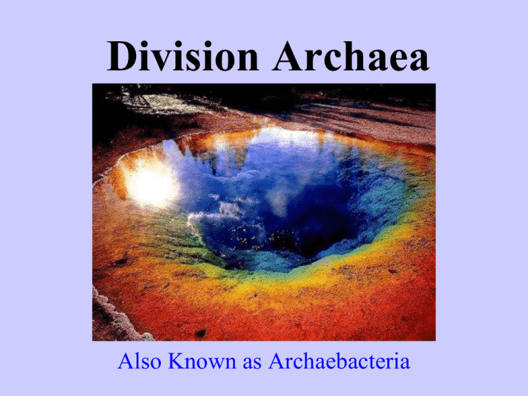
Division Archaea
Also Known as Archaebacteria
Typical conditions on Earth today are
comfortable for Typical Life Forms
Average Temperature −20 to 36°C (−4 to 97°F)
Pressure - 1 atm (sea level) to 0.5 atm (5500m)
Salinity - Oceans are 3.5% salt
Acidity - Neutral (pure water) to slightly alkaline (sea water)
Radiation - Low background: 0.003 Joules/kg/year
Location - Land or Sea
Introduction to Extremophiles
What
are they?
Microbes that live where conditions are extremely harsh.
Definition
- Lover of extremes
Extremophile archaea are members of 6 main physiological
groups.
Anaerobic Environments
High Salt Environments
Acid Environments
Alkali Environments
High Heat Environments
Extreme Cold Environments
General Characteristics of Archaea
Unicellular
Prokaryotes
First
appeared on Earth 3.5 GA, oldest living
organisms on the planet.
Many are Chemoautotrophic and are capable of
producing foods from inorganic molecules such
as ammonia or sulfur dioxide.
The Archaea are Extremophiles
The
Extremophiles
are found in
extremely harsh
environmental
conditions.
Life at High Temperatures
Extremophiles
Images from NASA, http://pds.jpl.nasa.gov/planets/
Methanogens (Anaerobes)
Found
in anaerobic
environments
Bacteria that produce
methane (CH4)
The methanogens are found in
mud, swamps (methane is
sometimes called “swamp
gas”) and in the intestines of
humans, cows, termites and
other animals.
Chemical Extremes
Chemical Extremes
Halophiles
– “salt lovers” High Salt Environments
– Natural salt lakes and manmade pools
– Sometimes occurs with extreme alkalinity
Alkaliphiles
– “base lovers” Alkaline Environments
pH 8 -14
Acidophiles
– “acid-lovers” Acid Environments
pH 1 - 6
Halophiles
Halophiles
are found in salt marshes, subterranean
salt deposits, dry soils, salted meats, hypersaline seas,
and salt evaporation pools.
Live in water with 10x the salinity of the oceans
Dead Sea & Great Salt Lake
Extra ions reduce osmotic pressure, stopping
desiccation
Implications: Salt seas on the early Mars & presentday Europa?
High salinity (too much salt) is bad for
most organisms
High
osmotic pressure draws out
water from the cells
Inhibits
Limits
Protein Function
the availability of oxygen
for respiration
Halophiles
solar salterns,
Owens Lake,
Great Salt Lake
The
Red Sea was named after
halobacterium that turns the water red
during massive blooms.
Facts
The
term “red herring” comes from the foul
smell of salted meats that were spoiled by
halophiles.
Alkaliphiles
”base-lovers”
pH 7.5 - 14
Some efficiently
neutralize
their cellular interiors
Other alkaliphiles
have evolved
base-stable proteins.
e.g. Mono Lake
alkaline soda lake, pH 9
salinity 8%
Acidophiles - “acid-lovers”
pH 0 -6.5
Some efficiently neutralize
their cellular interiors
Other acidophiles have
evolved
acid-stable proteins.
archaea recently discovered in a
mine drainage system high in acids.
Extreme Temperatures
Extreme Temperatures
– “heat and acid lovers” live
in high temperature, acidic environments
Thermoacidophiles
– Thermal vents and hot springs
– May go hand in hand with chemical extremes
– “cold lovers” live in low
temperature environments
Psychrophiles
– Arctic and Antarctic
Why High temperatures are bad for most
organisms
Degrades
chlorophyll, stopping photosynthesis
Decreases
the solubility of CO2 and
O2 in water
Denatures proteins, causing them to stop working
Have
proteins & enzymes that work at high
temperatures
Thermoacidophiles
Obsidian Pool,
Yellowstone National Park
Temp. 230 °F
Oceanic Hydrothermal Vents
Temp. 480 °F
Why Cold temperatures are bad for
most organisms
Freezing
damages cells
Increased viscosity, limiting mobility of nutrients &
waste
Proteins
and enzymes get stiff, inhibiting their
function
Found in glaciers, arctic ice, snow, & soil, and deep
oceans
Flexible enzymes and protein antifreeze
Psychrophiles
Alan Hills Ice Field: Antarctica
Some archaea have been found 2 miles deep in ice core samples
Division Bacteria
Includes Kingdom Monera
Quick Review
Remember from earlier this year that there
are two broad categories of organisms:
*Prokaryotes – have No membrane
bound organelle
*Eukaryotes – have membrane bound
organelle
General Characteristics
Prokaryotes
(No membrane-bound organelle)
Prokaryotes have a single, naked chromosome
Some Prokaryotes have Plasmids, circular DNA
Unicellular (single-cell)
Cell Walls can contain peptidoglycan, not cellulose
First appeared approximately 3.5 GA
Flagella, if present, made up of Flagellin.
Flagella do NOT have a 9 + 2 Structure
Typical Prokaryote Cell Structure
Internal Structure: Bacteria have a very simple
internal structure, and no membrane-bound
organelles.
Nucleoid
DNA in the bacterial cell is generally confined to this central region. Though it isn't bounded by
a membrane, it is visible.
Ribosomes
Ribosomes give the cytoplasm of bacteria a granular appearance in electron micrographs.
Though smaller than the ribosomes in eukaryotic cells, they have a similar function in
translating the genetic message in messenger RNA into the production of proteins.
Storage granules
Nutrients and reserves may be stored in the cytoplasm in the form of glycogen, lipids, and
sugars.
Endospore
Some bacteria, like Clostridium botulinum, form spores that are highly resistant to drought, high
temperature and other environmental hazards. Once the hazard is removed, the spore germinates
to create a new population.
Capsule
This layer of polysaccharide (sometimes proteins) protects the bacterial cell and is often
associated with pathogenic bacteria because it serves as a barrier against phagocytosis by white
blood cells.
Outer membrane
(not shown) This lipid bilayer is found in Gram negative bacteria and is the source of
lipopolysaccharide (LPS) in these bacteria. LPS is toxic and turns on the immune system of ,
but not in Gram positive bacteria.
Cell wall
Composed of peptidoglycan (polysaccharides + protein), the cell wall maintains the overall
shape of a bacterial cell. The three primary shapes in bacteria are coccus (spherical), bacillus
(rod-shaped) and spirillum (spiral). Mycoplasma are bacteria that have no cell wall and
therefore have no definite shape.
Plasma membrane
This is a lipid bilayer much like the cytoplasmic (plasma) membrane of other cells. There are
numerous proteins moving within or upon this layer that are primarily responsible for transport
of ions, nutrients and waste across the membrane.
Pili
These hollow, hairlike structures made of protein allow bacteria to attach to other
cells. A specialized pilus, the sex pilus, allows the transfer from one bacterial cell to
another. Pili (sing., pilus) are also called fimbriae (sing., fimbria).
Flagella
The purpose of flagella (sing., flagellum) is motility. Flagella are long appendages
which rotate by means of a "motor" located just under the cytoplasmic membrane.
Bacteria may have one, a few, or many flagella in different positions on the cell.
Bacteria
Escherichia coli
E. coli
Oxygen Preferences for Bacteria
obligate
aerobes must have oxygen
obligate anaerobes cannot live in oxygen
facultative anaerobes can grow with or
without oxygen
Nutrition
Autotrophs- manufacture organic compound
Photoautotrophs- use light energy H20 & CO2
Chemoautotrophs-use inorganic substances like
H2S, NH3, and other nitrogen compounds
Heterotrophs- obtain energy by consuming
organic compounds
– parasites- get energy from living organisms
– saprobes (saprophytes)- get energy from dead,
decaying matter; also called decomposers
Endospores
thick-walled
structures that are
highly resistant to
harsh environmental
conditions (high
temperature, drying,
oxygen, etc.);
generally formed only
by bacilli, and then
each cell only forms
one.
endospore
Locomotion (Methods of Movement)
Bacterial
Flagellum- lacks microtubules
Classification
Considerations
Gram-staining characteristics
Cell shapes and Groups
Methods of obtaining energy
Chemical Composition of the Cell Walls
Gram Staining
Gram-negative
cells lack the ability to
retain the deep violet dye because they have
little, if any, peptidoglycan in their cell
walls.
Gram-positive
cells have cell walls with
large amounts of peptidoglycan which
retain the deep violet dye and gives the cell
a purple color.
Gram Negative cell
Gram Positive Cells
Pink
Purple
Bacteria Photos
E. coli
Which of these cells are
Gram +, Gram - ?
Clostridium tetani
Bacteria Photos
Staphylococcus
aureus
Neisseria
gonorrhoeae
Cell Shapes and Groups
Spherical-shaped cells
Coccus (sng) , cocci (pl)
A Group of Two is referred to as:
Diplo…….. This is diplococccus
A Cluster of cells is referred to as:
Staphylo…. This is Staphylococcus
What a slide of Typical coccus
looks like in a microscope.
Streptococcus aurelius
Strep Throat
Staph Infection
Rod-shaped cells
Bacillus (sng) , Bacilli (pl)
Typical Bacillus
Bacillus
http://er1.org/docs/photos/Anthrax/bacillus%20anthracis%20-03.jpg
Typical Bacillus in a Microscope
Spiral-shaped cells
Spirillum (sng) , Spirlli (pl)
Spirochetes
Cyanobacteria
are
photosynthetic autotrophs that produce
carbohydrates and oxygen
tend
to cling together in filaments or colonies
The
“heterocysts contain enzymes that allow
them to “fix” atmospheric nitrogen
Filamentous: Chain of cells
http://www.spea.indiana.edu/joneswi/e455/Anabaena.jpg
Anabaena
_ http://www.bio.mtu.edu/~jkoyadom/algae_webpage/ALGAL_IMAGES/cyanobacteria/Anabaena_jason_dbtow17 2016.jpg
Some filamentous cyanobacteria have Heterocysts:
which are Nitrogen-fixing structures
http://www.people.vcu.edu/~elhaij/IntroBioinf/Scenarios/heterocyst2.JPG
Oscillatoria
http://botit.botany.wisc.edu:16080/images/130/Bacteria/Cyanobacteria/Oscillatoria/Oscillatoria_MC.jpg
Nitrogen-fixation
Some
soil bacteria live in the ground
and take in Nitrogen from the
surroundings
The
Nitrogen is combined with oxygen
to form nitrites and nitrates…. Plants
use the nitrates and nitrites to make
proteins…. (Grow !!)
Denitrification
Some
soil bacteria break down
the nitrogen compounds and
release the nitrogen back into
the environment.
Life
would not exist as we know it
without Nitrogen-fixing and
Denitrifying bacteria.
Asexual Reproduction
Binary
Fission – cells grow in size the split in
two…. Genetically identical
Sexual Reproduction in Bacteria
(methods of exchanging DNA)
Conjugation
Two bacteria join together and exchange portions of DNA
Transformation
DNA from the environment is simply taken in
by a bacterium
Transduction
A
virus obtains DNA from a host bacterium
Virus
Bacterium
Beneficial Uses
Chemical
recyclers (Nitrogen Cycle)
Used in the dairy industry to make cheese,
yogurts and sour cream.
Genetic Engineering of HGH, Insulin, Etc…
Oil spill cleanup
Synthesis of Vitamins in your intestines
Symbiotic Relationships
– E. coli in the intestines of
mammals aid in digestion.
Mutualism
– some bacteria are
parasites…. They live in a host and
eventually overpopulate…. As they do
they use the host’s food, water and
eventually starve the tissues.
Parasitism
Antibiotics
How Antibiotics Work Many Ways….
* Antibiotics can prevent bacteria from
making new cell walls
* Can disrupt Protein Synthesis
* Disrupt many cell metabolic reactions
Viruses (Virus?)
Most viruses are “pathogens”.
Pathogens are disease-causing agents and
are harmful to living organisms.
Virology
is the study of viruses.
A “Virologist”
is a person who
studies viruses.
Viruses are not Living Organisms
1. They are not made of cells
2. They are not capable of carrying out any
of the “life Functions” on their own such
as…..
3. Reproduction, metabolic reactions, grow
and develop and adapt.
4. Are only active inside living cells.
5. Require a “host” cell in order to
reproduce.
Viral History
Dimitri Iwanowski (1892) – discovered the unssen,
disease-causing agent in the filtrate of infected
tobacco leaves was “filterable”.
Martinus Beijerinck (1898) – coined the term “virus”
(poison) and confirmed they are filterable.
Wendell Stanley (1935) – isolated (with the advantage
of the newly developed Electron Microscope) the
particle causing tobacco mosaic disease. Discovered
even though he could crystallize it, it would still
destroy its host cells.
The First Vaccination
Dr. Edward Jenner's Inquiry, first published in 1798,
reported how, over a period of years, he had noticed
that an immunity to small pox was provided to anyone
who had once contracted cow-pox. He decided to
deliberately introduce cow-pox into a patient to see if
the immunity could be artificially produced.
Soon afterwards, he would again inoculate his patients,
this time with live smallpox virus to see if the cow-pox
had worked. A "healthy boy“, whom Jenner first
vaccinated with virus from the dairymaid, proved
Jenner's point by surviving repeated unsuccessful
attempts to infect him with smallpox.
Basic Viral Structures
All viruses have A Nucleic acid core of DNA or RNA
And a “capsid”. A capsid is a protective protein coat made
of protein units called capsomeres.
Some viruses also have An Envelope, which is a membrane-like structure outside
the capsid that is usually made of lipids.
Projections = protein containing sugar chains that attach
the virus to the host cell.
Viruses are 100’s to 1,000’s times smaller than bacteria
Variations of the Virus Theme
– RNA containing agents, smallest
particles known which have the ability to
replicate. Viroids destroy plants such as coconuts,
oranges and potatoes.
Viroids
– small infectious protein particles which
do not have a genome. Infections convert normal
brain proteins into prions and normal functions
stop. BSE, commonly known as Mad Cow
Disease, is caused by prions.
Prions
HIV(Human Immunodeficiency Virus)
Envelope
Projections
Capsid
RNA
(made up of capsomeres)
http://www.emc.maricopa.edu/faculty/farabee/BIOBK/HIV.gif
The T4 Bacteriophage
is a virus that destroys
(eats?) bacteria.
Viruses Destroy cells.
The Lytic Cycle is the step by step process by which a
virus destroys a cell. The Lytic Cycle has 5 steps.
a) Attachment- virus connects to host cell
b) Entry-nucleic acid is inserted into host cell
c) Replication-viral components are made
d) Assembly-new viruses are assembled
e) Release-host cell membranes are destroyed by viral
enzymes. New viruses are released and free to destroy
other cells.
The Lytic Cycle Pathway of the T4 Bacteriophage
2. Entry-nucleic acid is
inserted into host cell
3. Replication-viral
components are made
4. Assembly-new viruses
are assembled
1. Attachment: virus
connects to host cell
5. Release-host cell membranes
are destroyed by viral enzymes.
New viruses are released and
free to destroy other cells.
The Lytic Cycle
The Lysogenic Cycle Pathway
A) Attachment-virus connects to host cell
B) Injection-viral nucleic acid is inserted into
host cell and is incorporated into the host
cell’s DNA as a Prophage. It can remain
dormant for days, months, or even years.
C) Host cells replicates both the host cells DNA
and the Prophage.
D) The “new” host cells continue to survive.
Lysogenic Cycle
A) Attachmentvirus connects to
host cell
B) Injection-viral
nucleic acid is inserted
into host cell and is
incorporated into the
host cell’s DNA as a
Prophage. It can remain
dormant for days,
months, or even years.
C) Host cells
replicates both the
host cells DNA and
the Prophage.
D) The “new”
host cells
continue to
survive.
The Lysogenic Cycle
Radiation or chemicals can cause the lysogenic
cycle to change to the lytic cycle.
The Retrovirus Pathway
The
retrovirus has “RNA” as its nucleic
acid core, not DNA.
Retroviruses contain an enzyme called
reverse transcriptase. Reverse transcriptase
converts RNA to DNA
The viral DNA is then incorporated into the
host cell’s DNA
The Lytic Cycle can then take place.
HIV is a Retrovirus.
Examples of Viruses
Colds
Polio
Herpes
Chickenpox
Rabies
Warts
Measles
Mumps
Ebola
Smallpox
Hepatitis
Shingles
SARS
AIDS
Hantavirus
Flu
West Nile
Infectious Mononucleosis
HIV
Destroys
the Human Immune System by
destroying the White Blood cells.
HIV is a retrovirus
HIV uses the Lysogenic Pathway
Spread of Viral Diseases
is called transmission.
Viral diseases can be transmitted or transferred by:
1. Direct contact- touch or bites
2. Indirect contact- contaminated food, drinks or
air, or contact with objects that have viral
particles on them…. Doorknobs, utensils, etc..
Viruses can also be spread by Vectors
Vectors are “agents” which transfer viruses from one host to another.
Examples of vectors include:
Mosquitoes: Which carry yellow fever.
Mammals: Which carry rabies.
Rodents: Which carry hantavirus
Humans: HIV, HCV many others….
Dual Host Viruses
Dual host viruses are viruses that can exist
in two very different host cell types.
Example: Equine Encephalitis
Horse
Mosquito
vector
Humans
Viral Specificity
Most viruses require specific surface
receptors in order to attach to host cells.
They cannot infect any cell they cannot
attach to.
For example:
Rabies infects and destroys nerve cells.
Hepatitis infects and destroys liver cells.
HIV infects and destroys T4 lymphocytes.
Viral Disease Prevention
Vaccines = the injection of materials that
stimulate the immune system; many
contain inactive or altered viruses
Quarantine = the isolation of infected
individuals, keeping them away from
healthy individuals
Vector Control = vaccinations of some
vectors, and extermination of others
Treatment of Viral Diseases
Antiviral
Drugs interfere with the
the synthesis of viral parts during
the lytic or lysogenic cycle.
Antibiotics will NOT work against
viruses. They can only be used to
treat bacterial diseases.
Virus Origins
Probably
de-evolved from the first cells
The first viruses may have been naked bits
of nucleic acid that could travel from cell
to cell through damaged surfaces.
Viral Classifications
Viruses are grouped according to:
whether they contain DNA or RNA
their shape
Whether or not they have a membrane
the organisms they affect
size
etc……etc……etc………

