Tubular Processing - Guyton Ch. 27, 28
advertisement
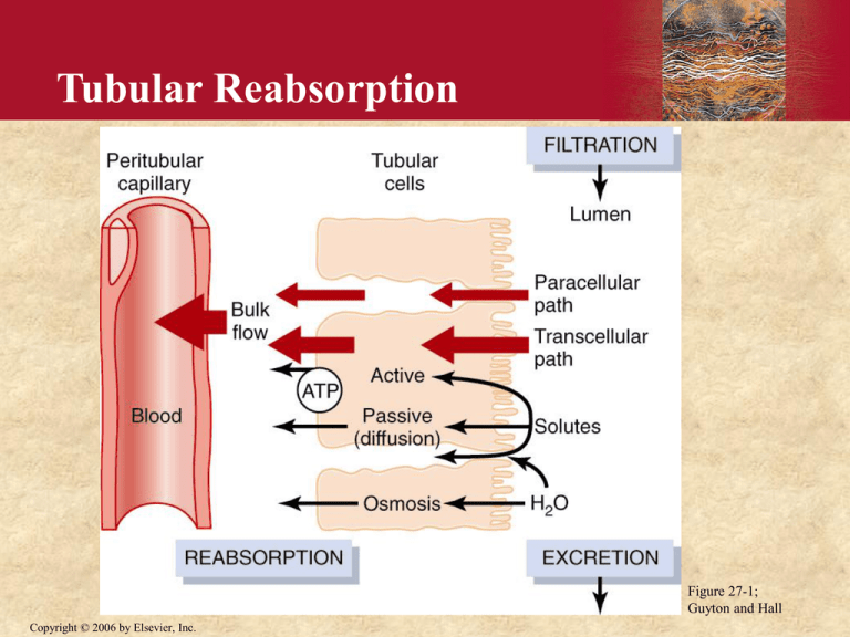
Tubular Reabsorption Figure 27-1; Guyton and Hall Copyright © 2006 by Elsevier, Inc. Primary Active Transport of Na+ Figure 27-2; Guyton and Hall Copyright © 2006 by Elsevier, Inc. Mechanisms of Secondary Active Transport Copyright © 2006 by Elsevier, Inc. Figure 27-3; Guyton and Hall Glucose Transport Maximum Figure 27-4; Guyton and Hall Copyright © 2006 by Elsevier, Inc. Reasorption of Water and Solutes is Coupled to Na+ Reabsorption Tubular Cells Interstitial Fluid - 70 mV Na + K+ ATP Na + ATP 0 mv Copyright © 2006 by Elsevier, Inc. K+ Tubular Lumen H+ Na + glucose, amino acids Na + Urea H20 Na + Cl- - 3 mV Mechanisms by which Water, Chloride, and Urea Reabsorption are Coupled with Sodium Reabsorption Figure 27-5; Guyton and Hall Copyright © 2006 by Elsevier, Inc. Cellular Ultrastructure and Primary Transport Characteristics of Proximal Tubule Figure 27-6; Guyton and Hall Copyright © 2006 by Elsevier, Inc. Cellular Ultrastructure and Transport Characteristics of Thin and Thick Loop of Henle very permeable to H2O) not permeable to H2O) Figure 27-8; Guyton and Hall Copyright © 2006 by Elsevier, Inc. Sodium Chloride and Potassium Transport in Thick Ascending Loop of Henle Figure 27-9; Guyton and Hall Copyright © 2006 by Elsevier, Inc. Countercurrent Multiplier System in the Loop of Henle Figure 28-3; Guyton and Hall Copyright © 2006 by Elsevier, Inc. • Countercurrent multiplier animation Copyright © 2006 by Elsevier, Inc. Recirculation of Urea Absorbed from Medullary Collecting Duct into Interstitial Fluid Figure 28-5; Guyton and Hall Copyright © 2006 by Elsevier, Inc. Net Effects of Countercurrent Multiplier 1. More solute than water is added to the renal medulla (i.e., solutes are “trapped” in the renal medulla). 2. Fluid in the ascending loop is diluted. 3. Horizontal gradient of solute concentration established by the active pumping of NaCl is “multiplied” by countercurrent flow of fluid. Copyright © 2006 by Elsevier, Inc. The Vasa Recta Preserve Hyperosmolarity of Renal Medulla • The vasa recta serve as countercurrent exchangers • Vasa recta blood flow is low (only 1-2 % of total renal blood flow) Figure 28-3; Guyton and Hall Copyright © 2006 by Elsevier, Inc. Sodium Chloride Transport in Early Distal Tubule Figure 27-10; Guyton and Hall Copyright © 2006 by Elsevier, Inc. Cellular Ultrastructure and Transport Characteristics of Early and Late Distal Tubules and Collecting Tubules • not permeable to H2O • not very permeable to urea • permeability to H2O depends on ADH • not very permeable to urea Figure 27-11; Guyton and Hall Copyright © 2006 by Elsevier, Inc. Sodium Chloride Reabsorption and Potassium Secretion in Collecting Tubule Principal Cells Figure 27-12; Guyton and Hall Copyright © 2006 by Elsevier, Inc. Cortical Collecting Tubules Intercalated Cells Tubular Cells Tubular Lumen H20 (depends on ADH) H+ K+ K+ ATP ATP Na + K+ H+ ATP ATP ATP Cl Copyright © 2006 by Elsevier, Inc. Cellular Ultrastructure and Transport Characteristics of Medullary Collecting Tubules Figure 27-13; Guyton and Hall Copyright © 2006 by Elsevier, Inc. Normal Renal Tubular Na+ Reabsorption 7% (16,614 mEq/day) 25,560 mEq/d (1789 mEq/d) 65 % 25 % 2.4% (6390 mEq/d) (617 mEq/day) 0.6 % (150 mEq/day) Copyright © 2006 by Elsevier, Inc. Summary of Water Reabsorption and Osmolarity in Different Parts of the Tubule • Proximal Tubule: 65% reabsorption, isosmotic • Desc. loop: 15-20% reabsorption, osmolarity increases • Asc. loop: 0% reabsorption, osmolarity decreases • Early distal: 0% reabsorption, osmolarity decreases • Late distal and coll. tubules: ADH dependent water reabsorption and tubular osmolarity • Medullary coll. ducts: ADH dependent water reabsorption and tubular osmolarity Copyright © 2006 by Elsevier, Inc. Concentration and Dilution of the Urine • Maximal urine concentration = 1200 - 1400 mOsm / L (specific gravity ~ 1.030) • Minimal urine concentration = 50 - 70 mOsm / L (specific gravity ~ 1.003) Copyright © 2006 by Elsevier, Inc. Obligatory Urine Volume The minimum urine volume in which the excreted solute can be dissolved and excreted. Example: If the max. urine osmolarity is 1200 mOsm/L, and 600 mOsm of solute must be excreted each day to maintain electrolyte balance, the obligatory urine volume is: 600 mOsm/d 1200 mOsm/L Copyright © 2006 by Elsevier, Inc. = 0.5 L/day Maximum Urine Concentration of Different Animals Animal Max. Urine Conc. (mOsm /L) Beaver Pig Human Dog White Rat Kangaroo Mouse Australian Hopping Mouse Copyright © 2006 by Elsevier, Inc. 500 1,100 1,400 2,400 3,000 6,000 10,000 Late Distal, Cortical and Medullary Collecting Tubules Principal Cells Tubular Lumen H20 (w/o ADH) Na + Na + K+ ATP K+ Cl - Aldosterone Copyright © 2006 by Elsevier, Inc. Abnormal Aldosterone Production • Excess aldosterone - Conn’s syndrome Na+ retention, hypokalemia, alkalosis, hypertension • Aldosterone deficiency - Addison’s disease Na+ wasting, hyperkalemia, hypotension, death if untreated Copyright © 2006 by Elsevier, Inc. Ang II Increases Tubular Na+ Transport Tubular Cells Interstitial Fluid Tubular Lumen Ang II Ang II Na+ Na+ ATP + K Copyright © 2006 by Elsevier, Inc. H+ Effect of Angiotensin II on Peritubular Capillary Dynamics Glomerular Capillary Ra Peritubular Re Capillary Arterial Pressure Ang II Re Pc (peritubular cap. press.) renal blood flow Copyright © 2006 by Elsevier, Inc. FF c Ang II Constriction of Efferent Arterioles Causes Na+ Retention and Maintains Excretion of Waste Products Na+ depletion Ang II Resistance efferent arterioles Glom. cap. press Prevents decrease in GFR and retention of waste products Copyright © 2006 by Elsevier, Inc. Renal blood flow Peritub. Cap. Press. Filt. Fraction Na+ and H2O Reabs. Angiotensin II Blockade Decreases Na+ Reabsorption and Blood Pressure • ACE inhibitors (captopril, benazipril, ramipril) • Ang II antagonists (losartan, candesartin, irbesartan) • decrease aldosterone • directly inhibit Na+ reabsorption • decrease efferent arteriolar resistance Natriuresis and Diuresis + Copyright © 2006 by Elsevier, Inc. Blood Pressure Formation of a Dilute Urine • Continue electrolyte reabsorption • Decrease water reabsorption Mechanism: decreased ADH release and reduced water permeability in distal and collecting tubules Figure 28-1; Guyton and Hall Copyright © 2006 by Elsevier, Inc. Formation of a Concentrated Urine • Continue electrolyte reabsorption • Increase water reabsorption Mechanism: • Increased ADH release which increases water permeability in distal and collecting tubules • High osmolarity of renal medulla • Countercurrent flow of tubular fluid Copyright © 2006 by Elsevier, Inc. Formation of a Concentrated Urine when Antidiuretic Hormone (ADH) Levels are High Figure 28-4; Guyton and Hall Copyright © 2006 by Elsevier, Inc. Osmoreceptor– antidiuretic hormone (ADH) feedback mechanism for regulating extracellular fluid osmolarity Copyright © 2006 by Elsevier, Inc. Figure 28-8; Guyton and Hall ADH synthesis in the neurons of hypothalamus, release by the posterior pituitary, and action on the kidneys Copyright © 2006 by Elsevier, Inc. Figure 28-9; Guyton and Hall Factors that Decrease ADH Secretion • Decreased osmolarity • Increased blood volume (cardiopulmonary reflexes) • Increased blood pressure (arterial baroreceptors) • Other factors: - alcohol - haloperidol (antipsychotic, tics,Tourette’s) Copyright © 2006 by Elsevier, Inc. Stimuli for Thirst • Increased osmolarity • Decreased blood volume (cardiopulmonary reflexes) • Decreased blood pressure (arterial baroreceptors) • Increased angiotensin II • Other stimuli: - dryness of mouth Copyright © 2006 by Elsevier, Inc. Factors that Decrease Thirst • Decreased osmolarity • Increased blood volume (cardiopulmonary reflexes) • Increased blood pressure (arterial baroreceptors) • Decreased angiotensin II • Other stimuli: -Gastric distention Copyright © 2006 by Elsevier, Inc. Atrial Natriuretic Peptide (ANP) Blood volume ANP Renin release aldosterone GFR Ang II Renal Na+ and H2O reabsorption Copyright © 2006 by Elsevier, Inc. Na+ and H2O excretion Late Distal and Collecting Tubules Tubular Cells Tubular Lumen H20 (depends on ADH) Aquaporin-3 H20 Aquaporins ADH cAMP V2 receptor Copyright © 2006 by Elsevier, Inc. Vesicle Aquaporin-2 H20 Sodium Chloride and Potassium Transport in Thick Ascending Loop of Henle Figure 27-9; Guyton and Hall Copyright © 2006 by Elsevier, Inc. Sodium Chloride Transport in Early Distal Tubule Figure 27-10; Guyton and Hall Copyright © 2006 by Elsevier, Inc. Sodium Chloride Reabsorption and Potassium Secretion in Collecting Tubule Principal Cells Figure 27-12; Guyton and Hall Copyright © 2006 by Elsevier, Inc.


