Slides - Philippe Le Fevre
advertisement
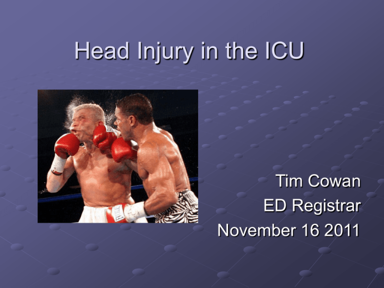
Head Injury in the ICU Tim Cowan ED Registrar November 16 2011 Overview Epidemiology Physiology, primary and secondary injury Initial care and resuscitation ICP control Surgical Medical Retrieval considerations Research focus Epidemiology Roughly 110/100 000 of hospital visits are for traumatic brain injury Males twice the rate of females Peaks at age 15-24 and greater than 75 years Estimated cost $4.8 billion every year in Australia MVA, falls and assaults account for the majority of severe brain injuries "Ute surfing": a novel cause of severe head injury Rodney S Allan, Peter J Spittaler and John G Christie MJA 1999; 171: 681-682 Impact at the cellular level • Excitotoxicity • Inflammatory changes • Cytotoxic oedema • Vasogenic oedema Monro-Kellie Doctrine CSF ~8% Blood ~ 12% Brain ~80% • Cranium is a rigid structure containing three components: brain, CSF and blood • Normal ICP is <15 mmHg • An increase in the volume of one of the components increases ICP unless there is a compensatory decrease in the volume of another component • These mechanisms are exhausted by about 25 mmHg Cerebral perfusion pressure = Mean arterial pressure Intracranial pressure The vicious circle of ICP and CBF Raised intra-cranial pressure Increased cellular injury, local hypoxia and hypercapnoea Increased cerebral blood flow Primary vs secondary brain injury Primary occurs at the time of impact and is usually refractory to medical treatment Secondary occurs subsequently and for the most part is preventable or treatable Secondary brain injury Major extracranial causes Hypoxia Hypo or hypercapnia Hypotension Why is hypoxia bad? Brain uses ~20% of total oxygen consumption Hypoxia leads to cell changes in seconds and quickly becomes irreversible Low arterial oxygen tension has profound effects on cerebral blood flow. When it falls below 50 mmHg, there is a rapid increase in CBF and arterial blood volume 0 50 100 150 What does CO2 do? Carbon dioxide causes cerebral vasodilation. As the arterial tension of CO2 rises, CBF increases. When PaCO2 is reduced vasoconstriction is induced. 100 Cerebral blood flow (ml/100g/min) 50 0 0 25 50 Arterial pCO2 (mmHg) 75 100 What’s wrong with hypotension? Brains like blood – receiving up to 15% of cardiac output Normally autoregulation maintains a constant blood flow between MAP of about 50 mmHg and 150 mmHg. Autoregulation is a poorly understood local vascular mechanism. In traumatised or ischaemic brain, CBF may become blood pressure dependent. Retrospective analysis of a large group of matched patients has suggested a single prehospital episode of hypotension doubled risk of mortality and was independent of age, GCS, pupillary status, GCS motor score and intracranial pathology Secondary brain injury Other extracranial causes Hyperthermia Hyponatraemia Hypo/perglycaemia Acidosis Secondary brain injury Intracranial causes Hemorrhage Oedema Infection Initial care ABCDE Usual ATLS guidelines Particular attention to avoiding secondary injury Don’t forget the C spine Airway and breathing Usually ETT required in severe TBI Useful to perform a limited neurological exam first if possible RSI Ketamine no longer contraindicated Opiates can blunt stress response to intubation Cervical spine injuries common Aim SaO2 >97%, pCO2 35-40 mmHg Circulation Hypotension clearly linked to poor outcome BTF guidelines suggest aim for SBP>90 JHH TBI protocol suggests MAP of 90 rather than SBP Consider permissive hypotension in multitrauma Correct coagulopathy Disability Limited exam GCS and pupils Monitor trends as well as initial Expedite CT brain Blown pupil Medial temporal lobe herniates Pressure on CN III interrupting parasympathetic input to the eye Transtentorial herniation also compresses the brainstem. Consider other causes (briefly!) ICP monitoring Clinically track GCS, pupillary responses Invasive operative insertion of an ICP monitor can be intraventricular or intraparenchymal Management of raised ICP in the resuscitative setting Simple things (if you don’t have a Black and Decker) Nurse 30 degrees head up if able Can tilt whole bed if spine not cleared Avoid impeding cranial venous drainage Tapes not too tight on ETT Subclavian or femoral CVC rather than IJ Sandbag and tape vs collar Mannitol Osmotic agent (sugar alcohol) 0.5gm/kg/dose Initially volume expander (decreases Hct, reduces viscosity, improves oxygen delivery) Osmotic effects take about 15-30 minutes, last from 90 minutes to several hours Filtered and not reabsorbed, thus net volume loss. Useful to ‘buy time’ Use with caution in hypotension Surgical intervention There is a clot. I must fix it. Different operations External ventricular drain (EVD) Burrholes Craniotomy (bone window replaced) Craniectomy (bone removed and sent to bone bank, stored in subcutaneous pouch, or discarded) Can leave a drain in situ EVD Medical intervention Mannitol Driving MAP Sedation Paralysis Hypertonic saline Barbiturates CPP = MAP-ICP CPP needs to be greater than 60mmHg Ideally preferable to reduce ICP rather than increase MAP If ICP optimised as possible, and CPP not satisfactory, use noradrenaline to drive MAP Sedation +/- paralysis Routinely sedated until ICP controlled >24h First line treatment of ICP ‘spike’ Bolus of M and M If that works, increase background rate Propofol reduces cerebral metabolism and blood flow Paralysis decreases straining, eg from coughing/ventilator dyssynchrony But can mask seizure, and causes complications if used long term Hypertonic saline “Current evidence is not strong enough to make recommendations on the use, concentration and method of administration of hypertonic saline for the treatment of traumatic intracranial hypertension.” Brain Trauma Foundation guidelines JHH ICU guideline – if serum Na+ <155, and CVP <12 give a dose (30ml 23.4% saline over 10 min) Barbiturates About 55% of the glucose and oxygen utilisation by the brain is meant for its electrical activity and metabolism Barbiturates depress cerebral metabolism and as a result, CBF needs are reduced Also inhibit free radical-mediated lipid peroxidation and excitotoxicity, cause alterations in vascular tone and resistance Improve ICP but not outcome - Cochrane Hypotension, myocardial depression, accumulation can all be associated problems. Other ICU care Nutrition and glycaemic control Analgesia DVT prophylaxis High risk patients TEDS and SCUDS Level III evidence supports the use of prophylaxis with low-dose heparin or LMWH for prevention of DVT in patients with severe TBI, but uncertainty remains around when to commence and what dose to use Ulcer prophylaxis Pressure care, chest physio Bowel care Retrieval considerations. Preventing secondary injury Mannitol Deep sedation +/- paralysis Consider intracranial air Expedite transfer to CT or surgery Complications Seizures Prophylactic anticonvulsants reduce the occurrence of post-traumatic seizures within the first week of injury, but they do not improve longterm outcome. Treat seizures immediately. Seizures raise ICP and can increase the volume of intraparenchymal and subarachnoid hemorrhage. Phenytoin load Salt and the injured brain Hyponatremia may lead to reduced levels of consciousness and even epileptic seizures. Hyponatremia after head injury is often due to SIADH, and less commonly to cerebral salt wasting syndrome. Hypernatraemia can also occur, especially if there is HP axis injury (causing diabetes insipidus) Monitor sodium, urine output and volume status, check osmolality if abnormal and don’t correct too quickly 10mmol/L/day is generally a safe rate to increase or reduce serum sodium Dysautonomia (“Storming”) Complex interactions between multiple areas of brain (cortex, hypothalamus, brainstem) Hypertension, fever, tachycardia, tachypnea, pupillary dilation, and extensor posturing. Diagnosis of exclusion NMS, serotonin syndrome, malignant hyperthermia, sepsis, withdrawal syndromes and thyroid storm can cause similar picture Propanolol and clonidine can reduce symptoms Hydrocephalus Acute or chronic post TBI Communicating or noncommunicating Blockage of outflow (blood in 3rd/4th ventricles) Impaired reabsorption (blood/proteins blocking arachnoid villi) Neurology varies May need LP, EVD or V-P shunt Research focus DECRA trial 2003-2010 Aust, NZ, Saudi Arabia Prospective randomised controlled trial 155 enrolments Randomized to craniectomy plus maximum medical care, versus maximum medical care only DECRA - background 1000 ICU admissions/year with severe TBI 50% have a focal surgical lesion 10% have diffuse injury and swelling refractory to drugs and drains Decompressive craniectomy was becoming more popular to manage these patients Inclusions and exclusions 15-59 years Non penetrating head trauma GCS 3-8 at admission Exclusions Not suitable for full active treatment as per treating clinician Fixed dilated pupils Mass lesion requiring surgery Spinal cord injury CA at scene ICP >20 mmHg for 15 minutes within one hour during first 72 hours of care and despite optimised first tier treatment DECRA n=73, standard care n=82 Interventions DECRA group underwent bifrontal decompressive craniectomy with dural opening, in addition to medical therapy as per control group Control group underwent medical therapy as per BTF clinical practice guidelines Both had scope for second tier interventions Controls could have compassionate craniectomy after 72 hours if ICPs remained high (15/82) DECRA findings No mortality difference – 19% in DECRA group vs 18% in standard care The craniectomy group had decreased intracranial pressure, markedly decreased medical therapies required for intracranial pressure, shortened mechanical ventilation time, and shortened stay in the intensive care unit by 5 days compared with the standard care group But at 6 month follow-up, 19% more patients had poor functional outcomes based on the extended Glasgow Outcome Score in the decompressive craniectomy group compared with patients who received standard care alone. (70% vs 51%) Why did the DECRA group do worse? ?surgical complications (including hydrocephalus), but surgical complications seem an unlikely explanation given that the rates overall were less than those reported in published case series. ?”axonal stretch” that occurred during swelling of the brain outside the skull through the craniectomy defect ?release of pressure, in and of itself, may have aggravated the development of brain edema that would otherwise have been self-limiting DECRA - criticisms 20mmHg low threshold for inclusion, and 15 minutes short duration of raised ICP Only 155 of 3500 potential patients enrolled, hard to generalise results Despite being randomized, more patients in the craniectomy arm had unreactive pupils (after randomization but before surgery) Some patients in the non-surgical arm went on to have craniectomy after 72 hours, yet were included in the medical group for analysis Didn’t evaluate cerebral hypoxia and blood flow What does it all mean? It’s really hard to do good research! Craniectomy will reduce ICP Maybe that is not the key issue (as suggested in barbiturate and hypothermia studies) and when considering outcome intracranial pressure cannot be used as a reliable short-term surrogate for the effectiveness of therapies for severe TBI. Need to consider ICP in the context of brain perfusion and oxygenation – further studies looking at ICP as well as transcranial dopplers, microdialysis catheters and jugular venous bulb oxygen saturations RESCUEicp trial 319/400 recruited so far Craniectomy versus standard care ICP >25mmHg for 1-12 hours Previous haematoma evacuation does not exclude entry Either bifrontal or wide unilateral decompression Outcome at discharge and 6 months Erythropoietin EPO receptors upregulated after TBI EPO crosses BBB, protects against further injury by restoring mitochondrial function, reducing glutamate exocytosis Activates anti-inflammatory, anti-oxidant and anti-apoptotic signalling. Also stimulates neurogenesis EPO-TBI trial Commenced May 2010 Randomised, double blinded, placebo controlled trial EPO 40 000 IU s/c weekly x 3 weeks vs normal saline placebo Primary outcomes – severe disability or death at 6 months as per GOSE Secondary outcomes – disability, quality of life at 6 months, cost, and rate of thrombotic events. Inclusion criteria Patients with non-penetrating moderate (GCS 9-12) or severe (GCS 3-8) TBI admitted to the ICU Are ≥ 15 to ≤ 65 years of age Are < 24 hours since primary traumatic injury Are expected to stay ≥ 48 hours Have a haemoglobin not exceeding the upper limit of the applicable normal Have written informed consent from legal surrogate Exclusion criteria GCS = 3 and fixed dilated pupils History of DVT, PE or other thromboembolic event A chronic hypercoagulable disorder, including known malignancy Treatment with EPO in the last 30 days First dose of study drug unable to be given within 24 hours of primary injury Pregnancy or lactation or 3 months post partum Uncontrolled hypertension (systolic blood pressure of >200 mm Hg or diastolic blood pressure of >110 mm Hg) Acute myocardial infarct within the past 12 months Past history of epilepsy with seizures in past 3 months Expected to die imminently (< 24 hours) Inability to perform lower limb ultrasounds Known sensitivity to mammalian cell derived products Hypersensitivity to the active substance or to any of the additives Pure red cell aplasia (PRCA) End stage renal failure (receives chronic dialysis) Severe pre-existing physical or mental disability or severe co-morbidity that may interfere with the assessment of outcome Spinal cord injury Treatment with any investigational drug within 30 days before enrolment The treating physician believes it is not in the best interest of the patient to be randomised to this trial Hypothermia isn’t cool. Yet. Prophylactic hypothermia is not associated with decreased mortality Does appear to have some impact on GOS compared to normothermia, BUT Limited good research, clarity needed in particular re: Target cooling temperature (32-33°C or >33°C) Cooling duration (<48 h, 48 h, or >48 h) Rate of rewarming (1°C per hour, 1°C per day, or slower) Steroids CRASH trial 10 008 patients GCS 14 or less Randomised to 48h of methylpred or saline Outcomes of death within 2 weeks of injury GOS at 6 months Steroids Placebo %Mortality at 2 weeks 21.1 17.9 %Mortality at 6 months 25.7 22.3 %Death and severe disability at 6 months 38.1 36.3 Steroids not recommended in management of head injury Free radical scavengers Oxidative stress and mitochondrial injury leads to the formation of free radicals These in turn can further damage neural cells and promote inflammation Scavengers ‘mop up’ free radicals Mannitol is a free radical scavenger Edaravone is being researched for stroke That is all. Make sure you wear a helmet. Extradural haematoma Subdural haematoma Traumatic subarachnoid haematoma Intracerebral contusions Trauma.org moulage

