iga-nephropathy
advertisement
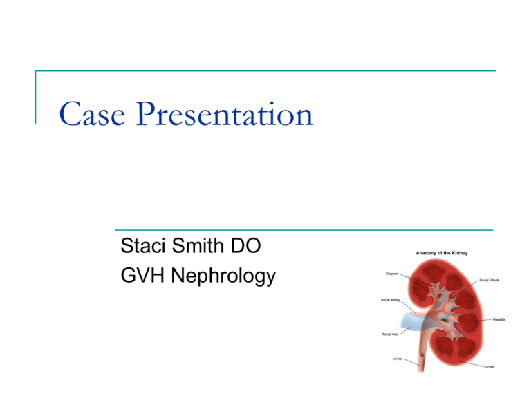
Case Presentation Staci Smith DO GVH Nephrology Case Presentation 44-year-old white right-handed male that presented to GVH with complaints of hematuria had associated abdominal pain and took a Zantac for it no history of known CKD and has not actually seen a doctor in 25 years no history of other hematuria,prostate trouble, dysuria, nocturia,incontinence not on any outpatient anticoagulation 20 years of tobacco abuse Case Presentation recent upper respiratory infection 2 weeks ago with fevers and chills foamy urine denies any recent known UTI, over-thecounter NSAIDs, history of nephrolithiasis, contrasted procedure, physical exercise Case Presentation PAST MEDICAL HISTORY: OA Tobacco abuse URI 2 weeks ago MEDS: SOCIAL HISTORY: PAST SURGICAL HISTORY: ALLERGIES: Right knee scope None FAMILY HISTORY: Htn No medical renal dz None Cleans gutters / construction work Positive tobacco 1-2 ppd x 20 yrs Social EtOh only Case Presentation Positive ROS : Recent URI two weeks ago 10 pound weight gain with LE edema Hematuria Foamy urine Abdominal pain - Zantac Case Presentation : Physical Exam VS:126/87- 97.9 F (T-max 99.2)- HR 84- RR 20- 96%sat GEN-Awake, alert and oriented x3. Family is at bedside and updated. No acute distress. HEENT: Poor dentition. Atraumatic, normocephalic. Extraocular muscles intact. Sclerae are anicteric. Mucous membranes are now moist CHEST: Respirations clear to auscultation bilaterally. No wheezing, rhonchi or crackles ABDOMEN: Soft, nontender, nondistended. Positive bowel sounds. No perotoneal signs or Foley EXTREMITIES: No clubbing, cyanosis. Plus 2 pitting edema. There is no asterixis SKIN: There is no rash or cellulitis NEUROLOGIC: Cranial nerves II-XII grossly intact. Case Presentation : Labs 133 109 13 5 22 2.0 122 CCa 9.6 Alb 1.6 No Phos or Mg UA is cloudy with positive nitrite, large blood, 300 protein, too numerous to count red cells, trace bacteria, 1-5 white cells Case Presentation : Labs CT of the abdomen and pelvis was negative for any stone, multiple loops of fluid-filled nondilated small bowel without obstruction or contracted GB Urine Pr/ Cr ratio- 10 CPK was negative at 148. LFTs normal. Amylase and lipase are normal. TSH is 3.03 Total cholesterol 277 total triglycerides 264, HDL 26, LDL 198 7.8 11.3 165 S 79-L 11-M 8 – E 2 33.6 Case Presentation : Labs All negative Complements ANA, dsDNA Hepatitis profile SPEP/ UPEP with IFE Rheumatoid factor/ Anti CCP Anti GBM P and C ANCA’s HIV ASO Differential Diagnosis Back to our case 44 yo with hematura, proteinuria, and lower extremity edema two weeks after an URI No previous know CKD or AKI Cr now 2.0 10 grams proteinuria Hyperlipidemia Differential Diagnosis hematuria proteinuria red blood cell casts glomerulonephritis Red Blood Cell Casts Glomerular hematuria Dysmorphic rbc’s glomerular damage rule out urologic causes Nephritic syndrome HTN RBC casts Proteinuria Edema Differential Diagnosis: Glomerulonephritis Postinfectious GN IgA nephropathy Thin basement membrane Henoch-Schönlein purpura Mesangial proliferative GN SLE Goodpasture’s dz Vasculitis Wegener’s, Churg-Strauss Cryoglobulinemia HIV Membranoproliferative glomerulonephritis Rapidly progressive GN Fibrillary glomerulonephritis Focal glomerulosclerosis Membranous nephropathy Amyloidosis Multiple Myeloma DM HUS Differential Diagnosis: Glomerulonephritis UA with C and S Cbc with differential Renal panel Urine Pr/Cr Renal US Renal Duplex Cystography Urine eosinophils SPEP / UPEP with IFE Differential Diagnosis: Glomerulonephritis ANA, dsDNA Anti-GBM dz Goodpasture’s ANCA’s c-ANCA: Wegener’s p-ANCA: PAN, Churg Strauss, MPA Complements Cryoglobulinemia, HCV Anti-GBM SLE Cryoglobulins, RF ASO MPGN HIV Post Strept GN Hepatitis profile C3, C4, CH50 HIV, FSGS Renal Biopsy Renal Biopsy : Ig A Nephropathy Light microscopy - Mesangial hypercellularity IgA is predominantly polymeric IgA1 mainly derived from the mucosal immune system IgA Nephropathy : Berger’s Disease the most common lesion found to cause primary glomerulonephritis peak incidence in the second and third decades of life 2:1 male to female greatest frequency in Asians and Caucasians relatively rare in blacks IgA Nephropathy large undiagnosed "latent" IgA nephropathy in the general population the process of mesangial IgA deposition is likely to be separate from the induction of glomerular injury IgA deposition does not necessarily need to be followed by nephritis IgA Deposition is Common IgA deposition in other forms of glomerulonephritis thin basement membrane nephropathy lupus nephritis minimal change disease diabetic nephropathy IgA Nephropathy Many patients are detected on routine urine screening asymptomatic hematuria and/or proteinuria higher prevalence active urine testing program low threshold for renal biopsy IgA Nephropathy IgA nephropathy is established only by kidney biopsy Immunofluorescence microscopy demonstrating large, globular mesangial IgA deposits also seen with HSP IgA often accompanied by C3 and IgG in the mesangium IgA Nephropathy EM electron dense deposits that are limited to the mesangial regions IgA Nephropathy mesangial glomerulonephritis showing segmental areas of increased mesangial matrix and cellularity Conditions associated with IgA nephropathy Idiopathic (most cases) Hepatic cirrhosis Gluten enteropathy HIV infection Minimal change disease Membranous GN Wegener’s granulomatosis Dermatitis herpetiformis Seronegative arthritis eg, ankylosing spondylitis Small cell carcinoma Disseminated tuberculosis Mycosis fungoides IgA Nephropathy Initiating event in the pathogenesis is the mesangial deposition of IgA Codeposits of IgG and complement commonly seen may contribute to disease severity Between episodes of gross hematuria persistent microhematuria, proteinuria, or both. Gross hematuria has also followed tonsillectomy, vaccinations, strenuous physical exercise, and trauma. Increased Plasma IgA Levels Not alone is not sufficient to produce mesangial IgA deposits Found in 50% of cases IgA is probably accumulated and deposited because of a systemic abnormality rather than a defect intrinsic to the kidney IgA Nephropathy Two common presentations episodic gross hematuria 40-50% upper respiratory tract infection, or, less often, gastroenteritis persistent microscopic hematuria 30-40% asymptomatic, with erythrocytes (RBCs), RBC casts, and proteinuria discovered on urinalysis IgA Nephropathy Nephrotic range proteinuria is uncommon occurring in only 5% of patients Indicates more advanced disease Approximately 1-2% of all patients with IgA nephropathy develop ESRD each year Hypertension seldom occurs at the time of initial presentation Ig A Pathophysiology Platelet Derived Growth Factors Made by mesangial cells Increased PDGG receceptors in glomerular dz Infusion of glomerular transfection with PDGF leads to mesangial proliferation TGF- beta Made by mesangial cells Pro fibrotic Antiinflammatory and immunosuppresive Morbidity and Mortality follow a benign course in most cases at risk for slow progression to ESRD approximately 15% of patients by 10 years 20% by 20 years Ig A Nephropathy Outcomes 20-30% progress to ESRD over 20 years 1-2% per year Clinical predictors of poor renal outcome Absence of gross hematuria Male Older onset age HTN Heavy proteinuria Elevated Cr >2-2.5 Ig A Nephropathy Outcomes Therapy remains to be defined Antibiotics Tonsillectomy Cyclophosphamide, dipyridamole High dose immunoglobulin therapy Statins Fish Oils Ace inhibition Cellcept (mycophenolate mofetil) Ig A Nephropathy Outcomes Ace inhibitors Effectively reduce proteinuria in glomerular dz ACE-I better than other anti – HTN meds to preserve GFR in Ig A Questionable addititive effect with ARBS Ig A Nephropathy Outcomes Fish Oil Meta analysis concluded there may be a minor benefit in heavy proteinuria Dillon 1997 JASN Low does omega 3 fatty acids as effective as high dose Cellcept Inhibits de novo guanosine nucleotide synthesis Established use for transplant Not that much improvement for Ig A Ig A Treatment Summary If Uprot <0.5 g/d and CrCl >70 If Uprot > 0.5 g/d and Cr Cl > 70 ACEI/ARB for target bp 125/75 If Uprot 1-3 g/d with Cr Cl >70 Observe and consider ACEI or ARB Maximal ACEI/ ARB Consider 6 months of high dose steroids and taper for 6 mo If Uprot >3 g/d and CrCl <70 or declining Steroids plus Cytotoxics Possible maintenance with AZA or MMF Transplant Recipients high recurrence rate in renal transplant recipients who have IgA nephropathy 25-60% Increased risk of allograft loss in living related donor disappearance of the deposits from donor kidneys with IgA nephropathy when transplanted into donors without the disease Patient Progression Cr continued to worsen with disease progression March Cr: 2.0 April Cr: 3.09 – 4.2 Initiation of Cytoxan and Steroids ( 2 cycles) ACEI caused hyperkalemia Fish Oil, BB, Lasix , Zaroxolyn, Statin, PPI, Oscal May Cr :5.2 June 18th: 8.76 ESRD with hemodialysis initiation Uncontrollable edema and pulmonary edema despite diuretics Question 1 Which of the following is the most predictive for progression of Ig A A- elevated levels of IgA B- elevated Cr at baseline diagnosis C- male gender D- absence of gross hematuria Question 1 Which of the following is the most predictive for progression of Ig A A- elevated levels of IgA B- elevated Cr at baseline diagnosis C- male gender D- absence of gross hematuria Question 2 You are seeing a 30yr Asian woman with bx proven Ig A. Her Uprotein is 3.5g/d despite maximal ACEI, Bp is 100/70, Cr stable at 1.6 for the past year. Diffuse foot process effacement is seen on EM. What is the next step for management? A- Add ARB B- Add MMF C- Add steroids D- Add fish oils E-Tonsillectomy Question 2 You are seeing a 30yr Asian woman with bx proven Ig A. Her Uprotein is 3.5g/d despite maximal ACEI, Bp is 100/70, Cr stable at 1.6 for the past year. Diffuse foot process effacement is seen on EM. What is the next step for management? A- Add ARB B- Add MMF C- Add steroids D- Add fish oils E-Tonsillectomy
