Budd-Chiari Syndrome: Ultrasound Diagnosis & Management
advertisement
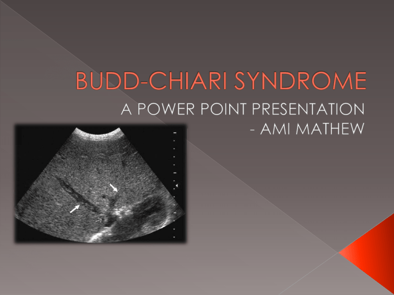
BCS is the manifestation of hepatic venous outflow obstruction Hepatic venous obstruction can occur at the level of the IVC or the hepatic veins BCS is a rare and life threatening condition if not treated promptly HCC Renal Carcinoma Pregnancy Congenital Abnormalities Coagulation Abnormalities › Polycythemia › Rubra vera › Chronic leukemia • In order to adequately visualise the middle and left hepatic veins – scan the liver in a transverse cross section at the level of the xiphoid process and angle the transducer cranially • The right hepatic vein can be evaluated from a right intercostal approach INTERCOSTAL SCAN PLANE From Ultrasoundpedia, 2014 From Ultrasoundpedia, 2014 Duplex ultrasound of the normal blood flow in the middle hepatic vein. Flow is phasic in response to both the cardiac and respiratory cycles From ImageArcade, 2014 B-mode image of the hepatic veins. Blood flows from the hepatic veins into the IVC immediately prior to the Right Atrium Chaubal et al 2006. J Ultrasound Med 25:1 373-379 White arrows represent thrombus within the left and middle hepatic veins whilst the thin arrows shows narrowing stenosis of the distal MHV as it drains into the IVC Chaubal et al 2006. J Ultrasound Med 25:1 373-379 White arrow shows intrahepatic collaterals with a enlarged caudate lobe (asterisk) Bargallo et al 2006. Am J Retrogenol; 187:W33-W42 Normal flow is seen in the middle hepatic vein. However, the right hepatic vein shows abnormal, inverted flow Kandpal et al 2008. RadioGraphics; 28: 669-689 Loss of cardiac pulsation is indicated by Duplex ultrasound showing reversed flow in the IVC There may be underestimation of the presence of thrombus and the characteristic ‘membranous webs’ may be inconclusive in cirrhotic patients with hepatic veins that are difficult to image BCS can be hard to distinguish from Hepatic veno-occlusive disease (progressive occlusion of hepatic venules). Ultrasound is highly operator dependent Using real time scanning, radiologists can evaluate the IVC and hepatic veins non invasively Using Duplex and Colour Ultrasound, the presence and direction of evaluation of hepatic venous flow can occur in patients with suspected BCS. Associated reversal of flow in portal vein can also be assessed. B-mode imaging can be used to draw conclusions on missing compressed or otherwise abnormal hepatic veins and IVC. The presence of epigastric collaterals can be readily assessed Ultrasound imaging is an important tool in BCS affecting patient management from diagnosis - follow up after treatment!! Traditionally › Surgical porto-systemic shunting › Liver transplant (for patients with end stage liver failure More recently › Recanalization of stenosed segments of hepatic vein/IVC › TIPS – Transjugular Intrahepatic Portosystemic shunts Image showing TIPS – an artificial connection is between the hepatic vein and portal vein is made via a stent in order to reduce portal venous pressure. From BayCare Clinic 2014 1. 2. 3. 4. 5. 6. 7. 8. 9. 10. 11. "Budd–Chiari Syndrome." Wikipedia. July 1, 2014. Accessed October 20, 2014. http://en.wikipedia.org/wiki/Budd–Chiari_syndrome. "Gallery For Hepatic Veins Ultrasound." Gallery For Hepatic Veins Ultrasound. January 1, 2014. Accessed October 30, 2014. "ULTRASOUND OF THE LIVER - Normal." Ultrasoundpaedia. January 1, 2013. Accessed October 13, 2014. Scoutt, Leslie. "Ultrasound Evaluation of Portal Hypertension." SONOWORLD. January 1, 2013. Accessed October 30, 2014. Kamath, Patrick S. "Budd-Chiari Syndrome: Radiologic Findings." Liver Transplantation 12, no. 1 (2006): S21-22. Rumack, Carol M.Diagnostic Ultrasound. 3rd ed. St. Louis: Elsevier Mosby, 2005. Bargallo, X., Rosa Gilabert, Carlos Nicolau, Juan Carlos García-Pagán, Juan Ayuso, and Concepció Bru. "Sonography of Budd-Chiari Syndrome." American Journal of Roentgenology187, no. 1 (2006): W33-41 Chaubal, Nitin, Vijay Hanchate, Manjir Dighe, Hemangini Thakkar, Hemante Deshmukh, and Krantikumar Rathod. "Sonography in Budd-Chiari Syndrome." J Ultrasound Med 25, no. 1 (2006): 37379. Knipe, Henry, and Frank Gaillard. "Budd-Chiari Syndrome." Radiopaedia Blog RSS. January 1, 2014. Accessed October 17, 2014. http://radiopaedia.org/articles/budd-chiari-syndrome-1. Haffar, Samir. "Doppler Ultrasound of Budd Chiari Syndrome & SOS." Doppler Ultrasound of Budd Chiari Syndrome & SOS. June 27, 2013. Accessed October 15, 2014. Mukund, Amar, and Shivanand Gamanagatti. "Imaging and Interventions in Budd-Chiari Syndrome." World J Radiol 3, no. 7 (2011): 169-77.
![Jiye Jin-2014[1].3.17](http://s2.studylib.net/store/data/005485437_1-38483f116d2f44a767f9ba4fa894c894-300x300.png)
