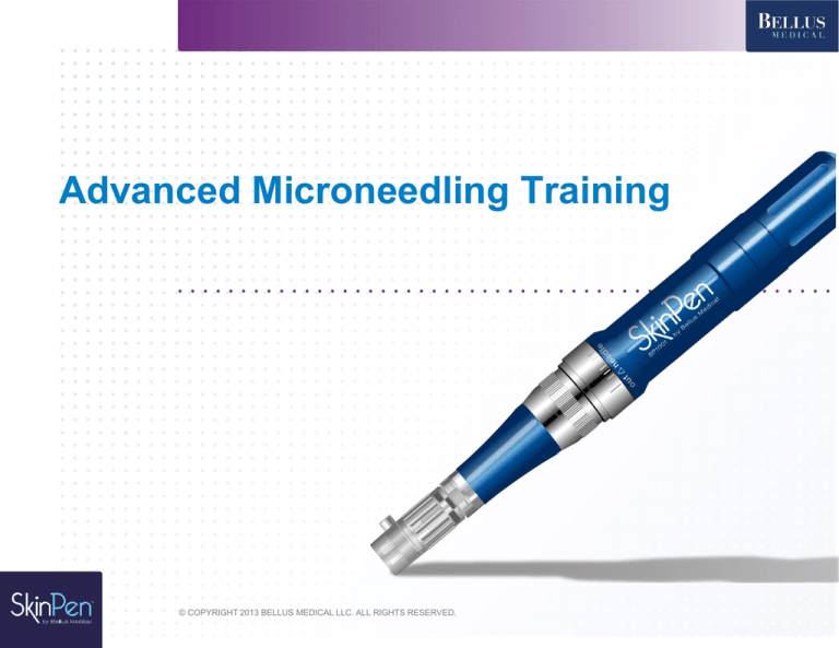
Advanced Microneedling Training
© COPYRIGHT 2013 BELLUS MEDICAL LLC. ALL RIGHTS RESERVED.
1.15
How Does Medical Needling Work?
•
0.5mm – 3.0mm
•
Uses very fine, surgical stainless steel needles to make channels into
the epidermis and dermis to release growth factors
•
Promotes scarless healing and deposition of normal woven collagen
rather than scar collagen
•
Similar to Fraxel, without the negative loss of dermal papillae,
potential destruction of melanocytes, abnormal collagen, coagulated
growth factors
•
Allows 80% more product into the skin (compared to 7-10% normally)
1.10
Collagen Induction Therapy
Microinjuries allow for release of serum
containing cytokines and growth factor*
*Fernandes, D. OralL Maxillofacial Surg Clin 2005; 17:51-63
1.19
Phases of Wound Repair
1.16
Most Effective Uses
of Dermal Needling
Wrinkles
Epidermal density and strength
Thin skin
Lax skin
Hyperpigmentation
UV damage
Rosacea
Stretch marks
Loss of Resiliency
Hair restoration
Premature aging
Scars
1.3
Features of Healthy Skin
Soft, compact stratum corneum, with strong skin barrier
Dense spinosum layer with consistent, strong cell-to-cell
adhesion
Even color, with melanocytes that distribute melanin uniformly
Resilience
Dermis rich with collagen and elastin fibers
Good dermal and epidermal hydration:
Extracellular matrix rich with glycosaminoglycans
1.4
The Three Layers of the Skin
1.7
Optimizing the Keratinocytes
Basic epidermal topical requirements:
Omega 3
Omega 6
Ceramide
Squalenes
Sphingolipid
Phospholipid
Epidermal Cell Requirements
Omega 3
• Kiwifuit seed oil
• Lecithin
• Hemp see oil
• Flax seed oil
• Camelina oil
Omega 6
• Hemp seed oil
• Borage oil
• Evening Primrose oil
• Rice bran oil
Ceramide
• Yeast (pichia anomala extract)
• Wheat extracts
Squalenes
• Rice bran oil
• Olive oil
Sphingolipid
• Yeast (pichia anamola extract)
Phospholipid
• Lecithin
Optimizing the Melanocyte
Unfortunately, melanocytes lie between
most anti-aging treatment modalities and the
targeted fibroblasts, and are often sacrificed in
overzealous attempts to obtain greater injury
through aggressive injury.
1.9
Optimizing the Melanocyte
1.8
Optimizing the Melanocyte
5 intervention points:
• Block UV radiation
• Block Melanin Stimulating Hormone (MSH) before it
stimulates the keratinocytes and melanocytes
• Inhibit tyrosinase, the enzyme needed to form
melanin in the melanosome
• Interfere with L-Dopa, the building blocks for
pigment in the melanosomes
• Interfere with transfer of pigment from the
melanosome to the keratinocyte
Product Ingredients Affecting Melanogenesis*
Active Ingredient
MSH
Tyrosinase
Magnesium ascorbyl phosphate
Ascorbyl tetra isopalmitate
Pigment
Granule
Niacinamide
Arbutin
Azelaic acid
Paper mulberry
Aloesin
Glabridin
Glucosamine
Ascorbic acid
*Florence Barrett-Hill. Secretions. Cosmetic Chemistry.
Melanosome
Transfer
Compounds Affecting Melanogenesis*
Compounds
MSH
Tyrosinase
Melanosome
Transfer
Lumixyl
Melanostat
Sulforawhite
Whitesphere
Lightocean
*Florence Barrett-Hill. Secretions. Cosmetic Chemistry.
Pigment
Granule
Ingredients TO AVOID* with microneedling
Substance
MSH
Tyrosinase
Kojic Acid**
Hydroquinone***
*Florence Barrett-Hill. Secretions. Cosmetic Chemistry.
**Banned in some countries. May cause dermatitis long term.
***Banned is some countries. Potential carcinogenic effect.
Pigment
Granule
Melanosome
Transfer
Medical Needling eliminates the risk
of melanocyte heat injury and
actually optimizes cell function,
making it the ideal treatment for all
skin types.
Lance Setterfield, Dermal Needling, Medical Edition, 2010
Optimizing the Fibroblast
Requires injury to stimulate:
•
•
•
•
•
•
•
chemical peels
Levulan and photodynamic therapy
IPL
Thermage
Fraxel
CO2
Laser
Ingredients for Optimal Fibroblast Function
Aids in
Collagen
Synthesis
Aids in
GAG
Synthesis
Prevents
Oxidative
Stress
Growth Factors
Magnesium ascorbyl phosphate
Ascorbyl tetra isopalmitate
Retinyl palmitate
Retinol
Copper peptides
Beta-carotene
Prevents
Lipid
Peroxidation
DMAE
Hyaluronic Acid
*Florence Barrett Hill. Secretions, Cosmetic Chemistry 2009
**all above have antioxidant and anti-inflammatory properties
and can be used on compromised and high-risk skins excepts
retinol.
Ingredients for Optimal Fibroblast Function
Aids in
Collagen
Synthesis
Aids in
GAG
Synthesis
Prevents
Oxidative
Stress
Prevents
Lipid
Peroxidation
Glucosamine
Super dismutase oxide
Resveratrol (Bioflavanoid)
Matrixyl®
Amino acid Proline
Amino acid Lysine
Ascorbic acid
Zinc
Calcium
*Florence Barrett Hill. Secretions, Cosmetic Chemistry 2009
Preserve the Epidermis
• Epidermis is complex, highly specialized organ
• 0.2mm thick
• Only protection from the environment
Preserve the Epidermis
Traditional Ablative Therapies
•
•
•
•
Damage the skin to cause fibrosis of the papillary dermis
Epidermis thinned
Dermal papillae destroyed
Severe changes in dermis
Preserve the Epidermis
Resultant Collagen from Ablative Therapies:
• Parallel (scar) orientation rather than normal, lattice network
• Scar collagen will be resorbed by the body over time – all
scar collagen is
• Fine wrinkles will be visible due to thinned epidermis and
lack of dermal papillae
1.12
Collagen Induction Therapy
CIT Promotes deposition of fresh new collagen
without scar formation*
Left, before CIT. Right, six months after CIT, more collagen (pink) and
elastin (brown) can be detected. Estimated > 400% more collagen and
improved epidermal/dermal thickness
*Fernandes, D. OralLMaxillofacial Surg Clin 2005; 17:51-63
Preserve the dermal papillae
• Exchanges oxygen, nutrients, and waste products
between the epidermis and dermis
• Provides strength between the epidermis and dermis to
prevent the deterioration and separation of the
dermal/epidermal junction, which presents as wrinkles
Break Down Scar Tissue
Allows the epidermis to lift and lay flat,
eliminating any shadowing
Induce Regenerative Healing
• Collagen forms from the base upwards
• Opposite of Regenerative Healing is “Cicatricial
healing”: leaves a scar when the formation of new
connecting tissue overlies a wound
1.10
Microneedling Meets All Goals
Optimize cell function
Preserve integrity of the epidermis
Strengthen dermal/epidermal junction
Preserve dermal papillae
Break down scar tissue
Release epidermal growth factors
Increase natural collagen: transforming growth factor ß3
(TGF-ß3)
Induce regenerative healing
1 . 11
Ablative vs. Non-Ablative Treatments
1.20
Comparison with Other Treatments
1.8
Introducing SkinPen
2013: SkinPen modernizes microneedling
•
•
•
•
Stainless steel, cordless design
Single, use disposable Advanced Microneedle Cartridge
Minimizes epidermal destruction while delivering over
1400 microchannels per second
Ideal for clinical practice
Fine lines or moderate
wrinkles
Diminished skin texture,
tone and color
Atrophic acne scars
Stretch marks
Traumatic scars
Photo aging
Dr. Des Fernandes – Topical A & C
Pre-treats patients with topical A and C three weeks to three month prior to
needling.
•
Vitamin A is essential for the normal physiology of the skin and for
collagen preservation; maximizes collagen production and the
skin will heal as rapidly as possible
• Vitamin C needs to replaced daily to ensure for natural protection and repair
of DNA; essential for the production of normal collagen
1.15
Microchannel Formation
For dermal rollers,
the number of
microchannels
increases as a
function of the
number of
passes made
1 pass
3 passes
5 passes
*Figure from: The AAPS Journal, Vol. 13, No. 3, September 2011
10 passes
15 passes
1.16
Microchannel Formation
For SkinPen, the number of microchannels depends on how quickly
you move the SkinPen across the surface of the skin.
► Needles cycle at 142 Hz or 142 “stamps”/second, potentially
creating 1704 microchannels/second when moving the skin pen
► Slower movement at a rate of 1 cm/second, you can create
roughly 4858 microchannels/ cm2 of skin*
► Faster movement at a rate of 3 cm/second, you can create
roughly 1621 microchannels/ cm2 of skin*
► The SkinPen produces significantly more microchannels with one
pass than created by a dermal rollers after many passes!
*Needles are ~1 mm apart (1000 microns) and the cartridge head has a diameter of 3.5 mm
3.4
Recommended Needle Depth
Needle depth is contingent on:
Thickness of dermis in area to be treated:
The dermis of the face is variable, typically no deeper
than 1.5 mm
Dermis in other areas of the body may be thicker or thinner,
with the dermis of the back typically the thickest (~3 mm)
Reason for Treatment:
Facial rejuvenation for improvement of skin texture of fine
lines will require less penetration
Improvement of scar tissue will require a more aggressive
treatment and therefore deeper penetration
Fabbrocini G, et al.. J Dermatolog Treat. 2012 Dec 8. [Epub ahead of print].
3.5
Recommended Needle Depth
Average Skin Thickness Measurements
Site
AVG ABC
Upper lip
Lower lip
Philtrum
Chin
Upper eyelid
Lower eyelid
Forehead
0.83 ± 0.17
0.82 ± 0.15
0.83 ± 0.10
1.15 ± 0.11
0.38 ± 0.09
0.82 ± 0.21
1.03 ± 0.15
Right cheek
1.07 ± 0.09
Plast Reconstr Surg. 2005 May;115(6):1769-73
Site
Left cheek
Malar eminence
Submental
Nasal tip
Nasal dorsum
Right neck
Left neck
AVG ABC
1.17 ± 0.08
1.05 ± 0.45
0.89 ± 0.19
1.22 ± 0.15
1.15 ± 0.11
0.52 ± 0.23
0.54 ± 0.20
Why SkinPen?
• Stainless steel, cordless design
• The most advanced microneedle cartridge on the market:
Bio-Sleeve technology eliminates cross-contamination and protect the pen
12 medical grade steel, 32 gauge needles to reduce epidermal destruction for
superior results and positive patient experience
Exhaust port to reduce suction and risk of broken capillaries
1.8
Treating Hyperpigmentation
5 intervention points:
1. Block UV radiation
2. Block Melanin Stimulating Hormone (MSH) before
it stimulates the kertinocytes and melanocytes
3. Inhibit tyrosinase, the enzyme needed to form
melanin in the melanosome
4. Interfere with L-Dopa, the building blocks for
pigment in the melanosomes
5. Interfere with transfer of pigment from the
melanosome to the keratinocyte
Topicals: Increase collagen synthesis
• Amino acids such as Proline and Lysine
• Copper peptides
• Peptides such as palmitoyl oligopeptide, Matrixyl 3000, palmitoyl
tetrapeptide 7, kinetin
• Zinc
• Bioflavinoids: plant-derived antioxidants such as grape seed
extract/resveratrol, green tea extract
• Stem cells
• Omega 3 & 6
2.22
Clinical Study: Scars
Microneedling offers a simple and safe modality
to improve the appearance of acne scars without risk
of dyspigmentation in patient of all skin types
60 patients of skin types phototype I to VI were treated with
microneedling for treatment of acne scars
Three treatments at monthly intervals.
Evaluated by using a Global Aesthetic Improvement Scale
(GAIS), and analyzed statistically by computerized image
analysis of the patients’ photographs.
Average reduction of 31% of scarring.
No short- or long-term dyschromia was observed.
Fabbrocini G, et al.. J Dermatolog Treat. 2012 Dec 8. [Epub ahead of print].
Stretch Marks
• Difficult to treat
•
•
Epidermis is atrophied and support structure beneath it is compromised
Melanocytes are sparse or absent, distance to cover is too great for migration
• Needling:
•
•
Improves density of the epidermis through release of epidermal growth factor
Considered to be the most effective solution for stretch marks
• Important to not overpromise and under-deliver results
Clinical Study: Stretch Marks
Treatment of striae distensae using needling therapy: a pilot study.
• 16 Korean volunteers
• 3 microneedling treatments at 4-week intervals
• Assessed by pre and post-treatment clinical photographs, skin biopsies,
and patient satisfaction scores.
Dermatological Surgery 2012 Nov:38(11): 1823-8
Clinical Study: Stretch Marks
Results:
7 patients (43.8%): marked to excellent improvement
9 patients: minimal to moderate improvement
Patient Satisfaction Scores:
6 patients (37.5%): highly satisfied
8 patients (50%): somewhat satisfied
2 patients (12.5%): unsatisfied
No significant side effects except mild pain, erythema, and spotty bleeding.
Dermatological Surgery 2012 Nov:38(11): 1823-8
Rosacea
• Stimulates EGF (epidermal growth factor) to increase density of
epidermis, lessening appearance of vessels under the skin and overall
redness
• Strengthens collagen in vessel walls and connective tissue that supports
the vessels
• Increases Platelet GF which attracts monocytes into the wound which
release interleukin 10 (anti-inflammatory cytokine)
• Increases availability of cell nutrients and antioxidants

