Contra-Indications
advertisement
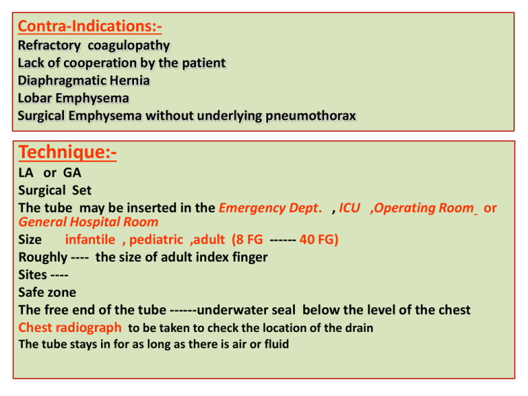
Contra-Indications:Refractory coagulopathy Lack of cooperation by the patient Diaphragmatic Hernia Lobar Emphysema Surgical Emphysema without underlying pneumothorax Technique:LA or GA Surgical Set The tube may be inserted in the Emergency Dept. , ICU ,Operating Room or General Hospital Room Size infantile , pediatric ,adult (8 FG ------ 40 FG) Roughly ---- the size of adult index finger Sites ---Safe zone The free end of the tube ------underwater seal below the level of the chest Chest radiograph to be taken to check the location of the drain The tube stays in for as long as there is air or fluid How long is a chest tube used ? The tube remains in place until the lung is re-expand or the fluid is drained. Occasionally the patient require more than one chest tube Indications for Removal Clinical Mechanical Radiological Complications:- 1-Minor Complications:Severe pain during placement Subcutaneous hematoma or seroma Anxiety Shortness of breath (Dyspnea) Cough ( Rapid drainage of fluid ) 2-Major Complications Hemorrhage ---haemothorax or haemoptysis Infection Reexpansion pulmonary edema Injury to the liver , spleen , diaphragm . Injury to the Thoracic aorta & the heart Bronchoscopy Bronchoscopy Looking into the living lungs (Chevalier Jackson 1928) Today with the major advance in technology View the fine details of the end bronchial anatomy Diagnosis of the disease Treating diseases It is the visualization of the air way using either rigid Bronchoscope (GA) or the flexible (Fiber optic Bronchoscope ) (LA) or both simultaneously .Through which we can remove FB , take BAL , brushing lesions &Trans bronchial Biopsy . HISTORY Gustav Killian (The father of bronchoscopy ) , was appointed professor of ENT at the university of Freiburg in 1892 . Gustav Killian in 1897 succeeded in removing aspirated pork bone from the bronchus of a 63 –year-old farmer under cocaine anesthesia .He used external light source , a head mirror , esophagoscope and forceps to remove the bone . He became famous & his clinic attracted patients from far and wide for his expertise in removing different kind of (FB) such as bones ,beans ,buttons ,coins & tin whistle . Gustav Killian Bronchoscope , external light source Bronchoscopy rapidly developed into a science (with the creation of a better instruments and techniques ) Chevalier Jackson Founded philadelphia school of bronchoesophagology Jackson‘s monograph first published in 1950 Chevalier Jackson’s Bronchoscope with a small distal bulb & built –in suction tube Early in the 1960s Shigeto Ikeda devised a means to replace the small electric bulb with glass fibers capable of transmitting brighter light from an outside source. He presented the first flexible bronchoscope at the 1966 International Congress on Diseases of the Chest in Copenhagen. H.H.Hopkins English physicist developed the rod-lens optical telescopes which could be used with the rigid bronchoscope At the end of the 1980s, Asahi Pentax replaced the fiberoptic bundle with a charge-coupled sensor at the tip of the scope. This videobronchoscope allowed the bronchoscopist to look at a monitor screen instead of through the eyepiece of the scope
