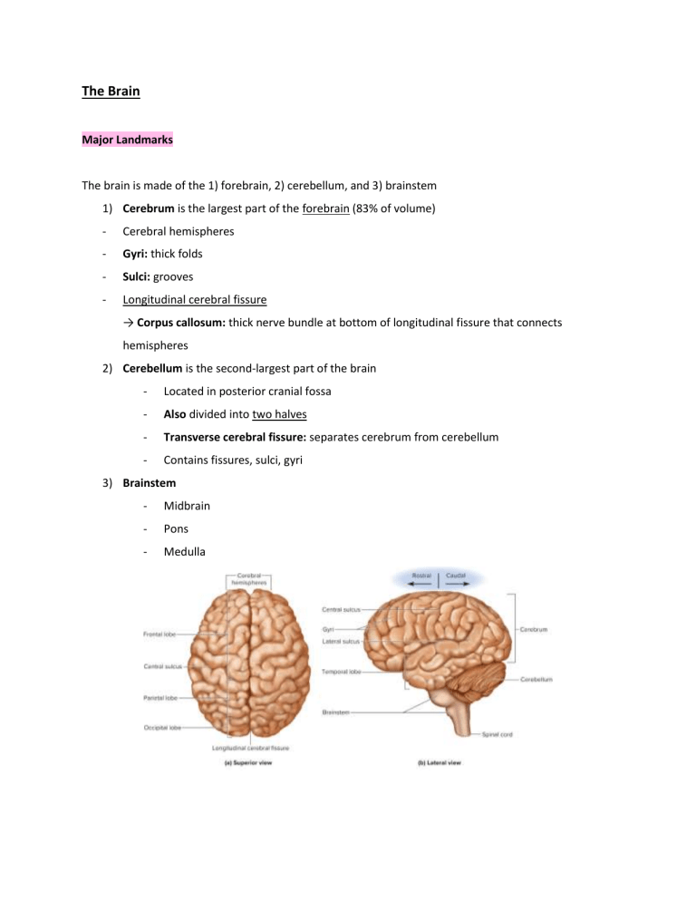
The Brain Major Landmarks The brain is made of the 1) forebrain, 2) cerebellum, and 3) brainstem 1) Cerebrum is the largest part of the forebrain (83% of volume) - Cerebral hemispheres - Gyri: thick folds - Sulci: grooves - Longitudinal cerebral fissure → Corpus callosum: thick nerve bundle at bottom of longitudinal fissure that connects hemispheres 2) Cerebellum is the second-largest part of the brain - Located in posterior cranial fossa - Also divided into two halves - Transverse cerebral fissure: separates cerebrum from cerebellum - Contains fissures, sulci, gyri 3) Brainstem - Midbrain - Pons - Medulla Gray and White Matter Gray Matter - Cell bodies, dendrites, synapses/ axon terminals - Myelin sheath - Cortex: the cerebrum and cerebellum have a surface of gray matter on the outside; thought processing occurs here in cerebrum - Nuclei: pockets of gray matter deep in the brain, surrounded by white matter White Matter - Composed of tracts; bundles of nerve fibers (axons) - Deep to cortical gray matter - Connect parts of the brain and to the spinal cord Gray matter is on the outside of the brain, white matter is on the inside of the brain - However, this flips when we get to the spinal cord because of processing/receiving differences Meninges Meninges are protective layers of connective tissue in the brain and are continuous all the way down the spinal cord 1) Dura Mater - tough, anchors, has connections to arachnoid mater - Outer periosteal layer: like periosteum - Inner meningeal layer: continues into vertebral canal and forms dural sheath around spinal cord → Dural sinuses: spaces between the separation of periosteal and meningeal layers that collect blood circulating through the brain; constantly producing CSF, part of vascular system 1) Superior sagittal sinus - return of CSF along median line 2) Transverse sinus - return of CSF from rear of head to each ear; between cerebellum and cerebrum Folds of Dura Mater - Falx cerebri: separate two cerebral hemispheres - Tentorium cerebelli: separate cerebrum from cerebellum - Falx cerebelli: separate right and left halves of cerebrum 2) Arachnoid Mater - transparent membrane over brain surface - Subarachnoid space separates it from the pia mater below; filled with CSF and blood vessels 3) Pia Mater - extremely thin membrane - Follows brain contours - Follows arteries as they penetrate into the cerebrum Ventricles - Aqueducts for CSF to flow ● Lateral ventricles: one in each cerebral hemisphere - CSF generated here; lined with ependymal cells ● Third ventricle: narrow medial space beneath corpus callosum ● Fourth ventricle: small chamber between pons and cerebellum Ventricles connected by… ● Interventricular foramen: pore connecting lateral ventricles to third ventricle ● Cerebral aqueduct: tube running through midbrain connecting third and fourth ventricles ● Central canal: tube connecting fourth ventricle and runs through spinal cord center - Largest passageway CSF leaves 4th ventricle to go through the central canal, to the spinal cord, to the pia and arachnoid mater, then flows back up Cerebrospinal Fluid - Clear, colorless fluid that fills ventricles of the CNS and bathes its external surface - Production of CSF begins with filtration of blood plasma through brain capillaries - Continuously flows CNS, driven by pressure, cilia, brain pulsations from heartbeat Choroid Plexus: spongy mass of blood capillaries on the floor of each ventricle Ependymal cells: neuroglia that line ventricles and cover the choroid plexus - Modify filtrate CSF has more Na+ and Cl- than blood plasma and less K+, Ca2+, glucose, and little protein Functions of CSF 1) Buoyancy - Brain attains size without being impaired by weight 2) Protection - Protects brain from striking cranium when head is jolted 3) Chemical Stability - Flow of CSF rinses away metabolic wastes from nervous tissue and regulates chemical environment The Hindbrain and Midbrain Medulla Oblongata Adult brain region that develops from the embryonic myelencephalon - Begins at foramen magnum - Basic functions; sends information to the heart Features: ● Pyramids: ridges on anterior surface; separated by anterior median fissure ● CN 8, 9, 10, 12 begin or end in medulla ● Olives: prominent bulges lateral to each pyramid ● Gracile (medial) and cuneate (lateral) fasciculi continue as two pairs of ridges on the posterior medulla - ● Contains sensory fibers Ascending and descending fibers connecting the brain and spinal cord pass through the medulla Ex: - Medial lemniscus: axons of gracile and cuneate nuclei decussate (cross over) and form ascending (sensory) tracts to the thalamus for communication - Corticospinal tracts: descending motor tracts in pyramids; carry signals to skeletal muscles ● Contains many nuclei - Inferior olivary nucleus: relay center for signals to cerebellum - Reticular formation: loose network of nuclei extending through entire brainstem; contains cardiac center, vasomotor center, and respiratory center; more posterior Cross Section of Brain Stem: Pons Adult brain region developed from embryonic metencephalon - Posteriorly, consists of thick stalks; cerebellar peduncles - Last area for finite control and communication Features: ● Cerebellar peduncles connect the pons and midbrain to the cerebellum on posterior side ● CN 5, 6, 7, 8 - Sensory roles: hearing, equilibrium, taste, facial sensation - Motor roles: eye movement, facial expression, chewing, swallowing, urination, secretion of saliva and tears ● Reticular formation in pons contains additional nuclei concerned with sleep, respiration, & posture Midbrain - Brain region that develops from the embryonic mesencephalon - Connects hindbrain to forebrain Anatomical Features: ● Cerebral aqueduct; surrounded by central (periaqueductal) gray substance involved in pain awareness ● Continuations of medial lemniscus and reticular formation ● Motor nuclei of CN III and IV that control eye movements ● Tectum: roof-like; posterior to cerebral aqueduct with four bulges: 1) Two superior colliculi: visual, tracking moving objects 2) Two inferior colliculi: relay signals from ear to thalamus and other parts of brain ● Cerebral peduncles: two anterior midbrain stalks that anchor the cerebrum to the brainstem 1) Tegmentum - Red nucleus - Inside - Connections run to and from cerebellum for motor control 2) Substantia Nigra - Nucleus within peduncle; dark, melanin - Relays inhibitory motor signals to thalamus and basal nuclei, suppressing unwanted body movement - Degeneration = Parkinson’s Disease 3) Cerebral Crus - Bundle of nerve fibers connecting communication from cerebrum to pons - Carries corticospinal tracts Reticular Formation - Loose web of gray matter running vertically through all levels of the brainstem and into the upper spinal cord - Occupies space between white fiber tracts and brainstem nuclei - Connects to many areas of cerebrum - Basic functions: heart rate, respiration - Sensory input and output; visual sent in from eye to the reticular formation Functions of Reticular Formation Nuclei 1) Somatic motor control - Adjusting muscle tension for tone, balance, posture - Relay signals from eye and ears to cerebellum - Integrate visual, auditory, balance, and motion stimuli into motor coordination - Gaze centers: allow eyes to track and fixate on objects - Central pattern generators: neural pools producing rhythmic signals to muscles of breathing and swallowing 2) Cardiovascular Control - Cardiac and vasomotor centers of medulla 3) Pain modulation - Some pain signals ascend through - Some descending analgesic pathways begin in the reticular formation and end in the spinal cord where they are blocked 4) Sleep and Consciousness - Crucial role - Injury = irreversible coma 5) Habituation - Reticular activating system modulates activity in the cerebral cortex; ignores repetitive, unimportant stimuli The Cerebellum The cerebellum is the largest part of the hindbrain, the second-largest part of the brain, contains more than half of the brain's neurons. - Granule cells: found in cerebellum; most abundant neuron in the brain - Purkinje cells: large cerebellar neurons; axons project deep into nuclei to synapse with neurons that lead to the brainstem; huge input side, small output side Anatomical Features: ● Right and left cerebellar hemispheres connected by the vermis ● There is a superficial cortex of gray matter with folia (folds), arbor vitae (branching white matter) ● Input to the cerebellum goes to the cortex, output comes from the deep nuclei ● Cerebellar peduncles: three pairs of stalks that connect the brainstem and cerebellum; their fibers carry signals to and from the cerebellum 1) Inferior peduncles: connect to the medulla, most spinal input enters the cerebellum through here 2) Middle peduncles: connected to pons, most input from rest of the brain enters through here 3) Superior peduncles: connected to midbrain, carries cerebellar output Functions: 1) Motor coordination and locomotor ability 2) Sensory, linguistic, emotional, and other nonmotor functions, including: ● Comparing textures of objects ● Perceiving space (as tested by pegboard puzzles) ● Recognizing objects from different views ● Keeping judge of elapsed time and maintaining tapping rhythm ● Directing eye movements to compensate for head movement ● Judging pitch of tones; distinguishing between spoken words ● Helping in verbal association tasks ● Planning, scheduling, and emotion control The Forebrain Two parts: 1) Diencephalon - Encloses third ventricle, most rostral part 2) Telencephalon - Develops into the cerebrum Diencephalon 1) Thalamus - Ovoid mass each side of brain - Joined by interthalamic adhesion - ⅘ of diencephalon - 23 nuclei in five groups: anterior, posterior, medial, lateral, ventral - Major TRANSMISSION area - Gateway to cerebral cortex; nearly all input to cerebrum synapses in thalamic nuclei - Not all information is passed along; screens out most of information - Motor control - Relays signals from cerebellum to cerebrum - Memory and emotion - Some of limbic system 2) Hypothalamus - Major CONTROL center - Extends anteriorly to optic chiasm, and posteriorly to mammillary bodies; each containing 3-4 mammillary nuclei that relay signals from the limbic system to thalamus - Infundibulum: stalk-like structure where the hypothalamus attaches to the pituitary - Contains many nuclei with a variety of visceral, emotional, and behavioral functions → Hormone secretion - Anterior pituitary: growth, metabolism, reproduction, stress response - Posterior pituitary: labor contractions, lactation, water conservation → Autonomic nervous system - Major integrator - Heart rate, BP, GI secretions → Thermoregulation - Hypothalamic thermostat monitors body temps → Food and water intake - Regulates hunger and satiety - Osmoreceptors; blood, ADH to conserve water → Sleep and circadian rhythms - Suprachiasmatic nucleus → Memory - Mammillary nuclei relay signals from hippocampus to thalamus → Emotional behavior and sexual response 3) Epithalamus ● Pineal gland - endocrine gland ● Habenula - relay info from limbic system to midbrain The Cerebrum ● Develops from the telencephalon and is the largest part of the brain ● Sensory perception, memory, thought, judgment, voluntary motor actions Anatomy ● Two hemispheres w/ longitudinal fissure ● Connected by corpus callosum; white fibrous tract ● Gyri and sulci increase amount of cortex for more information-processing Lobes ● Frontal - Voluntary motor, motivation, foresight, planning, memory, mood, emotion, social judgment, aggression ● ● ● ● - Cognition and conscious thought - Rostral to central sulcus Parietal - Between central sulcus and parieto-occipital sulcus - Integrates general senses, taste, and some visual - Where we receive sensory information/ somatosensory (coming in from periphery) Occipital - Caudal to parieto-occipital - Primary visual center Temporal - Below lateral sulcus - Broca and Wernicke’s areas - Hearing, smell, learning, memory, some vision and emotion Insula - Deep to lateral sulcus - Understanding spoken language, taste, pain, integration from visceral receptors Cerebral white matter is most of the volume of the cerebrum - Glia and myelinated fibers that transmit signals - Tracts: bundles of nerve fibers in CNS 1) Projection: vertically between higher and lower brain and spinal cord centers; dive down and in 2) Commissural: crossover cerebral hemispheres; most pass through corpus callosum 3) Association: contact different regions with the same cerebral hemisphere; long association fibers connect different lobes, short association connect gyri in a lobe; move laterally (ex: putting together words and processing them as a language) The cerebral cortex is a layer of gray matter covering the hemispheres - Billions of neurons - 40% of brain mass - Stellate cells: spheroidal cell bodies with short axons, and dendrites; receive sensory input and process information locally - Pyramidal cells: tall, conical with axons that project into white matter below, output neurons that connect cortex to other CNS parts - 90% is the neocortex: six-layered tissue that is evolutionary recent - Many dendrites at top and more axons towards bottom The limbic system is an important center of emotion and learning, sensation of joy; contains components in each hemisphere: - Cingulate gyrus: arches over corpus callosum in frontal and parietal lobes - Hippocampus: in medial temporal lobes - Amygdala: rostral to hippocampus Connected by fiber tracts for circular feedback patterns Reward and aversion Basal nuclei are masses of cerebral gray matter buried deep in white matter, lateral to the thalamus - Motor control - Processing and storage of information - Include: caudate nucleus, putamen, globus pallidus, collectively called corpus striatum - Putamen and globus are called lentiform nucleus Processing Cognition ● Sensory perception ● Thought ● Reasoning ● Judgment ● Memory ● Imagination ● Intuition Cognition is accomplished by association areas of the cerebral cortex, which is 75% of all brain tissue - Parietal: perceive and attend to stimuli → Lesions = Contralateral neglect syndrome: unaware of objects on opposite side of the body - Temporal: identify stimuli → Lesions = Agnosia: inability to recognize or identify familiar objects (ex: prosopagnosia - faces) - Frontal: think about world; plan and execute appropriate behaviors → Lesions = Personality disorders and socially inappropriate behaviors Memory Information management in the brain requires 1) Learning - acquiring info 2) Memory - info storage + retrieval 3) Forgetting - eliminating trivial info Anterograde amnesia: unable to store new information Retrograde amnesia: unable to recall known information before the injury The hippocampus is an important limbic system area for memory → Memory consolidation: teaching the cerebral cortex until a long term memory is established there; does not hold the memory, but organizes cognitive information into a unified long-term memory The cerebellum is involved in learning motor skills The amygdala is involved in emotional memory Emotion - Emotional feelings and memories involve interactions between the prefrontal cortex and diencephalon → Prefrontal cortex: judgment, intent, and control over emotional expression - Feelings arise from deep brain regions; hypothalamus and amygdala Amygdala - Fear - Food intake - Sexual behavior - Drawing attention to new stimuli - Receives input from sensory systems - Outputs to hypothalamus, influencing somatic and visceral motor systems → Heart races, blood pressure increases, hair stands, vomiting ensues - Outputs to prefrontal cortex, controlling expression of emotions → Expressing love, controlling anger, overcoming fear - Behavior shaping by learned associations between stimuli, our responses, and reward or punishment that results Sensation The primary sensory cortex is where sensory input is first received and when one becomes conscious of a stimuli - Association areas near the primary sensory areas process and interpret the sensory information Ex: primary visual cortex is boarded by visual association areas → Multimodal association areas: receive input from multiple senses and integrate into overall perception of surroundings Ex: orbitofrontal cortex receives taste, smell, and visual to provide desirability of food Our special senses are… ● Vision → Primary visual cortex: far posterior region of occipital lobe; receives visual signals → Visual association area: borders primary cortex anteriorly and occupies all the remaining occipital lobe → Temporal lobe deals with recognizing faces and familiar objects ● Hearing → Primary auditory cortex: superior region of temporal lobe; receives auditory signals → Auditory association area: temporal lobe, inferior to primary cortex; recognize spoken words, music, voices ● Equilibrium → Signals from inner ear project to cerebellum and brainstem nuclei ● Taste → Primary gustatory cortex: inferior end of postcentral gyrus; receives taste signals ● Smell → Primary olfactory cortex: medial cortex of temporal lobe; receives smell signals Our general (somatosensory, somesthetic, somatic) senses are… ● Touch ● Pressure ● Stretch ● Movement ● Heat & Cold ● Pain They are distributed over the entire body and involve simple receptors - Signals from head pass through CNs to brain - Signals from body ascend sensory tracts of spinal cord The thalamus processes input from the contralateral side - Relays signals to postcentral gyrus of parietal lobe → Primary somatosensory cortex: cerebral fold caudal to the central sulcus; processes awareness of stimuli → Somatosensory association area: caudal to the postcentral gyrus and in the roof of the lateral sulcus; makes cognitive sense of stimuli Sensory homunculus: diagram of sensory inputs to the primary somesthetic cortex in parietal lobe - Upside down sensory map of contralateral side of body Somatotopy: correspondence between an area of the body and an area of the CNS - Receptors in lower limbs projecting to superior and medial parts of the gyrus - Receptors from face projecting to the inferior and lateral parts of the gyrus Motor Control Voluntary motor control involves 1) Motor association (premotor) area - Intention/plan to contract muscle; found in frontal lobes - Neurons compile program required for action 2) Precentral gyrus (primary motor area) - Most posterior gyrus of frontal lobe - Send signals to brainstem and spinal cord for muscle contraction - Exhibits somatotopy, diagrammed as a motor homunculus - Neurons for toe movement are deep in the longitudinal fissure of the medial side of the gyrus - The summit of the gyrus controls the trunk, shoulder, and arm - The inferolateral region controls the facial muscles - Homunculus looks distorted because the amount of cortex devoted to a given body region is proportional to the number of muscles and motor units in that body region (not body region size) - Boundaries of cortical areas controlling body regions overlap, not sharply defined—a muscle is controlled by neurons at several points within a general area of gyrus Neurons involved in motor control: ● ● Upper motor neurons: pyramidal cells of the precentral gyrus - Synapse with lower motor neurons whose axons innervate skeletal muscles - End in nuclei of brainstem and/or form lateral corticospinal tracts - Decussate in medulla Basal nuclei - Onset and ceasing intentional movements - Hip and shoulder movements in walking - Practiced/learned behaviors; writing, typing, driving - Feedback circuit: Cerebrum & substantia nigra → basal nuclei → thalamus → back to cerebrum and substantia nigra ● Dyskinesias: movement disorders caused by lesions in basal nuclei (ex: Parkinson’s) Cerebellum - Learning motor skills - Muscle tone and posture - Smooths muscle contraction - Eye and body movements - Coordinates motions of joints - Lesions = ataxia: clumsy, awkward walking Language Wernicke Area: posterior to lateral sulcus, left hemisphere; recognition of spoken and written language - Fromulates phrases and transmits plan of speech to Broca Broca Area: inferior prefrontal cortex, left hemisphere; generates motor program for speech production - Transmits plan/program to primary motor cortex for commands to lower motor neurons supplying relevant muscles Emotional language aspects controlled by regions in opposite hemisphere - Affective language area: right - Lesions = prosody: flat, emotionless speech - Aphasia: language deficit from lesions in Wernicke or Broca areas Cerebral Lateralization Difference in structure and function between the two hemispheres; specialized for tasks and equally used Left - Categorical - Spoken and written language - Math and science - Information as fragments Right - Integrates information - Imagination and insight - Music and art - Patterns and spatial relationships - Comparison of sights, sounds, smells, taste Cerebral lateralization correlated with handedness, age, and sex - Lateralization develops with age, so children are more resilient to lesions on one side - Males exhibit more lateralization than females and suffer more functional loss with damage

