Genetics, Cell Division, DNA, RNA, and Mutations
advertisement
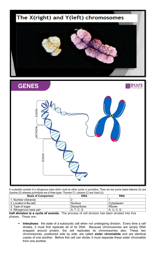
A nucleotide consists of a nitrogenous base which could be either purine or pyrimidine. There are two purine bases-Adenine (A) and Guanine (G) whereas pyrimidines are of three types- Thymine (T), cytosine (C) and Uracil (U). Basis of Comparison DNA RNA 1. Number of strands 2 1 2. Location in the cell) Nucleus Cytoplasam 3. Type of sugar Deoxyribose Ribose 4. Nitrogenous base pair A, T, C, G A, U, C, G Cell division is a cycle of events. The process of cell division has been divided into five phases. These are: Interphase: the state of a eukaryotic cell when not undergoing division. Every time a cell divides, it must first replicate all of its DNA. Because chromosomes are simply DNA wrapped around protein, the cell replicates its chromosomes also. These two chromosomes, positioned side by side, are called sister chromatids and are identical copies of one another. Before this cell can divide, it must separate these sister chromatids from one another. Prophase: The chromatin (The total collection of DNA and proteins in a chromosome.), diffuses in interphase, condenses into chromosomes. Each chromosome has duplicated and now consists of two sister chromatids. The centrioles form asters (ray-like structures) and move toward the opposite sides of the cell. At the end of prophase, the nuclear envelope breaks down into vesicles. Metaphase: The chromosomes align at the equatorial plate (center of the cell) and are held in place by microtubules attached to the mitotic spindle and to part of the centromere. Anaphase: The centromeres divide. Sister chromatids separate and move toward the corresponding poles. Telophase: Daughter chromosomes arrive at the poles and the microtubules disappear. The condensed chromatin expands and the nuclear envelope reappears. The cytoplasm divides, the cell membrane pinches inward ultimately producing two daughter cells (phase: Cytokinesis). Four stages can be described for each nuclear division. First division of meiosis Prophase 1: Each chromosome duplicates and remains closely associated. These are called sister chromatids. Crossing-over can occur during the latter part of this stage. Metaphase 1: Homologous chromosomes align at the equatorial plate. Anaphase 1: Homologous pairs separate with sister chromatids remaining together. Telophase 1: Two daughter cells are formed with each daughter containing only one chromosome of the homologous pair. Second division of meiosis: Gamete formation Prophase 2: DNA does not replicate. Metaphase 2: Chromosomes align at the equatorial plate. Anaphase 2: Centromeres divide and sister chromatids migrate separately to each pole. Telophase 2: Cell division is complete. Four haploid daughter cells are obtained. DNA Replication Here Are The Enzymes Of Dna Replication: The following are the events while DNA copies itself: Step 1: Initiation DNA replication starts at the Origin of Replication. The unzipping enzyme Helicase, causes the DNA strand separation, which leads to the formation of the replication fork. It breaks the hydrogen bond between the base pairs to separate the strand, thus separating the DNA into individua strands. Step 2: Elongation During elongation, DNA Polymerase III makes the new DNA strand by reading the nucleotides on the template strand and binding one nucleotide after the other to generate a whole new complementary strand. It helps in the proofreading and repairing the new strand. DNA Polymerase is able to identify and back track any mis paired nucleotides and corrects it immediately. The bases attached to each strand then pair up with the three nucleotides found in the cytoplasm. If it finds an Adenine (A) on the template, it will only add a Thymine (T). If it finds a Guanine (G) on the template, it will only add a Cytosine (C). Step 3: Termination In the previous steps of DNA replication, at the Origin, a Primer helps the DNA Polymerase to initiate the process. As the strand is created, the primer has to be removed. This is when DNA Polymerase I comes into the picture to replace the RNA nucleotides from the Primer with DNA nucleotides to make sure it is DNA all the way through. When DNA Polymerase III adds nucleotides to the lagging strand and forms Okazaki fragments, it leaves a gap or two between the fragments. These gaps are filled by the enzyme ligase and makes sure that everything else is connected. Transcription The following are the processes of Transcription: Step 1: Initiation Initiation is the start of transcription. It transpires when the enzyme RNA polymerase binds to a specific region of a gene which is called the promoter with the help of proteins called ‘transcription factors’. This signals the DNA double strand to unwind and open so the RNA polymerase enzyme can ‘‘read’’ the bases found in one of the DNA strands. With the open strands, one is considered as the template strand (anti-sense strand) and this will be used to generate the mRNA. The other is called the non-template strand (sense strand). After reading the bases, the RNA polymerase enzyme is now ready to make a strand of mRNA with a complementary sequence of bases. Step 2: Elongation Elongation is the adding of nucleotides to the mRNA strand. RNA polymerase reads the opened DNA strand and forms the mRNA molecule with the use of complementary base pairs. There is a short time during this process when the newly formed RNA is bound to the opened DNA. During this process of elongation, an adenine (A) in the DNA binds to an uracil (U) in the RNA. RNA polymerase does not need a primer during this process. It simply initiates the mRNA synthesis from the starting point and then moves downstream reading the anti-sense strand from 3’ to 5’ and generating the mRNA from the 5’ to 3’ end as it goes. Unlike helicase enzyme in DNA replication, RNA polymerase zips DNA back up as it goes keeping only 10-20 bases exposed one at a time. Step 3: Termination Termination is the last step of the transcription process. This happens when RNA polymerase enzyme reaches a stop or termination sequence in the gene. When the stop sequence or stop codon is reached, the enzyme detaches from the gene. The mRNA strand is now produced and it detaches from DNA. It carries with it the information encoded in the gene. By the end of transcription, the DNA segment is transcribed to form the mRNA molecule. The template strand shown below with the sequence T-A-C-T-A-G-A-G-C-A-T-T transcribes to form the mRNA A-U-G-A-U-C-U-CG-U-A-A. Translation The following are the processes of Translation: Step 1: Initiation After mRNA is formed in the nucleus, it leaves and moves to the cytoplasm where it finds the ribosome. Small ribosomal subunits then bind to mRNA. The initiator tRNA which is equipped with the anticodon (UAC) also binds to the start codon (AUG) of the mRNA. Let us say we have the mRNA codon AUG-UGC-AAG-UCCGGA-CAG, the tRNA anticodon would be UAC-ACG-UUC-AGG-CCU-GUC. The resulting large complex forms a complete ribosome and initiates protein synthesis. Each different tRNA is covalently linked to a particular amino acid. Step 2: Elongation Following initiation, a new tRNA-amino acid complex enters the codon next to the AUG codon. If the anticodon of the new tRNA matches the mRNA codon, base pairing occurs and the two amino acids are linked by the ribosome through a peptide bond. If the anticodon does not match the codon, base pairing cannot happen and the tRNA is rejected. Then, the ribosome moves one codon forward making space for a new tRNA-amino acid complex to enter. This process is repeated several times until the entire polypeptide has been translated. Step 3: Termination As the ribosome moves along the mRNA, it encounters one of the three stop codons for which there is no corresponding tRNA. Terminator proteins present at the stop codon bind to the ribosome and trigger the release of the newly synthesized polypeptide chain. The ribosome then disengages from the mRNA. On release from the mRNA, the small and large subunits of the ribosome dissociate and prepare for the next round of translation. The polypeptide chains produced during translation undergo some post-translational modifications, such as folding, before becoming a fully active protein. (14) Genetic Code Table Genetic Code Chart These amino acids are grouped as: essential and non-essential. Non-essential amino acids are those which the human body is capable of synthesizing, whereas essential amino acids must be obtained from the diet. Essential Amino Acids Symbol Non-Essential Amino Acids Symbol histidine His alanine Ala isoleucine Ile arginine Arg leucine Leu asparagine Asn lysine Lys aspartic acid Asp methionine Met cysteine Cys phenylalanine Phe glutamic acid Glu threonine Thr glutamine Gln tryptophan Trp glycine Gly valine Val proline Pro serine tyrosine Ser Tyr There are two types of mutations • Gene mutation is a permanent change in the DNA sequence that makes up a gene. • Chromosomal mutation occurs at the chromosome level resulting in gene deletion, duplication or rearrangement that may occur during the cell cycle and meiosis. It maybe caused by parts of chromosomes breaking off or rejoining incorrectly. Types of Gene Mutations Types Of Chromosome Mutation

