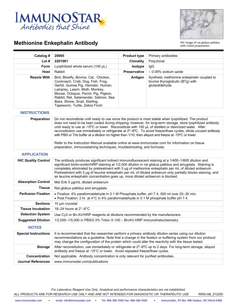
Methionine Enkephalin Antibody Catalog # IHC image of rat globus pallidus with nickel preparation. 20065 Product type Lot # 2201001 Form Lyophilized whole serum (100 µL) Host Rabbit Reacts With Clonality Isotype Preservative Bird, Blowfly, Bovine, Cat, Chicken, Cockroach, Crab, Dog, Fish, Frog, Gerbil, Guinea Pig, Hamster, Human, Lamprey, Leech, Moth, Monkey, Mouse, Octopus, Parrot, Pig, Pigeon, Rabbit, Rat, Salamander, Salmon, Sea Bass, Shrew, Snail, Starling, Tapeworm, Turtle, Zebra Finch Antigen Primary antibodies Polyclonal IgG ≤ 0.09% sodium azide Synthetic methionine enkephalin coupled to bovine thyroglobulin (BTg) with glutaraldehyde. INSTRUCTIONS Preparation Do not reconstitute until ready to use since the product is most stable when lyophilized. The product does not need to be kept cooled during shipping; however, for long-term storage, store lyophilized antibody until ready to use at -15ºC or lower. Reconstitute with 100 µL of distilled or deionized water. After reconstitution use immediately or refrigerate at 2º–8ºC. To avoid freeze/thaw cycles, dilute unused antibody with PBS or Tris buffer at a dilution no higher than 1/10, then aliquot and freeze at -15ºC or lower. Refer to the Instruction Manual available online at www.immunostar.com for information on tissue preparation, immunostaining techniques, troubleshooting, and formulas. APPLICATION IHC Quality Control The antibody produces significant indirect immunofluorescent staining at a 1/400–1/600 dilution and significant biotin-avidin/HRP staining at 1/2,000 dilution in rat globus pallidus and amygdala. Staining is completely eliminated by pretreatment with 5 µg of methionine enkephalin per mL of diluted antiserum. Pretreatment with 5 µg of leucine enkephalin per mL of diluted antiserum only partially blocks staining, and as leucine enkephalin concentration goes up, more diluted antiserum is blocked. Absorption Control Met Enk 5 µg/mL diluted antiserum Tissue Perfusion Fixation Sections Rat globus pallidus and amygdala • Fixative: 4% paraformaldehyde in 0.1 M Phosphate buffer, pH 7.4; 500 ml over 20–30 min. • Post Fixation: 2 hr. at 4°C in 4% paraformaldehyde in 0.1 M phosphate buffer pH 7.4. 10 µm cryostat Tissue Incubation 18–24 hours at 2°–8°C Detection System Use Cy3 or Bn-AV/HRP reagents at dilutions recommended by the manufacturers. Suggested Dilution 1/2,000–1/5,000 in PBS/0.3% Triton X-100 – Bn/AV-HRP immunohistochemistry NOTES Special Instructions It is recommended that the researcher perform a primary antibody dilution series using our dilution recommendations as a guideline. Note that a change in the fixation or buffering system from our protocol may change the configuration of the protein which could alter the reactivity with the tissue tested. Storage After reconstitution, use immediately or refrigerate at 2º–8ºC up to 2 days. For long-term storage, aliquot antibody and freeze at -15°C or lower. Avoid repeated freeze/thaw cycles. Concentration Journal References Not applicable. Antibody concentration is only relevant for purified antibodies. www.immunostar.com/publications For Laboratory Reagent Use Only. Analytical and performance characteristics are not established. ALL PRODUCTS ARE FOR RESEARCH USE ONLY AND ARE NOT INTENDED FOR DIAGNOSTIC OR THERAPEUTIC USE www.immunostar.com • email: info@immunostar.com • Tel: 866-386-3500 Fax: 866-386-4500 • RRID:AB_572250 ImmunoStar, PO Box 488 Hudson, WI 54016-0488
