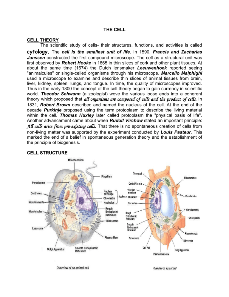
THE CELL CELL THEORY The scientific study of cells- their structures, functions, and activities is called cytology. The cell is the smallest unit of life. In 1590, Francis and Zacharias Janssen constructed the first compound microscope. The cell as a structural unit was first observed by Robert Hooke in 1665 in thin slices of cork and other plant tissues. At about the same time (1674) the Dutch lensmaker Leeuwenhoek reported seeing "animalcules" or single-celled organisms through his microscope. Marcello Malphighi used a microscope to examine and describe thin slices of animal tissues from brain, liver, kidney, spleen, lungs, and tongue. In time, the quality of microscopes improved. Thus in the early 1800 the concept of the cell theory began to gain currency in scientific world. Theodor Schwann (a zoologist) wove the various loose ends into a coherent theory which proposed that all organisms are composed of cells and the product of cells. In 1831, Robert Brown described and named the nucleus of the cell. At the end of the decade Purkinje proposed using the term protoplasm to describe the living material within the cell. Thomas Huxley later called protoplasm the "physical basis of life". Another advancement came about when Rudolf Virchow stated an important principle: All cells arise from pre-existing cells. That there is no spontaneous creation of cells from non-living matter was supported by the experiment conducted by Louis Pasteur. This marked the end of a belief in spontaneous generation theory and the establishment of the principle of biogenesis. CELL STRUCTURE I. CELL SURFACE 1. Cell wall - Plant cells are encased in an outer protective and supporting structure called the cell wall. It is composed of the polysaccharide cellulose in the form of threadlike structure called fibril. The first portion of the cell wall laid down by a young cell is the primary wall. Between the walls of adjacent cells is an intercellular layer called middle lamella which contains pectin. The middle lamella binds the cells together. The cells of the more woody portion of plants form an additional layer called secondary wall. This is located internal to the primary wall and contains lignin which adds stiffness and rigidity to cell walls. 2. Cell Membrane (plasma membrane or plasmalemma). This is the living boundary between the external and internal environment of the cell. It is a sturdy envelope that encloses the cell and behaves as a selective "gatekeeper" that determines what can and can not enter or leave the cell. The electron microscope together with biophysical and biochemical studies revealed that the cell membrane consists of a phospholipid bilayer sandwiched between layers of protein molecules that may penetrate or extend through the lipid membrane. The proteins may act as carrier molecules to allow passage of substances or as enzymes with biological functions such as converting nutrient molecules into ATP. These constantly moving and reorganizing protein molecules are responsible for the selective permeability of the cell membrane. The concept of fluidity refers to the fact that both lipids and proteins may have considerable lateral movement within the bilayer. Normally, these lipids are liquid at body temperature and the main consideration is the degree of saturation of the hydrocarbon chains. Functions: 1. protects the cell 2. seat of interaction of a cell with its surrounding 3. selective pathway for the transport of substances into and outside the cell 4. helps in cell adhesion and mobility 3. Cilia or flagella - Many cells bear specialized structures of locomotion called cilia or flagella. These are vibratile extensions of the cell surface. 2 II. NUCLEUS The most conspicuous protoplasmic structure is a spherical or ellipsoid body called the nucleus. It is the reproductive, metabolic, and dynamic center of the cell. A few unicellular organisms such as bacteria and blue-green algae do not show a distinctive separation of nuclear and cytoplasmic constituents. Cells without nuclei are called procaryotic cells in contrast to those with nuclei called eucaryotic cells. 1. Chromosomes- These are principal nuclear structures which consists of DNA and proteins. They are conspicuous only during cell division. at other times they appear as a network known as chromatin network. They bear in linear arrangement the basic units of heredity called genes Functions: 1. carry genetic information 2. play an important role in determining the phylogeny and taxonomy of the species 2. Nucleolus (little nucleus) - This is a highly compact and darkly staining spherical body which consists largely of RNA and proteins. It is now believed that the region of contact between the nucleolus and associated chromosome, the nucleolar organizer, is the site of large-scale production of RNA which is stored in the nucleolus and then sent into the cytoplasm, where it plays a key role in protein synthesis. Functions: 1. believed to be the site of ribosomal manufacture 2. collects freshly produced mRNA for cellular protein synthesis 3. plays an important role in cell division. 3. Nuclear Membrane - It has a double unit membrane structure. It communicates with an internal membrane network, the endoplasmic reticulum by minute pores. These pores may facilitate an exchange of materials between the nucleus and the cytoplasm. It is similar to the cell membrane in functions. 1. it bounds and protects the chromatin material in eukaryotic cells 2. gives definite shape to the nucleus 3. helps in the exchange of materials between the nucleus and cytoplasm 3 III. CYTOPLASM The largest volume of the cell is taken up by the cytoplasm, a transluscent, colorless, viscous mass where most of the organelles of the cell are situated. The soluble portion of the cytoplasm, the cytosol has a very high protein concentration exceeding 20%. Most of these are enzymes required in metabolism. 1. Mitochondria - These are round or filamentous structures with predominantly fatprotein composition. They are found in all cells. An outer layer covers the mitochondrial surface, whereas the inner surface is much folded and forms projections called cristae that extend into the interior fluid or matrix. In the matrix are a number of enzymes of the Krebs cycle, salts, water. The cristae is the site of protein synthesis for respiration. Research shows that the mitochondria are the principal sites for cellular respiration. The mitochondria, as the respiration centers have been aptly called the "powerhouse of the cell". Function: 1. production of energy during cell respiration 2. Golgi apparatus (Golgi bodies or dictyosomes) These structure were discovered by Camilo Golgi. They consist of flattened sacs which appear as parallel membranes, cluster of small, tightly packed vesicles and large clear vacuoles. Functions have not yet been completely defined but some investigators believe that they play an important role in secretion (cellulose) and provide a temporary storage area and repackaging center for substances like proteins and lipids. Lysosomes are believed to be derived from the golgi bodies. Functions: 1. Its main function is cell secretion not only of exportable proteins but also enzymes present in lysosomes and peroxisomes 2. Provide a temporary storage area and repackaging center for substances like proteins and lipids 3. Lysosomes- These organelles have been observed primarily in animal cells and protists. They are enclosed by single membrane and contain powerful digestive enzymes. These enzyme-filled sacs fuse with food vacuoles where intracellular digestion takes place. They also take part in the phagocytic activity of white blood cells, the degeneration of the tadpole's tail during metamorphosis and the digestion of certain bone cells. When the cell dies, the lysosomes rupture causing self4 digestion or autolysis of the cells. Thus these organelles are often referred to as "suicide bodies". Functions: 1. digestion of extracellular and intracellular substances 2. take part in phagocytic activity of white blood cells 3. lysis of organelles during cellular differentiation and metamorphosis (degeneration of the tadpole's tail) 4. when the cell dies, the lysosomes rupture causing self-digestion or autolysis of the cells. Thus these organelles are often referred to as suicide bodies 5. involved in cell aging 6. active in remodelling of tissues 4. Centrioles- These are two darkly stained bodies located just outside the nucleus in most animal cells. They determine the orientation of the plane of cell division. Functions: 1. determine the orientation of the plane of cell division 2. concerned with the movement of the chromosomes during cell division. The spindle is made up of microfilaments and microtubules. 5. Endoplasmic reticulum - This is an elaborate system of channels and cavities present in all nucleated cells. This double-layered system is continuous with the plasma membrane and nuclear membrane. It has often been described as a type of cytoskeleton that provides surface for chemical reactions, pathways for the transportation of cellular molecules and storage for synthesized molecules. Two types of endoplasmic reticulum have been observed: rough endoplasmic reticulum (RER) and smooth endoplasmic reticulum (SER). The rough endoplasmic reticulum has on its outer small granules called ribosomes. It is abundant in cells where protein synthesis is active. The smooth endoplasmic reticulum do not have ribosomes and occur in cells active in lipid synthesis. Functions: 1. it has often been described as a type of cytoskeleton that provides mechanical support to the cytoplasmic matrix 2. provides surfaces for chemical reactions, or as a transport system (circulatory system) that provides pathways for the transportation of cellular molecules and chemical reactions 3. mitochondria, golgi bodies, lysosomes are produced by endoplasmic reticulum 4. storage for synthesized molecules 5. transmits impulses in muscle and nerve cells 6. participates in excitation-contraction 7. involved in Cl- secretion in stomach cells 5 6. Ribosomes - These tiny spherical bodies contain relatively large amounts of RNA and are sites of protein synthesis. It has recently been discovered that protein synthesis involves the association of a molecule of messenger RNA sent out from the nucleus with a group of ribosomes called polyribosomes. Function: 1. site of protein synthesis 7. Plastids - These are organelles found in plants and serve as sites of synthesis of complex organic compounds and storage. They are classified according to color and function: a) Chloroplastids - are the green plastids containing the light-sensitive pigment chlorophyll. They are the centers of photosynthetic activity. b) Chromoplastids - these are yellow or red in color and owe their color to the presence of various carotenoids. They are responsible for the color of ripening fruits and autumn leaves. c) Leucoplastids- these are colorless plastids that serve as starch-forming and storage centers of plants. They are generally found in colorless leaf cells of stems, roots and other storage organs. 8. Microbodies - These are small cellular components possessing oxidases and catalases. One type of microbody is the peroxisome. The peroxisomes are membrane-bound organelles rich in peroxidase. They are abundant in liver and kidney cells and are concerned with peroxidation and beta oxidation of fatty acids. IV. CYTOSKELETON. The cytoskeleton is a network of filaments and fibers within the cytoplasm that helps maintain the shape of the cell, move substances within cells and anchors various structures in place. The cytoskeleton contains three types of fibers; the microfilament, the microtubule and the intermediate filaments. . 6 The microfilaments are the thinnest, twisted, double chain of protein molecules. They run parallel to the cell membrane in bundles. The microtubules are the thickest, consisting of a chain of proteins wrapped in a spiral to form a tube. They radiate from an area near the nucleus; they also provide intracellular support in nondividing cell. The intermediate filaments have “in-between size, thread-like protein molecules that wrap around one another to form “ropes” of protein. In a skin cell, these form thick, waxy bundles that probably provide structural support. 1. Microfilaments - These represent the active or motile part cytoskeleton consisting of actin, myosin, tropomyosin, and troponin. of the Functions: 1. cellular motility 2. cyclosis 3. ameboid movement 2. Microtubules - These are tiny tubules which are either free in the cytoplasm or forming part of centrioles, cilia and flagella. The main protein component is tubulin. Functions: 1. provide structural support to the cell 2. determine the polarity and direction of cell movement 3. play a role in the contraction of the spindle, movement of chromosomes and centrioles, as well as ciliary and flagellar movement. V. CELL INCLUSIONS - They are mostly organic in nature and are products of cell action. They may appear or disappear at different times in the life of the cell. a) Vacuoles (food or contractile). These are the most important inclusions of plant cells. The major components of vacuoles are water and some dissolved substances such as gases, salts, sugars, organic acids, soluble proteins and certain pigments called anthocyanin, responsible for the red, blue, and purple color of leaves, fruits and stems. Vacuoles facilitate exchange of gases with the surrounding medium. Contractile vacuoles found in numerous freshwater protozoans maintain water balance by pumping out excess water from the cell. Plants vacuoles contribute to cell turgor, contributing to the rigidity of plant parts. Food vacuoles enclose ingested food particles. Waste vacuoles serve as organs of excretion (metabolic trash cans). b) Other inclusions are starch granules, inorganic deposits in plant cells such as calcium oxalate crystals, crystals of calcium carbonate, calcium sulfate and silica, gluten and aleuron grains, and oil globules. 7
