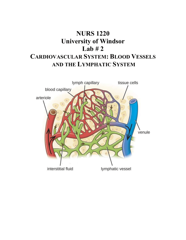
NURS 1220 University of Windsor Lab # 2 CARDIOVASCULAR SYSTEM: BLOOD VESSELS AND THE LYMPHATIC SYSTEM NURS 1220 – BLOOD VESSELS AND LYMPHATIC – Lab# 2 REFERENCE: any anatomy atlas/textbook and available references in lab; this includes the PowerPoint slideshow on BB. ACTIVITY ONE: Fetal Pig Identify the following structures on the fetal pig. o Ascending aorta o o Aortic arch o o Brachiocephalic artery o Coronary artery o o Right Subclavian artery o o Right Common Carotid artery o o Left Common Carotid artery Left Subclavian artery Descending Aorta (thoracic aorta) Diaphragm Abdominal aorta Renal artery (left and right) Hints for looking at the Fetal Pig: 1. Place the pig on the dissecting tray on its back. 2. Tie a piece of string around the “wrist” of one of the front legs. Run the string under the dissecting tray and tie it around the wrist of the other front leg. Pull the string so that the legs are spread apart. Don’t tie this too tight as adjustments may be necessary as the chest cavity is opened. Do the same with the hind legs. 3. Locate the heart. Turn the heart onto the dorsal side without removing it. 4. Locate the superior and inferior venae cavae (the two veins that bring blood to the heart from the body). 5. Locate the right and left pulmonary veins. (The veins that bring blood to the heart from the lungs). 6. Carefully dissect around the heart, picking away the connective tissue between the blood vessels. Locate the pulmonary artery, on the ventral side of the heart, as it leaves the right atrium. 7. Locate the aorta, the largest and most important artery; it can be found just beneath the pulmonary artery as it leaves the left ventricle. ACTIVITY THREE: Blood Vessel Slide Look at the prepared blood vessel slide set on 400x total magnification, identify the artery and vein. Be able to differentiate between the different vessels. Questions 1. Blood is carried away from the heart by ______________________, which divide into smaller vessels called _____________________. The vessels where gases/nutrients are exchanged between the blood and the body’s cells are the ___________________________. These drain into the venules, which unite to form increasingly larger vessels called ___________________. 2. Compare and contrast the blood vessel function and structure by completing the table below. Examples have been given. Blood Vessel Arteries Function Carries blood away from the heart Structure of Wall Capillaries Veins Three layers: all thinner than in arteries. 3. Label the following blood vessel figure using appropriate vocabulary (name of vessel, layers of walls) 4. List the general function of the following vessels. An example has been given. Structure Inferior vena cava Pulmonary arteries Superior vena cava Pulmonary veins Aorta Coronary arteries/veins Function Returns deoxygenated blood to heart from lower body 5. Label the following figure. 6. Match the following actions with the correct answer. Answers may be used more than once. a. lymph b. lymphatic capillaries c. lymphatic vessels ______ Contains macrophages ______ Begin blindly near blood capillaries ______ a.k.a. lymphatics ______ Fluid ‘lost’ to the interstitium ______ Empty into the right and thoracic ducts ______ Remove foreign materials from lymphatic fluid d. lymph nodes 7. Fill in the blanks regarding lymphatic vessels/organs Lymph from the left arm would return to the heart through the __________________________. The major job of the ____________________is to destroy worn-out red blood cells and return some of the products to the liver. The lymphoid tissues which trap and remove bacteria in the lymph are called ______________________.

