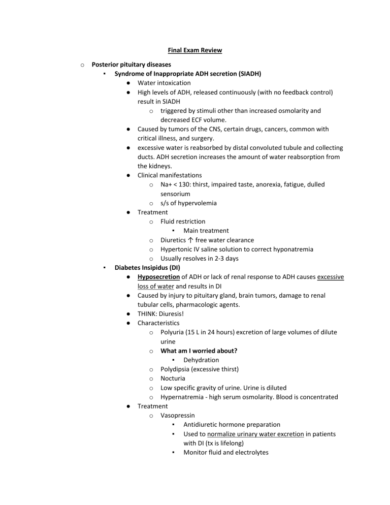
Final Exam Review o Posterior pituitary diseases ▪ Syndrome of Inappropriate ADH secretion (SIADH) ● Water intoxication ● High levels of ADH, released continuously (with no feedback control) result in SIADH o triggered by stimuli other than increased osmolarity and decreased ECF volume. ● Caused by tumors of the CNS, certain drugs, cancers, common with critical illness, and surgery. ● excessive water is reabsorbed by distal convoluted tubule and collecting ducts. ADH secretion increases the amount of water reabsorption from the kidneys. ● Clinical manifestations o Na+ < 130: thirst, impaired taste, anorexia, fatigue, dulled sensorium o s/s of hypervolemia ● Treatment o Fluid restriction ▪ Main treatment o Diuretics ↑ free water clearance o Hypertonic IV saline solution to correct hyponatremia o Usually resolves in 2-3 days ▪ Diabetes Insipidus (DI) ● Hyposecretion of ADH or lack of renal response to ADH causes excessive loss of water and results in DI ● Caused by injury to pituitary gland, brain tumors, damage to renal tubular cells, pharmacologic agents. ● THINK: Diuresis! ● Characteristics o Polyuria (15 L in 24 hours) excretion of large volumes of dilute urine o What am I worried about? ▪ Dehydration o Polydipsia (excessive thirst) o Nocturia o Low specific gravity of urine. Urine is diluted o Hypernatremia - high serum osmolarity. Blood is concentrated ● Treatment o Vasopressin ▪ Antidiuretic hormone preparation ▪ Used to normalize urinary water excretion in patients with DI (tx is lifelong) ▪ Monitor fluid and electrolytes ▪ o Monitor for water intoxication: ● Drowsiness ● Listlessness ● H/A ▪ Use caution in patients with CAD or PVD because it is a powerful vasopressin o DOC –DDAVP (Desmopressin) ▪ Administered twice daily as a nasal spray Anterior pituitary diseases ▪ GH deficiency ● Caused by decreased secretion of GHRF or GH, tumors, radiation, trauma ● Impairs normal growth and development in infants, children and adolescents (when GH is normally secreted in higher amounts). ● Treated with synthetic GH subcutaneously 3 to 7 days a week. Prior to closure of growth plates (epiphyseal plates) ● Goal: improved growth velocity and attainment of an adult height that is normal for the individual’s genetic background. ▪ GH excess ● Almost all cases caused by pituitary adenomas ● Gigantism– before puberty o Bones grow large, reach 7-9 ft tall ● Acromegaly – after puberty o Usually occurring in 4th or 5th decade of life. o Manifestations of Acromegaly ▪ Enlarged tongue ▪ Interstitial edema ▪ Coarse skin and body hair ▪ Enlargement facial bones, hands, feet ▪ Profusion of the jaw and forehead ▪ Barrel chest with arthralgia and arthritis ▪ Nerve damage: weakness, muscular atrophy, footdrop, and sensory changes ▪ Hypertension, left heart failure, CNS disturbances ▪ Enlarged & overactive sebaceous and sweat glands ▪ Impaired glucose tolerance o Treatment ▪ Remove Adenoma ● Resection of Anterior Pituitary tumor ▪ Possible radiation therapy ▪ Pharmacologic ● Octreotide (Sandostatin) ● Synthetic Somatostatin used to stop Growth Hormone Release ● o *Statin = stop* Thyroid ▪ Hypothyroidism ● Most common thyroid disorder ● Results from decreased levels of circulating thyroid hormone ● Primary – problem with thyroid o Results from pathologic process that destroys thyroid gland (high TSH, low thyroid hormone) ● Secondary – problem with pituitary o Caused by deficiency of pituitary TSH secretion (low TSH, low thyroid hormone) o May be med induced: ▪ Iodide, PTU ▪ Sulfonamides, Amiodarone, ▪ Interleukin 2, Interferon alpha ● If deficiency occurs during embryonic and neonatal life, called Cretinism (causes mental retardation and derangement of growth) ● Lowers BMR-results in a general slowing down of body processes (cold, dry skin) weight gain ● Lowers heat production, cold intolerance, lethargy, fatigue. Mentality may be impaired. ● Goiter if reduced levels of T3 and T4 promote excessive release of TSH ● Myxedema-altered composition of dermis and separation of connective fibers o Nonpitting, boggy edema around eyes, hands, feet o Thickened tongue, hoarseness, slurred speech o Myxedema coma – emergency!! (usually elderly women w chronic hypothyroidism)zx ▪ hypothermia ▪ hypoventilation ▪ hypotension ▪ bradycardia ● Diagnosis o Decreased levels of T3 and T4 o Serum TSH is high in primary and low in secondary ● Treatment o Synthetic Thyroxine (T4): ▪ Levothyroxine (Synthroid) is DOC (drug of choice) ● Usual dose: 100-150mcg/day for life ● Lifelong treatment; start low and work up ● Increases levels of T3 and T4 because most T4 is converted to T3 ● Also used for simple goiter and Hashimoto’s Disease o ▪ Other Pharmacologic options: ▪ Liothyronine (Cytomel) ● Synthetic T3 ● Not recommended for long-term use due to side effects ▪ Liotrix (Thyrolar) ● Combination of Levothyroxine and Liothyronine ● No advantage over Levothyroxine o If dosage is excessive, thyroid storm/thyrotoxicosis can occur Hyperthyroidism ● Increased thyroid hormone ● Caused by dysfunction of the thyroid gland, pituitary, or hypothalamus, or excessive intake of thyroid hormones ● Diagnosis o Excessive levels of circulating TSH ● Grave’s disease o Autoimmune disease in which developed antibodies stimulate TSH production and inappropriately activate production of thyroid hormones (T3 & T4) o Symptoms: Adrenergic stimulation- BMR ▪ Tachycardia and palpitations ▪ Heat intolerance-excessive sweating ▪ Nervousness ▪ Thin hair and skin ▪ Tremor ▪ Large and protruding eyeballs-exophthalmos ▪ Weight loss with hunger o Diffuse thyroid enlargement (goiter), may auscultate bruit o Because increased amounts of thyroid hormones reach the cells, all metabolic activities are increased; the BMR rises, energy expenditure is increased, and heat production rises ● Thyroid storm o Life-threatening complication o sudden increase in thyroid hormone levels o uncontrolled fever - 100 to 106 degrees o significant tachycardia, dysrhythmias o profuse diaphoresis o shock o vomiting o dehydration o CNS: hyperkinesis, anxiety, and confusion ● Treatment o Thiomides: ▪ Thyroid inhibitors (Antithyroid drugs) o o o ● Propylthiouracil (PTU) ● Methimazole (Tapazole) ▪ Stops the thyroid from making thyroid hormone! ▪ Does not destroy existing thyroid stores ▪ Overuse converts to hypothyroid state Monitor levels of T4 and T3 ▪ ▪ Goiter associated with prolonged use ▪ PTU is preferred treatment during pregnancy and breastfeeding Iodine Compounds ▪ Decrease the size and vascularity of the gland ▪ Radioactive Iodine (131I) – DOC for Graves Dx ● Used to destroy thyroid tissue (goal is to avoid destroying too much) ● Does not affect surrounding tissue ● Monitor bone marrow ● Usually 1-3 treatments, full effects may take 2-3 months ● Contraindicated with pregnancy ● Avoid sick people and children – body fluids are radioactive ▪ Lugol’s solution, SSKI (Potassium iodide) – nonradioactive ● Used preoperatively to decrease vascularity and decrease bleeding risk ● Dilute in fruit juice for taste, stains teeth ● Report symptoms of iodism: brassy taste, mouth burning, sore gum & teeth ● Report and discontinue if severe abdominal distress develops from toxicity Beta Blockers ▪ Inderal (Propranolol) ▪ Beta blockers won’t let you release catecholamines ▪ Decreases HR and BP ▪ Decreases anxiety ▪ Slows everything down, you remain calm Adrenals ▪ Addison’s Disease ● Hypofunction of Adrenals ● Chronic adrenal insufficiency o Caused by destruction of the adrenal glands o Autoimmune response – most common o ▪ Deficient cortisol secretion, may have ↓ aldosterone and androgen production ● Clinical manifestations: o Not enough aldosterone, will excrete Na+ and water and retain K+. Most of the S&S will initially come from HYPERKALEMIA o Cortisol insufficiency causes diminished gluconeogenesis, decreased liver glycogen, and increased sensitivity of peripheral tissues to insulin. o Blood sugar is going to go down o Symptoms are often vague & may not be apparent until 80-90% of the adrenals have been destroyed ● Commonly complain of: o Chronic fatigue, muscle weakness o N&V o Anorexia and weight loss o Occasional acute abdominal distress o Salt cravings (dt ↓ aldosterone and resulting hyponatremia) o Hypoglycemia o Hyperpigmentation o With persistent insufficient amts. of cortisol and aldosterone the body becomes: Weak, dehydrated, and unable to maintain BP ● Treatment o Combat the fluid volume deficit o Why are they losing volume? They are not producing enough aldosterone o Hormone replacement therapy ▪ Oral corticosteroids (Replace cortisol) ● Prednisone, Cortisone, Hydrocortisone ▪ Sometimes mineralocorticoids (Replace aldosterone) ● Fludrocortisone ● Maintains Na+/K+ balance o Increase salt in the diet o Treatment generally reverses symptoms o Lifelong treatment ● Addisonian Crisis o Adrenal crisis, acute adrenal insufficiency o Most commonly seen following abrupt withdrawal of long-term corticosteroid therapy Cushing’s Syndrome ● Corticosteroid excess o Result of pituitary or adrenal tumor o Excess intake of cortisol o or Corticosteroid drugs ● ● ● ● o ↑ Glucocorticoids o Breakdown of Fat and Protein ▪ Thin extremities, growth arrest o Increased risk of infection o Hyperglycemia o Depression to Psychosis ↑ Mineralocorticoids o Retain Na+ and Water, too much fluid o Lose K+ ↑ Sex hormones o Acne, oily skin o Hirsutism Treatment o Removal of tumor ▪ Will need replacement therapy for life o Adjunct to surgical removal of tumor o Drugs that ↓ corticosteroid production ▪ Aminoglutethimide (Cytadren) o Can expect resolution within one year after removal of tumor. ▪ Striae will persist Parathyroid ▪ Hyperparathyroidism ● Too much PTH in spite of Ca level ● Will see: o Increased Ca levels o Decreased Phos levels ● Bone becomes brittle and weak because calcium is being removed from bone and transported into serum ● Treatment o Remove parathyroid gland o Drug therapy may work initially, ▪ 1) Calcitonin (Miacalcin) – decreases serum calcium by driving Ca back into bones ▪ 2) oral phosphate - inhibit bone reabsorption, decreases calcium level (indirect relationship) ▪ 3) glucocorticoids - reduce intestinal absorption of calcium ▪ 4) several miscellaneous agents act directly on bone to prevent calcium removal o Surgical excision of parathyroid tumor ▪ Hypoparathyroidism ● Lack of normal feedback regulation ● Too little PTH in spite of Ca level ● Will see: ● ● o o Decreased Ca levels o Increased Phos levels Positive Chvostek and Trousseau’s sign Treatment o Calcitrol (Rocaltrol) ▪ Vitamin D analogue ▪ Promotes calcium secretion from the bone to bloodstream ▪ Promotes calcium uptake from the GI tract o Phosphorus binding drugs (PhosLo) ▪ Binds with Phosphorus ▪ Phosphorus decreases, Calcium increases o With continued use, assess for s/s of hypercalcemia Diabetes ▪ Type 1 DM ● Absolute lack of insulin ● Elevation in blood glucose ● Breakdown of body fats and proteins ● Diabetic ketoacidosis o Continued insulin deficiency results in lipolysis (breakdown) of body tissues. Both Pro and Fat are metabolized. As fat stores are metabolized, fatty acids are produced. The resulting fatty acids undergo transformation in the liver to ketoacids o Fruity breath o Ketones in urine ▪ Type 2 DM ● Caused by factors contributing to o 1) inadequate insulin secretion - the pancreas “gives out” o 2) insulin resistance - defect in the response of peripheral tissues to insulin o 3) increased hepatic glucose production ● HHNK o Characterized by extreme hyperglycemia (800-2000 mg/dL) and hyperosmolality (>350 mOsm/kg) ▪ Diagnosis ● OGTT > 200 after 2 hrs on at least 2 occasions = diabetes ● Fasting plasma glucose > 126 on at least 2 occasions = diabetes ● Casual plasma glucose test > 200 or higher suggests diabetes ● HA1C o 5% or less = negative o 5.7%-6.4% = pre-diabetes o 6.5% or more = diabetes ▪ S/S ● The 3 P’s o Polyuria, Polydipsia, Polyphagia Fatigue Glucosuria Blurred vision Dehydration / hyperkalemia/ hyponatremia with diuresis Hypovolemia Skin infections/yeast infections ▪ ▪ - ● ● ● ● ● ● Insulins ● Rapid-Acting Insulin o Lispro (Humalog), Novolog (Aspart), Apidra (Glulisine) o Onset: 10-30 minutes o Peak: 1-3 hours o Duration: 3-5 hours ● Short-Acting Insulin o Regular (Humulin R, Novolin R, Exubera) – unmodified human insulin o onset: 0.5-1 hour o peak: 2-4 hour o duration: 6-8 hours ● Intermediate-acting o NPH, Lente, Humulin N, Novolin N o Onset: 1-2 hr o Peak: 6-12 hr o Duration: 18-24 hr ● Long acting o Lantus (Glargine) ▪ Avoids peaks and valleys. ▪ Onset: 1 hour ▪ No peak ▪ Duration: 24 hours o Levemir (Detemir) ▪ Onset: 3-4 hours ▪ Peak: 6-8 hours ▪ Duration: 12-24 hours Hypoglycemia ● “Cold and clammy needs some candy” ● Blood glucose < 50 ● Caused by excess insulin Hematology o Anemias ▪ General s/s of anemia ● Pallor ● Tachycardia - reflect increased cardiac workload and output ● Angina - myocardial ischemia ▪ ▪ ● Dyspnea, SOB, and fatigue - decreased oxygen delivery ● Headache, dizziness, ringing in ears Iron deficiency anemia ● microcytic-hypochromic ● Causes o Increased iron demand during pregnancy (increase in blood volume and RBC synthesis by the developing fetus) o Inadequate iron intake, (i.e. children who only get milk first year of life) In most individuals, iron deficiency does not result from reduced iron uptake - need very little in diet to get sufficient amounts o Chronic blood loss - usually of GI or uterine origin o Hookworm infestation ● S/S o Tongue is shiny, smooth, beefy-red appearance, inflamed and sore o Cheilosis (cracks in the corners of the mouth) o Fine, brittle hair; thin nails o Pica (craving to eat unusual substances such as starch, ice chips) ● Lab values o Reduced RBC, hgb, hct level o MCV is decreased o Serum iron level is reduced o Total serum iron-binding capacity is increased ● Treatment o Iron is required for synthesis of hemoglobin o Inexpensive - drug of choice ▪ Oral – Ferrous sulfate (inexpensive) ▪ IM or IV – Iron Dextran (Imferon) beware of anaphylaxis o Adverse Effects: most significant involve the GI tract (dose dependent) ▪ nausea ▪ bloating ▪ constipation/diarrhea ▪ can aggravate peptic ulcers B12 deficiency ● Macrocytic-normochromic; can also be hyperchromic ● Pernicious anemia is the body’s inability to absorb Vitamin B12 d/t a lack of intrinsic factor secreted by the parietal cells of gastric mucosa ● S/S o Usually develops slowly because the body has a 3 year storage of Vitamin B12 o Deficiency of B12 causes demyelination of neurons, primarily in the spinal cord and brain - paresthesias and decreased DTR s o o In addition to other s/s of anemia, also experience severe glossitis (inflamed, painful beefy red tongue), diarrhea, and loss of appetite. o Proprioception (difficulty identifying one’s position in space) which may progress to difficulty with balance and spinal cord damage ● Treatment o Cyanocobalamin ▪ given SC or IM on a monthly basis o oral - not effective for pernicious anemia because not absorbed well o life-long treatment is required for pernicious anemia o Need to increase intake of foods such as eggs, meats, and dairy products ▪ Folic acid deficiency ● Macrocytic anemia; MCV high with low hgb ● Causes: poor nutrition, malabsorption syndrome (such as sprue – celiac disease), medications that impede absorption (oral contraceptives, anticonvulsants, alcohol abuse), anorexia, bulimia, and pregnancy ● S/S o Folate stores are limited to 3 mos. In the body, so symptoms are more pronounced than with pernicious anemia ▪ More severe diarrhea, glossitis, and cheilosis o Differentiated from B12 deficiency by lack of neurological symptoms ● Treatment – folic acid o Dietary - one fresh vegetable or one glass of fruit juice a day will usually correct the deficiency; foods such as green vegetables, broccoli, organ meats, eggs, and milk o Can be given orally - enough will be absorbed even if intestine is diseased o For severe deficiency may be given IM Anticoagulation therapy ▪ PT ● Responds to reduction of 3 of the 4 Vitamin K dependent clotting factors (II, VII, and, X) ● Used to monitor warfarin (Coumadin) therapy ● Measures the time blood takes to clot - common pathway ● Sensitive to alterations in vitamin K-dependent factors (Prothrombin) ● An increased PT refers to a longer time for clotting to occur, with clotting ability being less than normal ● Normal: 10-14 seconds ● Therapeutic warfarin is used to keep the PT at 1.5-2.0 times the normal level (15-28 seconds) ▪ INR ● ● ● ● ▪ - aPTT ● ● ● ● ● International Normalized Ratio (INR) – more consistent PT; used in lieu of PTs for some patients Because test results of PT can vary widely among labs and to ensure that test results among different labs are comparable, results are now reported in terms of an INR. INR = patient PT/mean normal PT An INR of 2-3 is appropriate for most patients on warfarin- although for some patients the target is 3 to 4.5. Examples: o Prevention of embolism in patients with atrial fib 2-3 o Prevention of recurrent DVT 2.5-4.0 o Prevention of arterial thrombosis, prevention of clots with mechanical heart valves - 3.0-4.5 Activated Partial Thromboplastin Time (aPTT) - more precise Used to assess the initial pathways (intrinsic/extrinsic) of clot formation Used to monitor heparin therapy Normal: 24-40 seconds Therapeutic heparin is used to keep the aPTT at 1.5 to 2 times the normal level (36-80 seconds) ▪ Coumadin (Warfarin) antidote ● Vitamin K ▪ Heparin antidote ● Protamine sulfate Immune system/Infections o Local signs of inflammation ▪ Warmth ▪ Redness ▪ Edema ▪ Loss of function ▪ Pain o Systemic signs of inflammation ▪ Fever ▪ Elevated WBC with a shift to the left ▪ Malaise ▪ Nausea ▪ Anorexia ▪ Tachycardia ▪ Tachypnea ▪ Lymphadenopathy o Complications of wound healing ▪ Adhesions- bands of scar tissue that forms around or between organs ▪ Contractions- shortening of muscle or scar tissue resulting in deformity ▪ Dehiscence- separation and disruption of joined wound edges o o o ▪ Evisceration- separation of wound edges where intestines extend through ▪ Fistula – abnormal passage ▪ Infection- microorganisms invade ▪ Hemorrhage- abnormal bleeding Antimicrobials ▪ Basic Principals ● Selective Toxicity: ability to injure target cell without injuring host cell ● Narrow spectrum: active against a few, selective microbes ● Broad spectrum: active against a wide variety of microbes ● Bacteriostatic: antibiotics that target protein synthesis ● Bactericidal: target bacterial cell wall or cell membrane ▪ Resistance – organisms have developed mechanisms to reduce or eliminate the effectiveness of the drug ● Prevention o Stop administering antibiotics to farm animals o Match the “BUG to the DRUG”—C&S testing o Stop unnecessary antibiotic use (virus), limit broad spectrum antibiotic use o Finish the complete prescription, don’t take leftovers o Use probiotics with antibiotic prescription to prevent suprainfection (elimination of normal flora) Types of immunizations ▪ Herd (community) immunity ● Resistance of a group to an infectious agent. ● Exists because a high proportion of people in the group are immune to an agent. ▪ Active immunity ● Resistance developed in response to an antigen and characterized by an antibody produced by the host. ● Natural: you get the disease ● Acquired: pharmaceutical (vaccine) ▪ Passive immunity ● Immunity conferred by an antibody produced in another host, ● Natural: breastfeeding ● Acquired: pharmaceutical (infusion of antibodies) Hypersensitivity reactions ▪ Type 1: Anaphylactic reaction – IgE mediated ● When exposed to an allergen the body produces IgE antibodies for that allergen ● The IgE antibodies attach to mast cell (tissue) and/or basophils (circulation). ● With re-exposure, the allergen attaches to the mast cell/basophil. ● ▪ ▪ ▪ ▪ The attachment signals the mast cell/basophil to release inflammatory mediators (histamine, serotonin, leukotrienes, kinins, bradykinins, heparin) from its vesicle ● Treatment o Antihistamines o Epinephrine o Corticosteroids o Mast cell stabilizers o Immunotherapy (allergy shots) Type 2: Cytotoxic reactions: Blood ● The direct binding of IgG or IgM antibodies to an antigen on the cell surface. ● Antigen-antibody complexes activate the complement system. ● Cellular tissue is destroyed in one of two ways o Activation of the complement system resulting in cytolysis or o Enhanced phagocytosis ● Typical cells destroyed include: erythrocytes, platelets, and leukocytes ● Disorders include: ABO incompatibility transfusion reaction, hemolytic anemias, leukopenias, thrombocytopenia, erythroblastosis fetalis. Type 3: immune complex reactions – chronic autoimmune diseases ● Soluble antigens combine with IgG and IgM antibodies to form complexes that are too small to be removed by the phagocytic system ● The complexes formed are then deposited into tissue and small blood vessels ● Complement system and inflammatory response is activated resulting in tissue destruction ● Common sites include: kidneys, skin, joints, blood vessels, lungs ● Disorders include: acute glomerulonephritis, rheumatoid arthritis, and systemic lupus erythematosus Type 4: Cell mediated or delayed hypersensitivity reactions ● Delay-type hypersensitivity o Sensitized T lymphocytes attack antigens or release cytokines o Macrophages invade and destroy tissue o Takes 24-72 after exposure to antigen for the reaction to occur o Types: ▪ Allergic Contact Dermatitis: Cosmetics, poison ivy, latex allergy ▪ Hypersensitivity Pneumonitis: exposure to inhaled organic dusts (mold, hay, bacteria). Experience labored breathing, dry cough, chills, fever. ▪ Transplant rejections Rheumatoid arthritis ● ▪ An autoimmune disease that results in a chronic, systemic inflammatory disorder that may affect connective tissue and organs, but principally attacks bilateral, flexible (synovial) joints. ● Diagnostic Tests (non-specific) o RF (Rheumatoid Factor) -Measures the amount of rheumatoid factor antibody in the blood. It is possible that the patient can have RF and not have rheumatoid arthritis o ACPA (Anti-citrullinated protein antibodies)-Are autoantibodies directed against one or more of an individual’s own proteins that are detected in the blood of RA patients. Better indicator of RA, but not definitive o ESR (Erythrocyte Sedimentation Rate)-Blood test that indicates acute or chronic inflammation and autoimmune disorders. Indicates inflammation in the body o CRP (C-Reactive Protein)-A protein that increases with inflammation. Indicates inflammation in the body ● Treatment o Immunomodulators ▪ Cyclophosphamide (Cytoxin) or Methotrexate ● Kills B & T cells undergoing proliferation. ● Toxic to all proliferating cells resulting in bone marrow suppression ▪ Cyclosporine: ● Acts on T cells to suppress production of interleukin 2, interferon gamma and other cytokines so immune system response suppressed or enhanced ● Side Effects: Nephrotoxicity, infection, hypertension, tremor, hirsutism ▪ Antibodies ● Directed against components of the immune system to suppress the immune response ● Ex: Rh Immune Globulin Systemic Lupus Erythematous (SLE) ● A multisystem inflammatory autoimmune disease. Affected systems include: skin, joints, serous membranes, renal, hematologic, and neurologic. ● Alternating periods of exacerbation and remissions ● Butterfly rash ● Lab Tests (non-specific) o RF levels elevated o ANA levels elevated in 97% of patients o Anti DNA levels elevated (more specific) ● Treatment o o o o o o HIV ▪ ▪ ▪ ▪ - - NSAIDs Antimalaria agents Corticosteroids Immunosuppressive drugs Topical immunomodulators HIV is a retrovirus that causes immunosuppression, which causes the person to be susceptible to infections that are normally controlled through the immune system. Replicates in a backward manner (RNA to DNA) HIV requires host cells with CD4 and chemokine receptors on the surface to replicate. These receptor sites are located on lymphocytes and monocytes/macrophages. Categories ● Cat 1: >500 CD4 cells, generally asymptomatic – this is where most people live today ● Cat 2: 200-499 cells, signs and symptoms of immune deficiency. ● Cat. 3: < 200 CD4 cells, AIDs defining illness Cancer o Hodgkin’s Lymphoma ▪ Proliferation of a specific abnormal B lymphocyte (Reed-Sternberg (RS) cells). Arises in a single node and spreads to anatomically contiguous nodes. ▪ Peaks at two ages – 15-20 and >50. Males > Females, whites > blacks ▪ Cause unknown: possibly carcinogens, viruses, genetic predisposition o Non-Hodgkin’s Lymphoma ▪ Malignant transformation of T cells, B cells, or NK cells. NHL does not have Reed-Sternberg (RS) Cells. ▪ Affects middle age to older adults primarily. More aggressive in children and young adults ▪ Causes ● Viral mutation of lymphocytes resulting in uncontrolled proliferation. ● Common viruses: HIV, EBV, Helicobacter pylori, Hepatitis C, Herpes. o Leukemia ▪ Clinical Manifestations ● Anemia/fatigue, Low grade fever, Night sweats, Weight loss ● Bleeding tendencies ● Bone pain ● Liver, spleen, lymph nodes enlargement ● Hyperuricemia ● Infection GI o Malabsorption syndrome ▪ Caused by ● Pancreatic insufficiency: o ● ● ▪ S/S ● ● ● ● ● o o lack of pancreatic enzymes needed for digestion of proteins, carbohydrates & fats. o Causes: pancreatitis, carcinoma, pancreatic resection, cystic fibrosis. Fat maldigestion is chief problem. o TX: pancreatic enzymes Lactase Deficiency: o lack of disaccharidase – congenital defect. o Unable to break down lactose – lactose ferments in intestines with bacteria where forms gas and increased osmotic gradient pulls water in which results in diarrhea Bile salt Deficiency: o Bile salts conjugated in liver – needed for fat absorption. o Causes: liver disease, Obstruction of bile duct diarrhea steatorrhea (stools contain excessive fat. The fat content causes bulky, yellow-gray malodorous that float in the toilet) flatulence/bloating abdominal pain and distention/cramps/wt loss failure to absorb fat-soluble nutrients/vitamins GERD ▪ Reflux of chyme and pepsin from stomach to esophagus (backward motion) ▪ S/S ● Heartburn, Upper abd. pain within an hour of eating that is worse if lying down. ● Often worse at night. Acidic foods, alcohol can cause pain on swallowing ▪ Eval/Tx ● endoscopy/ Antacids, weight reduction, stop smoking, sleep with HOB elevated ● Antireflux surgery = Nissen fundoplication “wrapping the fundus of the stomach around the lower portion of the esophagus ▪ Metoclopramide (Reglan) ● 1.Suppresses emesis by blocking dopamine/serotonin receptors n CTZ ● 2. Increases upper GI motility – enhances action of ACh ● SE: sedation as does goes up, extrapyramidal reactions. Gastritis ▪ Acute: ● erosions result of injury in protective mucosal barrier often by drugs, bacterial endotoxins, or chemicals ▪ SE: ● Abd. Discomfort, epigastric tenderness, bleeding ▪ Chronic: ● Autoimmune is the most severe type where gastric mucosa degenerates. ● ● ● o ▪ S/S: ▪ TX: PUD ▪ ▪ ▪ ▪ ▪ ▪ Decrease of chief and parietal cells decrease acid production, which impairs inhibition of gastrin. Intrinsic factor not available –B12 anemia. Also associated with chronic alcohol abuse, smoking, NSAIDs, and Helicobacter pylori most common cause of non-erosive ● vague – anorexia, fullness, N/V, pain ● small bland meals, B12 Break or ulcer in mucosal lining of lower esophagus, stomach, or duodenum (most common). Causes ● Helicobacter pylori (Most common cause), ● NSAIDs (2nd most common), ● Smoking, alcohol, Chronic disease, ● Duodenal – greatest frequency ● Greater among men. ● Hemorrhage maybe 1st sign S/S: ● burning, gnawing pain, cramp-like – when stomach is empty. ● Relieved with food/antacid. ● Worsening: anorexia, vomiting, wt. Loss Testing: ● Biopsy thru endoscopy, Breath radiolabeled urea, Blood – antibodies to H pylori or stool for antigens Complications: ● hemorrhage, obstruction, perforation with peritonitis Treatment ● Histamine receptor antagonist: o suppress secretion of gastric acid o Cimetidine (Tagamet): ▪ H2 receptor in parietal cells which secrete gastric acid are suppressed. Reduces vol. And H ion concentrations. ▪ SE: Low. CNS in elderly. Can cause levels of other drugs to rise. Antacids can decrease absorption o Ranitidine (Zantac) ▪ similar to Cimetidine – more potent with fewer SE ● Proton Pump Inhibitors: o Most effective for decreasing gastric acid secretion. o Omeprazole (Prilosec) ▪ converted to active form in parietal cells. Inhibits H+, K+, and ATPase – the enzyme that generates gastric acid. ▪ ● o o o SE: (short term) H/A, dizziness, N/V (long term): may have increased cancer risk. Others o Sucralfate (Carafate): ▪ creates a protective barrier against acid and pepsin. Forms a viscous and sticky gel that adheres to the ulcer crater that lasts up to 6 hr. ▪ SE: constipation o Misoprostol: (Cytotec) ▪ Prevents NSAID induced ulcers by replacement of prostaglandin E1. ▪ Category X – prostaglandins cause uterine contractions. ● Antacids: o alkaline compounds neutralize stomach acids o SE: constipation or diarrhea depending on type. Increase sodium load, effect of absorption of many drugs – take one hour before or after other meds. o Four groups of Antacids: ▪ Magnesium compounds ▪ Aluminum (Amphojel) compounds ▪ Calcium (TUMS) compounds ▪ Sodium (Na HCO3) compounds Ulcerative colitis ▪ Inflammatory disease of the rectum and colon ▪ Primarily affects the mucosal layer of the large intestine. ▪ The mucosa and submucosa are inflamed, become edematous, and ulcerations develop (and spread up the colon) ▪ Ulcerations also destroy the mucosal epithelium, causing bleeding and diarrhea (30 - 40 a day). ▪ Colitis goes through stages of exacerbation and remissions. ▪ With severe ulceration, severe diarrhea ▪ Major symptoms are pain, bloody diarrhea, and abdominal pain. Crohn’s disease ▪ Chronic, nonspecific relapsing, granulomatous, inflammatory bowel disorder of unknown origin (thought to be autoimmune currently) that can affect any part of the GI tract (small or large intestine). Can affect anywhere from the mouth to the rectum ▪ Inflammation occurs in a characteristic distribution called “skip lesions” with affected segments clearly separated by areas of normal tissue. ▪ Highly associated with formation of fistulae &/or obstructions ▪ Affects individuals 15-30 years of age, family history ▪ Typically, ulcerations are deep and longitudinal and penetrate between islands of inflamed edematous mucosa, causing a cobblestone appearance. Treatment: Ulcerative Colitis and Chron’s ▪ o o o S-Aminosalicylates: Sulfasalazine (Azulfidine) ● metabolized by bacteria into a component that decreases inflammation. ● Most effective against acute episodes of ulcerative colitis. ● SE: nausea/fever/ rash/ hematologic disorders ▪ Glucocorticoids: ● used for induction of remission, not long-term ▪ Immunomodulators/Immunosuppressants. ● Azathiaprine (Imuran) & Mercaptopurine (Purinethol). o Long term therapy. Used to maintain remission. Effects may be delayed up to 6 months. More toxic than the above drugs. o SE: pancreatitis, neutropenia ● Cyclosporine: acute/severe cases. o SE: immunosuppression ● Infliximab (Remicade). o Monoclonal antibody that binds with and inactivates TNF-alpha with is believed to play a role in Chron’s. o SE: fever/chills, CV- chest pain. Appendicitis ▪ The appendix becomes inflamed, swollen, and gangrenous, and it eventually perforates if not treated. ▪ Associate with intraluminal obstruction with fecalith. ▪ Most common 5-30 yo but can occur at any age. ▪ S/S ● Pain associated with inflammatory process. Localized in lower right quadrant. Rebound. Nausea, possible diarrhea, temperature, elevated WBC, polymorphonuclear cells (segs) ● Rebound tenderness ▪ Complications ● peritonitis, localized peri-appendiceal abscess formation, septicemia Diverticular disease ▪ Diverticulosis ● refers the presence of diverticula ▪ Diverticulitis ● refers to the inflammation or infection of diverticula ▪ Diagnosis ● confirmed with barium enema x-ray studies or colonoscopy ▪ Complication ● peritonitis if ruptures, hemorrhage, bowel obstruction ▪ S/S: pain lower left quadrant, accompanied by nausea and vomiting, tenderness in the lower left quadrant, a slight fever, and elevation in WBC. Peritonitis ▪ Inflammatory response of the serous membrane that lines the abdominal cavity and covers the visceral organs ▪ Rigid abdomen is hallmark sign o o o Cirrhosis ▪ Condition of diffuse liver scarring and fibrosis. Normal liver tissue is replaced by hard fibrous nodules. ▪ End stage of chronic liver disease ▪ Liver may be larger or smaller and have a cobbly appearance. Feel firm on palpation ▪ Leading causes ● Alcohol induced liver disease ● Chronic Hep C ▪ Stages ● Fatty liver: o The first change in the liver caused by alcohol is the gradual accumulation of fat within the liver cells (fatty infiltration). This is reversible if ingestion ceases ● Alcoholic hepatitis: o Grossly the liver is enlarged, fragile, and greasy in appearance, and may be functionally deficient because of large accumulation of fat. Inflammation causes a hepatitis. Common in “spree” drinkers. Leads to necrosis ● Cirrhosis: o Small and large nodules form. Nodules may compress the hepatic veins, curtailing blood flow out of the liver and producing portal hypertension. o In the final stages, the liver is shrunken, hard, and almost devoid of normal parenchyma, which results in portal hypertension and hepatic failure. ▪ Latent for many years ▪ Late s/s is liver failure, portal hypertension, varices, ascites, hepatic encephalopathy Cholelithiasis ▪ Gallstones ▪ Gallstones are essentially precipitates of one or more components of bile: ● cholesterol, bilirubin, bile salts, calcium, protein, fatty acids, and phospholipids. Cholesterol and bilirubin are poorly soluble in water. ▪ Affects 20% of population in U.S. ▪ Risk Factors: obesity, middle age, female, Am. Indian. Cholesterol stones assoc. obesity, multiple pregnancy, oral contraceptives ▪ 4 F’s (female, forty, fertile, fat) Cholecystitis ▪ Inflammation of the gallbladder ▪ Acute cholecystitis ● Acute RUQ pain (pos murphy’s sign) ● Sweating, walk the floor ● N/V o ● Fever ▪ Chronic cholecystitis – ● History of vague dyspepsia ● Fat intolerance ● Heartburn ● Flatulence ● Jaundice ▪ Treatment ● Laparoscopic cholecystectomy is preferred method. ● Oral bile acids may dissolve cholesterol gallstones ● Lithotripsy - fragmented by shock waves of laser Pancreatitis ▪ Acute pancreatitis ● Severe life threatening acute inflammatory process: Toxic mediators released into blood stream & cause injury to vessels (massive hemorrhage) and other organs – esp. lungs (pleural effusions) & kidneys – shock can develop 2nd to vasoactive peptides. Multiple organ system failure associated with increased mortality. ● Most often caused by alcohol (not sure why) – also gallstones (cause reflux) ● Characterized by severe (incapacitating) abdominal pain, distention, sudden and continuous ● Loss of large fluid volume into retroperitoneal cavity and abdominal cavity. Causes tachycardia, decreased b/p, fever. ● N/V, cool and clammy skin accompany the pain. ● Elevated amylase and lipase help diagnosis – markers for pancreas function ● Hypocalcemia (calcium precipitates in fat necrosis areas). ● Risk for abscess and pseudocyst development. ● Treatment of acute pancreatitis - “put the pancreas to rest” o Pain relief o NPO - gastric suctioning o IV. Fluids—TPN o Decrease Gastric acid production (cimetidine) also decreases pancreatic secretion o Antibiotic treatment to minimize risk of secondary infections (fluid collects in pancreas) ▪ Chronic Pancreatitis ● recurrent episodes leading to permanent cell damage ● Alcohol abuse most common cause ● leads to steatorrhea (fat in stools), malabsorption, weight loss, and diabetes ● Treatment is difficult: o pain management - require large doses o o o low fat diet, oral admin of fat-soluble vitamins and pancreatic enzymes May have insulin dependent diabetes Cyst formation common which may require repeated surgery

