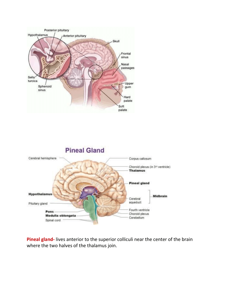
Pineal gland- lives anterior to the superior colliculi near the center of the brain where the two halves of the thalamus join. Interpeduncular cistern: Caudal (posterior) to the mammillary bodies and between the cerebral peduncles is a substantial indentation called the interpeduncular cistern (p. 13 dissection). This is part of the network of ventricles that circulates CSF through the brain. Cerebrospinal fluid/cerebrospinal fluid: is a fluid that is derived from blood. It's made specifically by cells in tissues called choroid plexus --we're going to see that each one of the brain ventricles actually has a colloid plexus. Colloid plexus: These are clusters of cells that take blood (arterial blood). The basically selectively take certain parts of the blood and move it into the ventricles as a new fluid called cerebrospinal fluid. The interpeduncular cistern is a cone-shaped cistern situated between the cerebral peduncles and the mesencephalic and diencephalic leaflets of the LM at the confluence of the supra- and infratentorial compartments of the subarachnoid space Circulation of CSF Written Out The brain’s cerebral cortex is the outermost layer that gives the brain its characteristic wrinkly appearance. The cerebral cortex is divided lengthways into two cerebral hemispheres connected by the corpus callosum. Traditionally, each of the hemispheres has been divided into four lobes: frontal, parietal, temporal and occipital. Although we now know that most brain functions rely on many different regions across the entire brain working in conjunction, it is still true that each lobe carries out the bulk of certain functions. In humans, the lobes of the brain are divided by a number of bumps and grooves. These are known as gyri (bumps) and sulci (groves or fissures). The folding of the brain, and the resulting gyri and sulci, increases its surface area and enables more cerebral cortex matter to fit inside the skull. Two major sulci located on the lateral, or side, surface of each hemisphere distinguish these lobes. The central sulcus, or fissure of Rolando, separates the frontal and parietal lobes, and the deeper lateral sulcus, or fissure of Sylvius, forms the boundary between the temporal lobe and the frontal and parietal lobes. The frontal lobe, the largest of the cerebral lobes, lies rostral to the central sulcus (that is, toward the nose from the sulcus). One important structure in the frontal lobe is the precentral gyrus, which constitutes the primary motor region of the brain. When parts of the gyrus are electrically stimulated in conscious patients (under local anesthesia), they produce localized movements on the opposite side of the body that are interpreted by the patients as voluntary. Injury to parts of the precentral gyrus results in paralysis on the contralateral half of the body. Parts of the inferior frontal lobe (close to the lateral sulcus) constitute the Broca area, a region involved with speech (see below Functions of the human nervous system: Language). The parietal lobe, posterior to the central sulcus, is divided into three parts: (1) the postcentral gyrus (2) the superior parietal lobule (3) the inferior parietal lobule. The postcentral gyrus (middle ?) receives sensory input from the contralateral half of the body. The sequential representation is the same as in the primary motor area, with sensations from the head being represented in inferior parts of the gyrus and impulses from the lower extremities being represented in superior portions. The superior parietal lobule, located caudal to (that is, below and behind) the postcentral gyrus, lies above the intraparietal sulcus. This lobule is regarded as an association cortex, an area that is not involved in either sensory or motor processing, although part of the superior parietal lobule may be concerned with motor function. The inferior parietal lobule (composed of the angular and supramarginal gyri) is a cortical region involved with the integration of multiple sensory signals. In both the parietal and frontal lobes, each primary sensory or motor area is close to, or surrounded by, a smaller secondary area. The primary sensory area receives input only from the thalamus, while the secondary sensory area receives input from the thalamus, the primary sensory area, or both. The motor areas receive input from the thalamus as well as the sensory areas of the cerebral cortex. The temporal lobe, inferior to the lateral sulcus, fills the middle fossa, or hollow area, of the skull. The outer surface of the temporal lobe is an association area made up of the superior, middle, and inferior temporal gyri. Near the margin of the lateral sulcus, two transverse temporal gyri constitute the primary auditory area of the brain. The sensation of hearing is represented here in a tonotopic fashion—that is, with different frequencies represented on different parts of the area. The transverse gyri are surrounded by a less finely tuned secondary auditory area. A medial, or inner, protrusion near the ventral surface of the temporal lobe, known as the uncus, constitutes a large part of the primary olfactory area. The occipital lobe lies caudal to the parieto-occipital sulcus, which joins the calcarine sulcus in a Y-shaped formation. Cortex on both banks of the calcarine sulcus constitutes the primary visual area, which receives input from the contralateral visual field via the optic radiation. The visual field is represented near the calcarine sulcus in a retinotopic fashion—that is, with upper quadrants of the visual field laid out along the inferior bank of the sulcus and lower quadrants of the visual field represented on the upper bank. Central vision is represented mostly caudally and peripheral vision rostrally. Not visible from the surface of the cerebrum is the insular, or central, lobe, an invaginated triangular area on the medial surface of the lateral sulcus; it can be seen in the intact brain only by separating the frontal and parietal lobes from the temporal lobe. The insular lobe is thought to be involved in sensory and motor visceral functions as well as taste perception. The limbic lobe is a synthetic lobe located on the medial margin (or limbus) of the hemisphere. Composed of adjacent portions of the frontal, parietal, and temporal lobes that surround the corpus callosum, the limbic lobe is involved with autonomic and related somatic behavioral activities. The limbic lobe receives input from thalamic nuclei that are connected with parts of the hypothalamus and with the hippocampal formation, a primitive cortical structure within the inferior horn of the lateral ventricle. Cerebral ventricles Deep within the white matter of the cerebral hemispheres are cavities filled with cerebrospinal fluid that form the ventricular system. These cavities include a pair of C-shaped lateral ventricles with anterior, inferior, and posterior “horns” protruding into the frontal, temporal, and occipital lobes, respectively. Most of the cerebrospinal fluid is produced in the ventricles, and about 70 percent of it is secreted by the choroid plexus, a collection of blood vessels in the walls of the lateral ventricles. The fluid drains via interventricular foramina, or openings, into a slitlike third ventricle, which, situated along the midline of the brain, separates the symmetrical halves of the thalamus and hypothalamus. From there the fluid passes through the cerebral aqueduct in the midbrain and into the fourth ventricle in the hindbrain. Openings in the fourth ventricle permit cerebrospinal fluid to enter subarachnoid spaces surrounding both the brain and the spinal cord.
