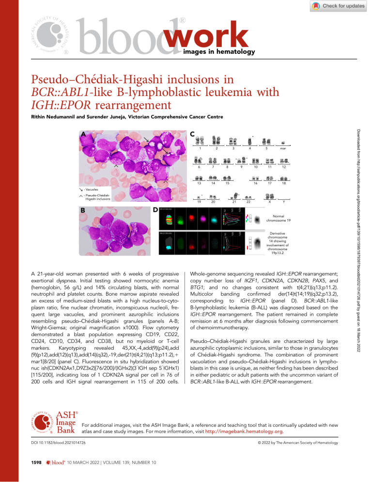
work images in hematology Pseudo–Chediak-Higashi inclusions in BCR::ABL1-like B-lymphoblastic leukemia with IGH::EPOR rearrangement Rithin Nedumannil and Surender Juneja, Victorian Comprehensive Cancer Centre C 1 2 3 6 7 8 13 14 15 19 20 4 9 5 mar 10 11 12 16 17 18 X Y - Vacuoles - Pseudo-ChédiakHigashi inclusions 22 100% 0% D 21 50% B MB-G213026.006 Normal chromosome 19 100% 0% A 21-year-old woman presented with 6 weeks of progressive exertional dyspnea. Initial testing showed normocytic anemia (hemoglobin, 56 g/L) and 14% circulating blasts, with normal neutrophil and platelet counts. Bone marrow aspirate revealed an excess of medium-sized blasts with a high nucleus-to-cytoplasm ratio, fine nuclear chromatin, inconspicuous nucleoli, frequent large vacuoles, and prominent azurophilic inclusions resembling pseudo–Ch ediak-Higashi granules (panels A-B; Wright-Giemsa; original magnification x1000). Flow cytometry demonstrated a blast population expressing CD19, CD22, CD24, CD10, CD34, and CD38, but no myeloid or T-cell markers. Karyotyping revealed 45,XX,-4,add(9)(p24),add (9)(p12),add(12)(q13),add(14)(q32),-19,der(21)t(4;21)(q13;p11.2),1 mar1[8/20] (panel C). Fluorescence in situ hybridization showed nuc ish(CDKN2Ax1,D9Z3x2)[76/200]/(IGHx2)(39 IGH sep 59 IGHx1) [115/200], indicating loss of 1 CDKN2A signal per cell in 76 of 200 cells and IGH signal rearrangement in 115 of 200 cells. 50% MB-G213026.006 Derivative chromosome 14 showing involvement of chromosome 19p13.2 Whole-genome sequencing revealed IGH::EPOR rearrangement; copy number loss of IKZF1, CDKN2A, CDKN2B, PAX5, and BTG1; and no changes consistent with t(4;21)(q13;p11.2). Multicolor banding confirmed der(14)t(14;19)(q32;p13.2), corresponding to IGH::EPOR (panel D). BCR::ABL1-like B-lymphoblastic leukemia (B-ALL) was diagnosed based on the IGH::EPOR rearrangement. The patient remained in complete remission at 6 months after diagnosis following commencement of chemoimmunotherapy. Pseudo–Ch ediak-Higashi granules are characterized by large azurophilic cytoplasmic inclusions, similar to those in granulocytes of Ch ediak-Higashi syndrome. The combination of prominent vacuolation and pseudo–Ch ediak-Higashi inclusions in lymphoblasts in this case is unique, as neither finding has been described in either pediatric or adult patients with the uncommon variant of BCR::ABL1-like B-ALL with IGH::EPOR rearrangement. For additional images, visit the ASH Image Bank, a reference and teaching tool that is continually updated with new atlas and case study images. For more information, visit http://imagebank.hematology.org. DOI 10.1182/blood.2021014726 1598 blood® 10 MARCH 2022 | VOLUME 139, NUMBER 10 © 2022 by The American Society of Hematology Downloaded from http://ashpublications.org/blood/article-pdf/139/10/1598/1879397/bloodbld2021014726.pdf by guest on 18 March 2022 A
