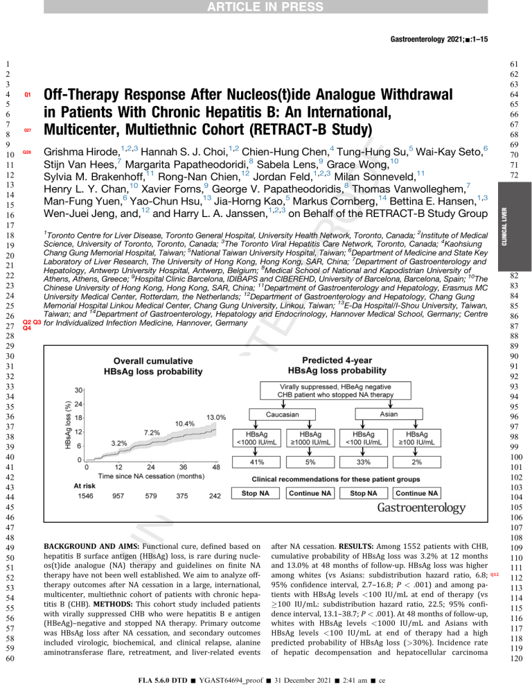
Gastroenterology 2021;-:1–15 Q1 Q27 Q28 Off-Therapy Response After Nucleos(t)ide Analogue Withdrawal in Patients With Chronic Hepatitis B: An International, Multicenter, Multiethnic Cohort (RETRACT-B Study) Grishma Hirode,1,2,3 Hannah S. J. Choi,1,2 Chien-Hung Chen,4 Tung-Hung Su,5 Wai-Kay Seto,6 Stijn Van Hees,7 Margarita Papatheodoridi,8 Sabela Lens,9 Grace Wong,10 Sylvia M. Brakenhoff,11 Rong-Nan Chien,12 Jordan Feld,1,2,3 Milan Sonneveld,11 Henry L. Y. Chan,10 Xavier Forns,9 George V. Papatheodoridis,8 Thomas Vanwolleghem,7 Man-Fung Yuen,6 Yao-Chun Hsu,13 Jia-Horng Kao,5 Markus Cornberg,14 Bettina E. Hansen,1,3 Wen-Juei Jeng, and,12 and Harry L. A. Janssen,1,2,3 on Behalf of the RETRACT-B Study Group CLINICAL LIVER 1 2 3 4 5 6 7 8 9 10 11 12 13 14 15 16 17 18 19 20 21 22 23 24 25 26 27 28 29 30 31 32 33 34 35 36 37 38 39 40 41 42 43 44 45 46 47 48 49 50 51 52 53 54 55 56 57 58 59 60 1 Q2 Q3 Q4 Toronto Centre for Liver Disease, Toronto General Hospital, University Health Network, Toronto, Canada; 2Institute of Medical Science, University of Toronto, Toronto, Canada; 3The Toronto Viral Hepatitis Care Network, Toronto, Canada; 4Kaohsiung Chang Gung Memorial Hospital, Taiwan; 5National Taiwan University Hospital, Taiwan; 6Department of Medicine and State Key Laboratory of Liver Research, The University of Hong Kong, Hong Kong, SAR, China; 7Department of Gastroenterology and Hepatology, Antwerp University Hospital, Antwerp, Belgium; 8Medical School of National and Kapodistrian University of Athens, Athens, Greece; 9Hospital Clinic Barcelona, IDIBAPS and CIBEREHD, University of Barcelona, Barcelona, Spain; 10The Chinese University of Hong Kong, Hong Kong, SAR, China; 11Department of Gastroenterology and Hepatology, Erasmus MC University Medical Center, Rotterdam, the Netherlands; 12Department of Gastroenterology and Hepatology, Chang Gung Memorial Hospital Linkou Medical Center, Chang Gung University, Linkou, Taiwan; 13E-Da Hospital/I-Shou University, Taiwan, Taiwan; and 14Department of Gastroenterology, Hepatology and Endocrinology, Hannover Medical School, Germany; Centre for Individualized Infection Medicine, Hannover, Germany BACKGROUND AND AIMS: Functional cure, defined based on hepatitis B surface antigen (HBsAg) loss, is rare during nucleos(t)ide analogue (NA) therapy and guidelines on finite NA therapy have not been well established. We aim to analyze offtherapy outcomes after NA cessation in a large, international, multicenter, multiethnic cohort of patients with chronic hepatitis B (CHB). METHODS: This cohort study included patients with virally suppressed CHB who were hepatitis B e antigen (HBeAg)–negative and stopped NA therapy. Primary outcome was HBsAg loss after NA cessation, and secondary outcomes included virologic, biochemical, and clinical relapse, alanine aminotransferase flare, retreatment, and liver-related events after NA cessation. RESULTS: Among 1552 patients with CHB, cumulative probability of HBsAg loss was 3.2% at 12 months and 13.0% at 48 months of follow-up. HBsAg loss was higher among whites (vs Asians: subdistribution hazard ratio, 6.8; 95% confidence interval, 2.7–16.8; P < .001) and among patients with HBsAg levels <100 IU/mL at end of therapy (vs 100 IU/mL: subdistribution hazard ratio, 22.5; 95% confidence interval, 13.1–38.7; P < .001). At 48 months of follow-up, whites with HBsAg levels <1000 IU/mL and Asians with HBsAg levels <100 IU/mL at end of therapy had a high predicted probability of HBsAg loss (>30%). Incidence rate of hepatic decompensation and hepatocellular carcinoma FLA 5.6.0 DTD YGAST64694_proof 31 December 2021 2:41 am ce Q12 61 62 63 64 65 66 67 68 69 70 71 72 73 74 75 76 77 78 79 80 81 82 83 84 85 86 87 88 89 90 91 92 93 94 95 96 97 98 99 100 101 102 103 104 105 106 107 108 109 110 111 112 113 114 115 116 117 118 119 120 2 Gastroenterology Vol. was 0.48 per 1000 person-years and 0.29 per 1000 personyears, respectively. Death occurred in 7/19 decompensated patients and 2/14 patients with hepatocellular carcinoma. CONCLUSIONS: The best candidates for NA withdrawal are virally suppressed, HBeAg- negative, noncirrhotic patients with CHB with low HBsAg levels, particularly whites with <1000 IU/mL and Asians with <100 IU/mL. However, strict surveillance is recommended to prevent deterioration. Keywords: HBV; seroconversion. CLINICAL LIVER 121 122 123 124 125 126 127 128 129 130 131 132 133 134 135 136 137 138 139 140 141 142 143 144 145 146 147 148 149 150 151 152 153 154 155 156 157 158 159 160 161 162 163 164 165 166 167 168 169 170 171 172 173 174 175 176 177 178 179 180 Hirode et al Q13 H Discontinuation; Antiviral; HBsAg epatitis B virus (HBV) infection remains a major public health concern with significant morbidity and mortality, affecting 292 million individuals globally.1 Currently approved agents for management include (pegylated [PEG]-)interferon and nucleos(t)ide analogue (NA) therapies.2–5 Despite the advent of effective oral antiviral agents with a good safety profile,6,7 the majority of the patients with chronic hepatitis B (CHB) require long-term management and treatment. NAs have been shown to reduce progression toward cirrhosis, liver failure, and hepatocellular carcinoma (HCC),8–11 however, even with sustained HBV DNA suppression, the risk of long-term complications, particularly HCC, remains.12,13 Hepatitis B surface antigen (HBsAg) loss, which is considered the functional cure, is rare on NA therapy.2,4,14–22 Long-term adherence, compliance, drug safety, and financial and emotional burdens for patients and caregivers present additional challenges.23,24 Finite NA therapy has been proposed as an alternative to long-term treatment. Because virologic relapse is nearly universal, even after prolonged viral suppression,25 the rationale for stopping NAs is to ultimately induce a durable remission in the form of an inactive carrier state or, ideally a functional cure.12 However, the occurrence of a combined virologic and biochemical relapse can range from mild alanine aminotransferase (ALT) elevations to clinically significant ALT flares, which may even result in hepatic decompensation.17,26–31 Because such findings raise concerns on whether the current criteria for stopping NAs are applicable to all patients with CHB, the safe cessation of NA therapy remains one of the most controversial topics in the clinical management of CHB with discordance between guidelines.2,4,19,32–34 Existing studies included small, single-center cohorts with different study-specific endpoints. Based on the patient population and study design, studies on finite NA therapy have reported off-therapy HBsAg loss rates with wide variability ranging from 0%–55% over follow-up periods spanning 0.5–8 years.17,35–42 Thus, a large cohort with individual patient-level data with sufficient statistical power to analyze the safety and efficacy of NA cessation in patients with CHB is required. The main objective of this study was to investigate factors associated with HBsAg loss, and describe virologic, biochemical, and clinical responses after cessation of NA therapy with the hope to improve current -, No. - WHAT YOU NEED TO KNOW BACKGROUND AND CONTEXT Functional cure, or hepatitis B surface antigen loss is rare on nucleos(t)ide analogue therapy. Nucleos(t)ide analogue withdrawal as a therapeutic alternative remains elusive in clinical practice because current knowledge is mainly based on small and single-center studies. NEW FINDINGS In this global study of individual patient-level data on 1552 patients with chronic hepatitis B who stopped nucleos(t) ide analogue therapy, cumulative off-therapy hepatitis B surface antigen loss probability was 3.2% at 1 year, and 13.0% at 4 years (annual incidence: 2.9 per 1000 person-years). The predicted probability was >30% among whites with hepatitis B surface antigen <1000 IU/mL and Asians with hepatitis B surface antigen <100 IU/mL at nucleos(t)ide analogue withdrawal, while controlling for other factors. LIMITATIONS Some bias may persist due to heterogeneity across centers despite adjusting for potential confounders and accounting for differences in retreatment criteria. IMPACT These findings identify factors associated with off-therapy hepatitis B surface antigen loss that help in the selection of patients for nucleos(t)ide analogue withdrawal. patient management and help in the design of prospective HBV cure studies. Methods This is a large, global, multicenter, multiethnic cohort study of patients with CHB who stopped NA therapy between 2001 and 2020 from 13 participating centers across Asia, Europe, and North America (Figure 1, Supplementary Table 1).17,21,38,41,43–45 A standardized case report form was used to capture data. All data cleaning, data quality assessments, and analyses were centralized at the Toronto Centre for Liver Disease (University Health Network, Canada). After anonymized and deidentified individual patient-level longitudinal data were received from the participating centers, meticulous data queries were sent to each center to ensure accuracy. According to local rules, the study was approved by the research ethics board of each participating center and performed in concordance with Good Clinical Practice guidelines and the Declaration of Helsinki 1964 as modified by the 59th WMA General Assembly, Seoul, South Korea, in October 2008, and the local national laws governing the conduct of clinical research studies. Abbreviations used in this paper: ALT, alanine aminotransferase; antiHBs, hepatitis B surface antibody; CHB, chronic hepatitis B; CI, confidence interval; ETV, entecavir; HBeAg, hepatitis B e antigen; HBsAg, hepatitis B surface antigen; HBV, hepatitis B virus; HCC, hepatocellular carcinoma; NA, nucleos(t)ide analogue; PEG, pegylated; TDF, tenofovir disoproxil fumarate; ULN, upper limit of normal. © 2021 by the AGA Institute 0016-5085/$36.00 https://doi.org/10.1053/j.gastro.2021.11.002 FLA 5.6.0 DTD YGAST64694_proof 31 December 2021 2:41 am ce Q14 181 182 183 184 185 186 187 188 189 190 191 192 193 194 195 196 197 198 199 200 201 202 203 204 205 206 207 208 209 210 211 212 213 214 215 216 217 218 219 220 221 222 223 224 225 226 227 228 229 230 231 232 233 234 235 236 237 238 239 240 241 242 243 244 245 246 247 248 249 250 251 252 253 254 255 256 257 258 259 260 261 262 263 264 265 266 267 268 269 270 271 272 273 274 275 276 277 278 279 280 281 282 283 284 285 286 287 288 289 290 291 292 293 294 295 296 297 298 299 300 2021 Finite NA Therapy is Effective for HBsAg Loss Study Population and Variables Q15 Adult patients (aged 18 years) with CHB (HBsAg positive >6 months) were included if they were virally suppressed and hepatitis B e antigen (HBeAg)–negative at the end of therapy (Figure 1). Stopping criteria and retreatment criteria varied by center location as listed in Supplementary Table 1. Patients who had previously been diagnosed with HCC, patients with coinfection (hepatitis C virus, hepatitis delta virus, and/or human immunodeficiency virus), and patients who received (PEG)interferon treatment within 12 months prior to NA cessation were excluded from this study. NA therapy duration refers to the duration of continuous NA therapy including consolidation. Follow-up refers to time since NA cessation while the patient remained off-therapy. The patient was defined as being cirrhotic if cirrhosis had been diagnosed before cessation. Cirrhosis was diagnosed based on histologic findings or ultrasonographic evidence with or without splenomegaly. Hepatic decompensation was defined based on development of a serum total bilirubin level 2 mg/dL, an increased INR, appearance of clinical jaundice, onset of ascites, variceal bleeding, or hepatic encephalopathy. Laboratory Assays Quantitative or qualitative HBsAg, HBeAg, and HBV DNA was determined using in-house or commercially available assays as described in Supplementary Table 2. The upper limit of normal (ULN) for ALT values as defined by each participating center was used. Off-Therapy Outcome Measures The main outcome analyzed in this study was HBsAg loss after NA cessation, with or without seroconversion to hepatitis B surface antibody (anti-HBs).32,46 Secondary outcomes after NA cessation included virologic, biochemical, and clinical relapse, ALT flare, retreatment, liver-related events including hepatic decompensation and HCC, and mortality. Virologic relapse was defined as a single elevation of HBV DNA 2000 IU/mL, biochemical relapse was defined as a single elevation of ALT 2x ULN, and clinical relapse was defined as elevations of HBV DNA 2000 IU/mL and ALT 2x ULN at the same visit. An ALT flare was defined as ALT 5x ULN with or without virologic relapse. Hepatic decompensation was considered related to NA cessation if diagnosed off-therapy or within 6 months of starting retreatment. HCC was only considered to have occurred off-therapy if diagnosed at least 6 months after NA cessation, and within 6 months of starting retreatment if retreated. Statistical Analysis Q16 Clinical and demographic characteristics of the study cohort were presented as frequencies and proportions for categorical variables, and mean ± standard deviation or median (range), as appropriate, for continuous variables. Cumulative probabilities were estimated using Kaplan–Meier analysis and compared between groups using the log-rank test. All outcomes were analyzed while the patient remained off-therapy. Patients were censored at the last recorded visit date, date lost-to-follow-up, or at retreatment if retreated. While analyzing retreatment as an outcome, patients were censored at the last recorded visit date, date lost-to-follow-up, or at HBsAg loss. Competing risks 3 regression using the Fine-Gray subdistribution method was used to analyze factors associated with HBsAg loss, modeled with retreatment as a competing risk.47 Variables were entered into the multivariable model a priori based on the hypothesized effect on the outcome and clinical relevance. To develop a clinically meaningful rule, the predicted probability of HBsAg loss in different patient subgroups was calculated. These probabilities are estimates calculated at the mean of all other covariates in the multivariable model. Incidence rates were calculated over an off-therapy follow-up period of 120 months for all outcomes except hepatic decompensation for which a follow-up period of 48 months was used. For Kaplan-Meir and competing risks regression analyses, the latest time under which patients were both under observation and at risk was 48 months. A two-tailed P value < .05 was considered statistically significant. Statistical analyses used STATA Version 15.1 (StataCorp, College Station, TX). Results Characteristics of the Study Cohort Of 1726 patients with CHB who stopped NA therapy, 1552 met the inclusion and exclusion criteria for this study (Figure 1). Patient characteristics have been described in Table 1. Mean age at end of therapy was 52.9 ± 11.3 years, and 72.3% were male, 87.6% were Asian, and 11.3% were white. Genotype B (42.7%) was the most prevalent genotype followed by genotype C (11.0%); however, genotype was unavailable for 42.7% of the cohort due to low or undetectable levels of HBV DNA. Most patients received either entecavir (ETV; 63.2%) or tenofovir disoproxil fumarate (TDF; 27.1%) therapy before cessation. The median followup duration was 18.4 (range, 7.9–39.4) months. At end of therapy, 11.9% had been previously diagnosed with cirrhosis, mean HBsAg was 2.6 ± 0.8 log10 IU/mL, and median ALT ULN was 0.6 (range, 0.4–0.8). Outcomes After NA Cessation HBsAg loss. Overall, 114 patients achieved HBsAg loss with an incidence rate of 2.9 per 1000 person-years. The cumulative probability of HBsAg loss increased from 1.3% (95% confidence interval [CI], 0.8%–2.1%) at 6 months to 3.2% (95% CI, 2.3%–4.4%) at 12 months and reached 13.0% (95% CI, 10.5%–16.0%) at 48 months of follow-up (Figure 2A). No HBsAg reversions were reported. When stratified by baseline characteristics, there were statistically significant differences in the cumulative probability of HBsAg loss by age at end of therapy (P ¼ .03), race/ ethnicity (P < .001), NA type before cessation (P ¼ .01), and HBsAg levels at end of therapy (P < .001) (Figure 3). At 48 months of follow-up, the cumulative probability of HBsAg loss was higher among patients aged 50 years at end of therapy (16.8%; 95% CI, 12.9%–21.7%) compared with those aged <50 years (8.7%; 95% CI, 6.0%–12.5%) (Figure 3A), among whites (36.5%; 95% CI, 26.0%–49.5%) compared with Asians (10.6%; 95% CI, 8.1%–13.7%) (Figure 3C), among patients treated with TDF before cessation (18.1%; 95% CI, 12.2%–26.5%) compared with ETV-treated patients (10.5%; 95% CI, 7.8%–14.2%) FLA 5.6.0 DTD YGAST64694_proof 31 December 2021 2:41 am ce CLINICAL LIVER - 301 302 303 304 305 306 307 308 309 310 311 312 313 314 315 316 317 318 319 320 321 322 323 324 325 326 327 328 329 330 331 332 333 334 335 336 337 338 339 340 341 342 343 344 345 346 347 348 349 350 351 352 353 354 355 356 357 358 359 360 4 Gastroenterology Vol. Europe (N = 248) Belgium, Germany, Greece, Netherlands, Spain Asia (N = 1,233) North America (N = 245) Hong Kong, Taiwan Canada -, No. - Received data on patients who stopped NA therapy (N = 1,726) Patients excluded from this study (N = 174) CLINICAL LIVER 361 362 363 364 365 366 367 368 369 370 371 372 373 374 375 376 377 378 379 380 381 382 383 384 385 386 387 388 389 390 391 392 393 394 395 396 397 398 399 400 401 402 403 404 405 406 407 408 409 410 411 412 413 414 415 416 417 418 419 420 Hirode et al • • • • • 9 (PEG-)interferon within 12 months prior to NA cessation 11 Coinfection (HCV, HDV) or prior HCC diagnosis 22 HBsAg loss during NA therapy 50 HBeAg positive at NA cessation 82 HBV DNA positive or unknown at NA cessation Patients that met inclusion/exclusion criteria (N = 1,552) Patients excluded from all time-to-event analysis (N = 6) • 6 Lost to follow-up at or soon after NA cessation Patients with complete follow-up data (N = 1,546) Figure 1. Flow diagram of patient inclusion and exclusion. HCV, hepatitis C virus. (Figure 3D), and among patients with HBsAg <100 IU/mL at end of therapy (43.0%; 95% CI, 34.4–52.7%) compared with patients with HBsAg levels between 100–1000 IU/mL at end of therapy (7.4%; 95% CI, 4.6–11.7%) or HBsAg >1000 IU/mL at end of therapy (1.1%; 95% CI, 0.3–3.5%; Figure 3F). Univariate competing risks regression yielded results similar to those of the Kaplan-Meir analysis. Rate of HBsAg loss was significantly higher among whites compared with Asians (subdistribution hazard ratio, 4.9; 95% CI, 3.2–7.4; P < .001) and patients treated with TDF before cessation compared with ETV-treated patients (subdistribution hazard ratio, 1.8; 95% CI, 1.1–2.7; P ¼ .01) (Table 2). HBsAg levels at the end of therapy were strongly associated with HBsAg loss, and patients with HBsAg <100 IU/mL at the end of therapy had the highest rate of HBsAg loss. Longer NA duration and prior (PEG-)interferon treatment were also significantly associated with HBsAg loss (Table 2). On adjusted multivariable competing risks regression, rate of HBsAg loss was 6.8 times higher (95% CI, 2.7–16.8; P < .001) among whites compared with Asians, and 22.5 times higher (95% CI, 13.1– 38.7; P < .001) among patients with HBsAg levels <100 IU/mL at the end of therapy compared with patients with HBsAg levels 100 IU/mL at the end of therapy. Start of therapy HBeAg status was not significant on univariate or multivariable analyses. There were no interactions included in the multivariable model presented in Table 2. When exploring interactions between race and HBsAg levels at the end of therapy, we analyzed 3 thresholds for HBsAg levels: 10 IU/mL (1 log10), 100 IU/mL (2 log10), and 1000 IU/mL (3 log10) (Figure 4). In this cohort, the average predicted probabilities of HBsAg loss at 48 months of follow-up among patients with low HBsAg levels of <10 IU/ mL at the end of therapy were comparable and >75% among whites and Asians (P ¼ not significant [NS]; Q17 Figure 4A); however, the predicted probabilities of HBsAg loss were considerably higher among whites with HBsAg levels <100 IU/mL (84.1%; Figure 4B) or <1000 IU/mL (40.9%; Figure 4C) at end of therapy compared with Asians using the same cut-points (<100 IU/mL: 32.5%; <1000 IU/ mL: 9.7%) (P < .01 for both comparisons). Patient characteristics based on race/ethnicity have also been described in Supplementary Table 3. Virologic and biochemical responses. Virologic relapse occurred in 1207 patients, and cumulative probabilities increased from 47.8% (95% CI, 45.3%–50.3%) at 6 months to 68.9% (95% CI, 66.5%–71.2%) at 12 months and reached 83.4% (95% CI, 81.2%–85.5%) at 48 months of follow-up (Figure 2B). Biochemical relapse occurred in 757 FLA 5.6.0 DTD YGAST64694_proof 31 December 2021 2:41 am ce 421 422 423 424 425 426 427 428 429 430 431 432 433 434 435 436 437 438 439 440 441 442 443 444 445 446 447 448 449 450 451 452 453 454 455 456 457 458 459 460 461 462 463 464 465 466 467 468 469 470 471 472 473 474 475 476 477 478 479 480 481 482 483 484 485 486 487 488 489 490 491 492 493 494 495 496 497 498 499 500 501 502 503 504 505 506 507 508 509 510 511 512 513 514 515 516 517 518 519 520 521 522 523 524 525 526 527 528 529 530 531 532 533 534 535 536 537 538 539 540 2021 Finite NA Therapy is Effective for HBsAg Loss patients, and cumulative probabilities increased from 22.3% (95% CI, 20.2%–24.5%) at 6 months to 38.1% (95% CI, 35.5%–40.7%) at 12 months of follow-up and reached 61.1% (95% CI, 58.0%–64.2%) at 48 months (Figure 2C). Clinical relapse occurred in 658 patients, and cumulative probabilities increased from 17.2% (95% CI, 15.4%–19.3%) at 6 months to 31.9% (95% CI, 29.4%–34.4%) at 12 months of follow-up and reached 54.6% (95% CI, 51.5%–57.7%) at 48 months (Figure 2D). An ALT flare occurred in 359 patients, and cumulative probabilities increased from 10.5% (95% CI, 9.0%–12.2%) at 6 months to 18.6% (95% CI, 16.6%–20.8%) at 12 months of follow-up and reached 30.8% (95% CI, 27.9%–33.9%) at 48 months (Figure 2E). Retreatment. After NA cessation, 729 patients were retreated, and the cumulative probability of retreatment increased from 16.2% (95% CI, 14.4%-18.2%) at 6 months to 29.8% (95% CI, 27.5%–32.2%) at 12 months of follow-up and reached 54.7% (95% CI, 51.7%-57.7%) at 48 months (Figure 2F). There were statistically significant differences 5 in the cumulative probability of retreatment by age group (P < .001), start of therapy HBeAg status (P ¼ .02), and HBsAg levels at end of therapy (P < .001) (Supplementary Figure 1). Liver-related events and mortality. There were 19 patients who developed hepatic decompensation after NA cessation (8/184 [4.3%] patients with cirrhosis vs 11/1368 [0.8%] patients without cirrhosis; P < .001) with an incidence rate of 0.48 per 1000 person-years. No decompensating events occurred after 48 months of follow-up. Among patients who developed hepatic decompensation, 1/19 (5.3%) had subsequent HBsAg loss, and 16/19 (84.2%) started retreatment. Death occurred in 7 (36.8%) of the 19 decompensated patients, of whom 6 died after starting retreatment. In 4/7 (57.1%), death was reported to be related to a hepatitis B–associated flare. Among the other 3/7 (42.9%) deaths, 1 patient died due septic shock caused by urosepsis, 1 died due to lymphoma, and 1 died due to cholangiocarcinoma. Table 1.Characteristics of the Patients Who Stopped NA Therapy Q23 Total, N 1552 Age at end of therapy, y, mean ± SD 52.9 ± 11.2 Male sex, n (%) 1122 (72.3) Race/ethnicity: white/Asian/black/other, n (%) 175 (11.3)/1359 (87.6)/13 (0.8)/5 (0.3) HBV genotype: A/B/C/D/other/missing, n (%) 9 (0.6)/662 (42.7)/170 (11.0)/45 (2.9)/4 (0.3)/662 (42.7) Prior (PEG-)interferon, n (%) 133 (8.6) NA-naïve, n (%) 1292 (83.3) NA type before cessation: ETV/TDF/other, n (%) 981 (63.2)/421 (27.1)/150 (9.7) Minimum consolidation, y: <1/1–2/3 83 (5.4)/1129 (72.7)/340 (21.9) a NA duration, y, median (range) 3.0 (3.0–4.0) Number of follow-up visits, median (range) Follow-up duration between visits, months, median (range) Total follow-up duration, months, median (range) 6 (3–9) 2.8 (2.0–5.0) 18.4 (7.9–39.4) b At start of therapy HBeAg-negative, n (%) 1306 (84.2) HBV DNA, log10 IU/mL, mean ± SD ALT x ULN, median (range) 5.9 ± 1.6 3.0 (1.9–7.3) At end of therapy (NA cessation) c HBsAg, log10 IU/mL, mean ± SD HBsAg, IU/mL: <100/100–1000/>1000, n (%) 2.6 ± 0.8 225 (14.5)/682 (43.9)/463 (29.8) d Cirrhosis, n (%) ALT x ULN, median (range) 184 (11.9) 0.6 (0.4–0.8) a NA duration was unknown for 15 (1%) patients. At start of therapy, HBeAg status was unavailable for 11 (0.7%), HBV DNA levels were unavailable for 190 (12%), and ALT levels were unavailable for 376 (24%) patients. c At end of therapy, HBsAg levels were unavailable for 182 (12%), and ALT levels were unavailable for 47 (3%) patients. d Patient was defined as cirrhotic at end of therapy if cirrhosis had been diagnosed at any time before NA cessation. b FLA 5.6.0 DTD YGAST64694_proof 31 December 2021 2:41 am ce CLINICAL LIVER - 541 542 543 544 545 546 547 548 549 550 551 552 553 554 555 556 557 558 559 560 561 562 563 564 565 566 567 568 569 570 571 572 573 574 575 576 577 578 579 580 581 582 583 584 585 586 587 588 589 590 591 592 593 594 595 596 597 598 599 600 6 Gastroenterology Vol. -, No. - CLINICAL LIVER 601 602 603 604 605 606 607 608 609 610 611 612 613 614 615 616 617 618 619 620 621 622 623 624 625 626 627 628 629 630 631 632 633 634 635 636 637 638 639 640 641 642 643 644 645 646 647 648 649 650 651 652 653 654 655 656 657 658 659 660 Hirode et al Figure 2. Cumulative probability of outcomes during off-therapy follow-up: (A) HBsAg loss, (B) virologic relapse (HBV DNA 2000 IU/mL), (C) biochemical relapse (ALT 2x ULN), (D) clinical relapse (HBV DNA 2000 IU/mL and ALT 2x ULN), (E) ALT flare (ALT 5x ULN), and (F) retreatment. FLA 5.6.0 DTD YGAST64694_proof 31 December 2021 2:41 am ce 661 662 663 664 665 666 667 668 669 670 671 672 673 674 675 676 677 678 679 680 681 682 683 684 685 686 687 688 689 690 691 692 693 694 695 696 697 698 699 700 701 702 703 704 705 706 707 708 709 710 711 712 713 714 715 716 717 718 719 720 - Finite NA Therapy is Effective for HBsAg Loss 7 < ≥ CLINICAL LIVER 721 722 723 724 725 726 727 728 729 730 731 732 733 734 735 736 737 738 739 740 741 742 743 744 745 746 747 748 749 750 751 752 753 754 755 756 757 758 759 760 761 762 763 764 765 766 767 768 769 770 771 772 773 774 775 776 777 778 779 780 2021 < ≥ < > < > Figure 3. Cumulative probability of HBsAg loss by patient characteristics: (A) age at NA cessation, (B) sex, (C) race/ethnicity, (D) NA type before cessation, (E) start of therapy HBeAg status, and (F) end of therapy HBsAg levels. FLA 5.6.0 DTD YGAST64694_proof 31 December 2021 2:41 am ce 781 782 783 784 785 786 787 788 789 790 791 792 793 794 795 796 797 798 799 800 801 802 803 804 805 806 807 808 809 810 811 812 813 814 815 816 817 818 819 820 821 822 823 824 825 826 827 828 829 830 831 832 833 834 835 836 837 838 839 840 8 Gastroenterology Vol. There were 14 patients who developed HCC at least 6 months after NA cessation (4/184 [2.2%] patients with cirrhosis vs 10/1368 [0.7%] patients without cirrhosis; P ¼ NS) with an incidence rate of 0.29 per 1000 person-years. Among patients who developed HCC, 1/14 (7.1%) had subsequent HBsAg loss whereas 1/14 (7.1%) had HBsAg loss before HCC diagnosis, and 6/14 (42.9%) started retreatment. Death occurred in 2 (14.3%) of the 14 patients with HCC, of whom 1 died after starting retreatment. Two additional cases of HCC were reported off-therapy within 6 months after cessation. No patients included in this study developed both hepatic decompensation and HCC. There were 5 other deaths among patients who did not develop liver-related CLINICAL LIVER 841 842 843 844 845 846 847 848 849 850 851 852 853 854 855 856 857 858 859 860 861 862 863 864 865 866 867 868 869 870 871 872 873 874 875 876 877 878 879 880 881 882 883 884 885 886 887 888 889 890 891 892 893 894 895 896 897 898 899 900 Hirode et al -, No. - complications off-therapy, of whom 3 died after starting retreatment. Discussion In this study of 1552 patients with CHB who stopped NA therapy, the cumulative probability of HBsAg loss at year 1 of follow-up was 3.2%, which more than quadrupled to 13.0% by year 4. As would be expected, by year 4 of followup, most of the cohort had virologic relapse (83.4%) while the rates of clinical relapse were lower (54.6%), and 54.7% of the cohort had started retreatment. This study is unique in that, to our knowledge, it is the first study to use individual patient-level data to analyze outcomes after cessation Table 2.Fine-Gray Competing Risks Regression Models for HBsAg Loss Univariate Multivariable SHR (95% CI) P SHR (95% CI) P Age at end of therapy, y 1.01 (0.99–1.02) .55 0.99 (0.97–1.01) .24 Age at end of therapy, y <50 y 50 y 1.00 (reference) 1.28 (0.83–1.96) .26 Sex Female Male 1.00 (reference) 1.45 (0.88–2.37) .14 1.00 (reference) 0.98 (0.57–1.70) .96 Race/ethnicity Asian White 1.00 (reference) 4.86 (3.19–7.41) <.001 1.00 (reference) 6.80 (2.75–16.8) <.001 Prior (PEG-)interferon No Yes 1.00 (reference) 2.18 (1.28–3.73) .004 NA type ETV TDF Other 1.00 (reference) 1.76 (1.14–2.73) 2.02 (1.13–3.59) .01 .02 1.00 (reference) 1.29 (0.81–2.05) 0.48 (0.17–1.36) .29 .17 NA duration, y 1.16 (1.10–1.23) <.001 1.05 (0.96–1.16) .29 HBeAg at start of therapy Negative Positive 1.00 (reference) 1.07 (0.62–1.84) .81 1.00 (reference) 1.57 (0.69–3.57) .28 HBsAg levels at end of therapy, log10 IU/mL 0.24 (0.19–0.30) <.001 HBsAg level at end of therapy 100 IU/mL <100 IU/mL 1.00 (reference) 15.6 (9.75–25.0) <.001 1.00 (reference) 22.5 (13.1–38.7) <.001 HBsAg level at end of therapy >1000 IU/mL 100–1000 IU/mL <100 IU/mL 1.00 (reference) 4.74 (1.41–15.9) 50.4 (15.7–161) .01 <.001 Cirrhosis at end of therapy Noncirrhotic Cirrhotic 1.00 (reference) 1.01 (0.56–1.84) .96 ALT x ULN at end of therapy log10 ULN 1.28 (0.58–2.82) .54 SHR, subdistribution hazard ratio. FLA 5.6.0 DTD YGAST64694_proof 31 December 2021 2:41 am ce 901 902 903 904 905 906 907 908 909 910 911 912 913 914 915 916 917 918 919 920 921 922 923 924 925 926 927 928 929 930 931 932 933 934 935 936 937 938 939 940 941 942 943 944 945 946 947 948 949 950 951 952 953 954 955 956 957 958 959 960 - Finite NA Therapy is Effective for HBsAg Loss 9 < ≥ < ≥ < ≥ < ≥ < ≥ < ≥ Figure 4. Predicted probability of HBsAg loss after multivariable competing risks regression by race for 3 thresholds of HBsAg levels at end of therapy: (A) 10 IU/mL (1 log10), (B) 100 IU/mL (2 log10), and (C) 1000 IU/mL (3 log10). FLA 5.6.0 DTD YGAST64694_proof 31 December 2021 2:41 am ce CLINICAL LIVER 961 962 963 964 965 966 967 968 969 970 971 972 973 974 975 976 977 978 979 980 981 982 983 984 985 986 987 988 989 990 991 992 993 994 995 996 997 998 999 1000 1001 1002 1003 1004 1005 1006 1007 1008 1009 1010 1011 1012 1013 1014 1015 1016 1017 1018 1019 1020 2021 1021 1022 1023 1024 1025 1026 1027 1028 1029 1030 1031 1032 1033 1034 1035 1036 1037 1038 1039 1040 1041 1042 1043 1044 1045 1046 1047 1048 1049 1050 1051 1052 1053 1054 1055 1056 1057 1058 1059 1060 1061 1062 1063 1064 1065 1066 1067 1068 1069 1070 1071 1072 1073 1074 1075 1076 1077 1078 1079 1080 10 CLINICAL LIVER 1081 1082 1083 1084 1085 1086 1087 1088 1089 1090 1091 1092 1093 1094 1095 1096 1097 1098 1099 1100 1101 1102 1103 1104 1105 1106 1107 1108 1109 1110 1111 1112 1113 1114 1115 1116 1117 1118 1119 1120 1121 1122 1123 1124 1125 1126 1127 1128 1129 1130 1131 1132 1133 1134 1135 1136 1137 1138 1139 1140 Hirode et al Gastroenterology Vol. of NA therapy in a large, ethnically diverse, global cohort of patients with CHB. Although there remains heterogeneity between participating centers, individual patient-level data provide robust estimates with the ability to adjust for potential confounders ,which was not possible in any of the prior studies.35,48,49 Modeling retreatment as a competing risk accounts for differences in stopping and retreatment criteria by center location and policies. This study affirms the favorable outcomes associated with lower HBsAg levels at the time of NA cessation.14,17,22,33,40,48,50 This may be associated with patient status with respect to rates of viral replication at the time of NA cessation.2,12,35,51,52 These data reiterate the importance of HBsAg quantification during regular clinical follow-up.53 Comparison of results from different prior studies have suggested that HBsAg loss is typically higher among whites compared with Asians.17,20,41,45,54 Nevertheless, most of these studies were rather small and were single-center studies in populations that were predominantly one race. In this study, although whites had relatively higher rates even when adjusted for potential confounders, off-therapy HBsAg loss among Asians was also considerably higher than known on-therapy rates.36,55 Thus, contrary to speculations in prior studies, Asians may also benefit from stopping NA therapy. The disparities by race and age at end of therapy may stem from differences in confounding variables such as HBV genotype, mode of transmission, and duration of infection.56–58 Virologic and biochemical responses are typically used to define retreatment criteria (Supplementary Table 1), and thus retreatment can be thought of as a composite outcome with respect to relapse and flares. In a systematic review, Papatheodoridis et al59 reported no significant differences in virologic response between groups by start of therapy HBeAg status and numerically higher durable biochemical response rates in start of therapy HBeAg-positive cases, however, retreatment rates were not evaluated in their study. Other studies have reported conflicting results pertaining to rates of retreatment by start of therapy HBeAg status.20–22,33,59–61 In our cohort, there were no significant differences in HBsAg loss by start of therapy HBeAg status, however, it affected the magnitude of associations in the competing risks multivariable model, which may be attributable to the lack of standardized definitions and criteria in the current guidelines.2–4 Contrary to findings by Jeng et al,17 HBsAg loss appeared higher among patients treated with TDF prior to NA cessation compared with ETV-treated patients. Nevertheless, similar to their study, there were no significant differences between the 2 groups in the multivariable model. With respect to virological relapse, our results are comparable to other studies in that the TDFtreated patients experienced earlier and higher rates of relapse compared with ETV-treated patients.62–64 This study highlights that even though all patients with low HBsAg levels would benefit from NA withdrawal with respect to HBsAg loss, the HBsAg level cut-point at NA withdrawal for beneficial outcomes vary by race/ethnicity (Figures 4, 5). We arbitrarily chose those with a predicted probability of HBsAg loss of at least 30% to be good -, No. - candidates for NA withdrawal. Thus, we recommend NA withdrawal in Caucasian patients with HBsAg levels <1000 IU/mL and Asian patients with HBsAg levels <100 IU/mL (Figure 5). These results also agree with the recommendations provided by Berg et al.34 The results from our study suggest that higher rates of HBsAg loss can be achieved during shorter follow-up periods with finite NA therapy. To date, there have been three randomized controlled trials comparing HBsAg loss on- and off-NA therapy.20–22 The trials showed minimal to absent HBsAg loss in those who continued NA therapy, and they also suggested that NA withdrawal is more effective among whites compared with Asians. In our study, one could question whether a control group of patients who continued NA therapy would solidify the proven efficacy of NA withdrawal. However, considering the complexity of such an approach at multiple sites across the globe, and given the ample evidence in the literature showing that HBsAg loss on NA is extremely low, we did not pursue such a study design. Results from a prospective cohort study by Jeng et al17 showed a 1.78% annual HBsAg loss rate for those who stopped NA therapy versus 0.15% among those who continued. Chan et al65 recently reported approximately 1% HBsAg loss at 5 years among 1248 patients treated with tenofovir alafenamide or TDF followed by tenofovir alafenamide in prospective registration randomized controlled trials. Comparing on- and off-NA therapy rates of HBsAg loss across cohort studies, such as the current study, will not yield meaningful information due to differences in the included patient population and baseline criteria. Larger prospective randomized controlled trials with a diverse patient population are necessary to fully determine the differences in outcomes between those who stopped and continued NA therapy. Although we may be able to discern which patient is more likely to achieve functional cure based on end of therapy profiles, it is still unclear what, and when, preemptive measures need to be taken to prevent severe hepatic flares, which often lead to severe or even fatal outcomes. Distinguishing between beneficial and detrimental flares in clinical practice at the time of occurrence is challenging.66–68 The cumulative probability of patients who developed hepatic decompensation reached 1.7% with significant mortality, and 1.5% developed HCC by year 4 of follow-up.69 It is unclear whether HCC incidence was related to treatment cessation in this cohort. None of the patients decompensated after HBsAg loss, and only 1 patient developed HCC. Thus, HBsAg loss remains the most important endpoint,19,70 however, it is difficult to predict patient outcome after a relapse, and hepatic decompensation remains a threat to patient safety. The cumulative probability of HBsAg loss continued to increase over time regardless of the type of relapse, however, there were no statistically significant differences in rates of HBsAg loss between patients who had biochemical relapse and those who did not (data not shown). Moreover, the majority of the patients was retreated soon after the occurrence of either relapse and, thus, it is hard to ascertain whether these patients would have had subsequent HBsAg loss or hepatic FLA 5.6.0 DTD YGAST64694_proof 31 December 2021 2:41 am ce 1141 1142 1143 1144 1145 1146 1147 1148 1149 1150 1151 1152 1153 1154 1155 1156 1157 1158 1159 1160 1161 1162 1163 1164 1165 1166 1167 1168 1169 1170 1171 1172 1173 1174 1175 1176 1177 1178 1179 1180 1181 1182 1183 1184 1185 1186 1187 1188 1189 1190 1191 1192 1193 1194 1195 1196 1197 1198 1199 1200 - Finite NA Therapy is Effective for HBsAg Loss 11 Virally suppressed, HBeAg negative CHB patient on NA therapy Race/ethnicity End of therapy HBsAg levels 4-year off-therapy HBsAg loss probability Clinical recommendations Caucasian Asian HBsAg <1000 IU/mL HBsAg ≥1000 IU/mL HBsAg <100 IU/mL HBsAg ≥100 IU/mL 41% 5% 33% 2% Stop NA Continue NA Stop NA Continue NA Figure 5. Clinical recommendations on NA withdrawal based on the predicted 4-year HBsAg loss probability by patient groups. These predicted probabilities are estimates calculated for a patient of average age irrespective of sex, NA type before cessation, and start of therapy HBeAg status. decompensation. Ghany et al71 and Liaw et al66 suggested that early retreatment may lower the probability of HBsAg loss by dampening the host immune response, and, provided that virologic relapse is almost certain in the majority of the patients, it may not be a suitable criterion for retreatment.72 Although this study may not provide strong evidence for or against certain retreatment criteria, these results emphasize the need for standardization of retreatment criteria and monitoring frequency after cessation.42 Prior studies seem to agree on restarting NA therapy in cases of persistent clinical relapse, ALT flares, progression of fibrosis, or signs of decompensation.19–21,66 In current clinical practice, the final decision on whether to retreat is typically left to the discretion of the treating physician. Most guidelines recommend continued NA therapy in cirrhotic patients in the absence of HBsAg loss,2–4,20 however, the exclusion of finite therapy as an option for cirrhotic patients alone may not be sufficient. In this study, cirrhosis status before NA cessation did not appear to be associated with off-therapy HBsAg loss, but patients with a cirrhosis diagnosis had higher rates of liver-related complications, and more specifically, hepatic decompensation. Thus, our results support the current guidelines in that patients with documented cirrhosis should continue NA therapy. An indepth analysis of predictors of hepatic decompensation and HCC after cessation is necessary to understand the role of cirrhosis, while accounting for differences in the diagnostic methods used to define cirrhosis. Comparative studies analyzing on- and off-therapy rates of hepatic decompensation are sparse. Of the few published studies analyzing rates among cirrhotic patients, some report no difference,73 some report low rates of hepatic decompensation after NA cessation,59,74 whereas others report fatal outcomes.17,43,75,76 With respect to HCC in HBeAg-negative patients with CHB, most studies concluded that there was no difference between on- and off-therapy rates of HCC but higher rates among cirrhotics.17,26,36 One major limitation of comparing on- and off-therapy rates of hepatic decompensation and HCC is the differences in patient profile at baseline because those who stop usually have milder disease. Given that potent NAs are highly effective, affordable, well-tolerated with proven safety, and shown to improve long-term prognosis, the costs associated with more frequent postcessation monitoring, increased laboratory testing, and the development of liver-related complications may attribute to higher patient burden compared with continued therapy and should be taken into consideration where appropriate. Nonadherence and issues with compliance persist on- as well as off-therapy whereas the risk of potentially fatal outcomes is higher with finite therapy. Therefore, strict long-term postcessation surveillance is critical if stopping NA therapy is pursued to aim for HBsAg loss or necessary due to socioeconomic concerns or local policies. This study has limitations. First, the frequency of followup visits and length of follow-up varied by center. Nonetheless, the median time between visits was short (2.8 months) in this cohort and the majority of patients started retreatment before the end of follow-up at centers with shorter follow-up durations. Second, there may have been misclassification bias, for cirrhosis and HCC in particular, based on the center-specific monitoring and surveillance policies. Because there were no biopsies performed at NA withdrawal, patients who were considered noncirrhotic and who developed complications may have had undiagnosed underlying cirrhosis. Third, a small minority of this cohort had been previously treated with (PEG-)interferon, however, it was discontinued at least 12 months before NA cessation and, therefore, the likelihood of an on-going effect would be low.77 Prior (PEG-)interferon use was not a significant predictor in the multivariable model and, hence, it was excluded to avoid overfitting.78 Lastly, HBV genotyping could not be performed for many patients because of viral suppression before NA cessation. In conclusion, the findings from this study in a large cohort of patients with CHB suggest that stopping NA therapy may be beneficial to achieve functional cure in FLA 5.6.0 DTD YGAST64694_proof 31 December 2021 2:41 am ce CLINICAL LIVER 1201 1202 1203 1204 1205 1206 1207 1208 1209 1210 1211 1212 1213 1214 1215 1216 1217 1218 1219 1220 1221 1222 1223 1224 1225 1226 1227 1228 1229 1230 1231 1232 1233 1234 1235 1236 1237 1238 1239 1240 1241 1242 1243 1244 1245 1246 1247 1248 1249 1250 1251 1252 1253 1254 1255 1256 1257 1258 1259 1260 2021 1261 1262 1263 1264 1265 1266 1267 1268 1269 1270 1271 1272 1273 1274 1275 1276 1277 1278 1279 1280 1281 1282 1283 1284 1285 1286 1287 1288 1289 1290 1291 1292 1293 1294 1295 1296 1297 1298 1299 1300 1301 1302 1303 1304 1305 1306 1307 1308 1309 1310 1311 1312 1313 1314 1315 1316 1317 1318 1319 1320 12 CLINICAL LIVER 1321 1322 1323 1324 1325 1326 1327 1328 1329 1330 1331 Q18 1332 1333 1334 1335 1336 1337 1338 1339 1340 1341 1342 1343 1344 1345 1346 1347 1348 1349 1350 1351 1352 1353 1354 1355 1356 1357 1358 1359 1360 1361 1362 1363 1364 1365 1366 1367 1368 1369 1370 1371 1372 1373 1374 1375 1376 1377 1378 1379 1380 Hirode et al Gastroenterology Vol. virally suppressed, HBeAg-negative, noncirrhotic patients with low HBsAg levels provided that close and frequent postcessation monitoring is feasible. NA withdrawal may be particularly effective among white patients with HBsAg levels <1000 IU/mL and among Asian patients with HBsAg levels <100 IU/mL regardless of their start of therapy HBeAg status. These results have important implications not only in aiding decision making for regular clinical practice and providing evidence to promote uniformity across guidelines, but also in the design of prospective studies and randomized trials analyzing novel treatment options and biomarkers focused on a HBV cure. Supplementary Material Note: To access the supplementary material accompanying this article, visit the online version of Gastroenterology at www.gastrojournal.org, and at http://doi.org/10.1053/ j.gastro.2021.11.002. References 1. Polaris Observatory Collaborators. Global prevalence, treatment, and prevention of hepatitis B virus infection in 2016: a modelling study. Lancet Gastroenterol Hepatol 2018;3:383–403. 2. Lampertico P, Agarwal K, Berg T, et al. EASL 2017 Clinical Practice Guidelines on the management of hepatitis B virus infection. J Hepatol 2017;67:370–398. 3. Terrault NA, Lok ASF, McMahon BJ, et al. Update on prevention, diagnosis, and treatment of chronic hepatitis B: AASLD 2018 hepatitis B guidance. Hepatology 2018; 67:1560–1599. 4. Sarin SK, Kumar M, Lau GK, et al. Asian-Pacific clinical practice guidelines on the management of hepatitis B: a 2015 update. Hepatol Int 2016;10:1–98. 5. Campenhout MJH, Brouwer WP, Xie Q, et al. Long-term follow-up of patients treated with entecavir and peginterferon add-on therapy for HBeAg-positive chronic hepatitis B infection: ARES long-term follow-up. J Viral Hepat 2019;26:109–117. 6. Buti M, Tsai N, Petersen J, et al. Seven-year efficacy and safety of treatment with tenofovir disoproxil fumarate for chronic hepatitis B virus infection. Dig Dis Sci 2015; 60:1457–1464. 7. Papatheodoridis GV, Sypsa V, Dalekos G, et al. Eightyear survival in chronic hepatitis B patients under longterm entecavir or tenofovir therapy is similar to the general population. J Hepatol 2018;68:1129–1136. 8. Chang TT, Liaw YF, Wu SS, et al. Long-term entecavir therapy results in the reversal of fibrosis/cirrhosis and continued histological improvement in patients with chronic hepatitis B. Hepatology 2010;52:886–893. 9. Lai CL, Yuen MF. Prevention of hepatitis B virus-related hepatocellular carcinoma with antiviral therapy. Hepatology 2013;57:399–408. 10. Marcellin P, Gane E, Buti M, et al. Regression of cirrhosis during treatment with tenofovir disoproxil fumarate for chronic hepatitis B: a 5-year open-label follow-up study. Lancet 2013;381:468–475. -, No. - 11. Liu K, Choi J, Le A, et al. Tenofovir disoproxil fumarate reduces hepatocellular carcinoma, decompensation and death in chronic hepatitis B patients with cirrhosis. Aliment Pharmacol Ther 2019;50:1037–1048. 12. Lok ASF. Hepatitis B treatment: what we know now and what remains to be researched. Hepatol Commun 2019; 3:8–19. 13. Dusheiko G, Wang B, Carey I. HBsAg loss in chronic hepatitis B: pointers to the benefits of curative therapy. Hepatol Int 2016;10:727–729. 14. Yeo YH, Ho HJ, Yang HI, et al. Factors associated with rates of HBsAg seroclearance in adults with chronic HBV infection: a systematic review and meta-analysis. Gastroenterology 2019;156:635–646.e9. 15. Kim GA, Lim YS, An J, et al. HBsAg seroclearance after nucleoside analogue therapy in patients with chronic hepatitis B: clinical outcomes and durability. Gut 2014; 63:1325–1332. 16. Cornberg M, Siederdissen CH. HBsAg seroclearance with NUCs: rare but important. Gut 2014; 63:1208–1209. 17. Jeng WJ, Chen YC, Chien RN, Sheen IS, Liaw YF. Incidence and predictors of hepatitis B surface antigen seroclearance after cessation of nucleos(t)ide analogue therapy in hepatitis B e antigen–negative chronic hepatitis B. Hepatology 2018;68:425–434. 18. Yip TCF, Wong GLH, Chan HLY, et al. HBsAg seroclearance further reduces hepatocellular carcinoma risk after complete viral suppression with nucleos(t)ide analogues. J Hepatol 2019;70:361–370. 19. Terrault NA, Bzowej NH, Chang KM, Hwang JP, Jonas MM, Murad MH. AASLD Guidelines for Treatment of Chronic Hepatitis B. Hepatology 2016;63:261–283. 20. Berg T, Simon KG, Mauss S, et al. Long-term response after stopping tenofovir disoproxil fumarate in noncirrhotic HBeAg-negative patients – FINITE study. J Hepatol 2017;67:918–924. 21. Liem KS, Fung S, Wong DK, et al. Limited sustained response after stopping nucleos(t)ide analogues in patients with chronic hepatitis B: results from a randomised controlled trial (Toronto STOP study). Gut 2019; 68:2206–2213. 22. van Bömmel F, Stein K, Heyne R, et al. Response to discontinuation of long-term nucleos(t)ide analogue treatment in HBeAg negative patients: results of the Stop-NUC trial. J Hepatol 2020;73:S118–S119. 23. Ford N, Scourse R, Lemoine M, et al. Adherence to nucleos(t)ide analogue therapies for chronic hepatitis B infection: a systematic review and meta-analysis. Hepatol Commun 2018;2:1160–1167. 24. Shin JW, Jung SW, Lee SB, et al. Medication nonadherence increases hepatocellular carcinoma, cirrhotic complications, and mortality in chronic hepatitis B patients treated with entecavir. Am J Gastroenterol 2018; 113:998–1008. 25. Lampertico P, Berg T. Less can be more: a finite treatment approach for HBeAg-negative chronic hepatitis B. Hepatology 2018;68:397–400. 26. Chen YC, Peng CY, Jeng WJ, Chien RN, Liaw YF. Clinical outcomes after interruption of entecavir therapy in FLA 5.6.0 DTD YGAST64694_proof 31 December 2021 2:41 am ce 1381 1382 1383 1384 1385 1386 1387 1388 1389 1390 1391 1392 1393 1394 1395 1396 1397 1398 1399 1400 1401 1402 1403 1404 1405 1406 1407 1408 1409 1410 1411 1412 1413 1414 1415 1416 1417 1418 1419 1420 1421 1422 1423 1424 1425 1426 1427 1428 1429 1430 1431 1432 1433 1434 1435 1436 1437 1438 1439 1440 1441 1442 1443 1444 1445 1446 1447 1448 1449 1450 1451 1452 1453 1454 1455 1456 1457 1458 1459 1460 1461 1462 1463 1464 1465 1466 1467 1468 1469 1470 1471 1472 1473 1474 1475 1476 1477 1478 1479 1480 1481 1482 1483 1484 1485 1486 1487 1488 1489 1490 1491 1492 1493 1494 1495 1496 1497 1498 1499 1500 2021 27. 28. 29. 30. 31. 32. 33. 34. 35. 36. 37. 38. 39. 40. Finite NA Therapy is Effective for HBsAg Loss HBeAg-negative chronic hepatitis B patients with compensated cirrhosis. Aliment Pharmacol Ther 2015; 42:1182–1191. Chen CH, Hung CH, Hu TH, et al. Association between level of hepatitis B surface antigen and relapse after entecavir therapy for chronic hepatitis B virus infection. Clin Gastroenterol Hepatol 2015;13:1984–1992.e1. Chen CH, Hung CH, Wang JH, Lu SN, Hu TH, Lee CM. Long-term incidence and predictors of hepatitis B surface antigen loss after discontinuing nucleoside analogues in noncirrhotic chronic hepatitis B patients. Clin Microbiol Infect 2018;24:997–1003. Chen CH, Hsu YC, Lu SN, et al. The incidence and predictors of HBV relapse after cessation of tenofovir therapy in chronic hepatitis B patients. J Viral Hepat 2018;25:590–597. Jeng WJ, Chen YC, Sheen IS, et al. Clinical relapse after cessation of tenofovir therapy in hepatitis B e antigennegative patients. Clin Gastroenterol Hepatol 2016; 14:1813–1820.e1. Jeng WJ, Sheen IS, Chen YC, et al. Off-therapy durability of response to entecavir therapy in hepatitis B e antigennegative chronic hepatitis B patients. Hepatology 2013; 58:1888–1896. Cornberg M, Lok ASF, Terrault NA, et al. Guidance for design and endpoints of clinical trials in chronic hepatitis B - report from the 2019 EASL-AASLD HBV Treatment Endpoints Conference. J Hepatol 2020; 72:539–557. Papatheodoridi M, Papatheodoridis GV. Emerging diagnostic tools to decide when to discontinue nucleos(t)ide analogues in chronic hepatitis B. Cells 2020; 9:493. Berg T, Lampertico P. The times they are a-changing – a refined proposal for finite HBV nucleos(t)ide analogue therapy. J Hepatol Published online 2021. Liem KS, Gehring AJ, Feld JJ, Janssen HLA. Challenges with stopping long-term nucleos(t)ide analogue therapy in patients with chronic hepatitis B. Gastroenterology 2020;158:1185–1190. Hung CH, Wang JH, Lu SN, Hu TH, Lee CM, Chen CH. Hepatitis B surface antigen loss and clinical outcomes between HBeAg-negative cirrhosis patients who discontinued or continued nucleoside analogue therapy. J Viral Hepat 2017;24:599–607. Ge GH, Ye Y, Zhou XB, et al. Hepatitis B surface antigen levels of cessation of nucleos(t)ide analogs associated with virological relapse in hepatitis B surface antigennegative chronic hepatitis B patients. World J Gastroenterol 2015;21:8653–8659. Seto WK, Hui AJ, Wong VWS, et al. Treatment cessation of entecavir in Asian patients with hepatitis B e antigen negative chronic hepatitis B: a multicentre prospective study. Gut 2015;64:667–672. Yao CC, Hung CH, Hu TH, et al. Incidence and predictors of HBV relapse after cessation of nucleoside analogues in HBeAg-negative patients with HBsAg 200 IU/mL. Sci Rep 2017;7:1–10. Hadziyannis SJ, Sevastianos V, Rapti I, Vassilopoulos D, Hadziyannis E. Sustained responses and loss of HBsAg 41. 42. 43. 44. 45. 46. 47. 48. 49. 50. 51. 52. 53. 54. 13 in HBeAg-negative patients with chronic hepatitis B who stop long-term treatment with adefovir. Gastroenterology 2012;143:629–636.e1. Papatheodoridis GV, Rigopoulou EI, Papatheodoridi M, et al. Daring-B: discontinuation of effective entecavir or tenofovir disoproxil fumarate long-term therapy before HBsAg loss in non-cirrhotic HBeAg-negative chronic hepatitis B. Antivir Ther 2018;23:677–685. Höner Zu Siederdissen C, Rinker F, Maasoumy B, et al. Viral and host responses after stopping long-term nucleos(t)ide analogue therapy in HBeAg-negative chronic hepatitis B. J Infect Dis 2016;214:1492–1497. Van Hees S, Bourgeois S, Van Vlierberghe H, et al. Stopping nucleos(t)ide analogue treatment in Caucasian hepatitis B patients after HBeAg seroconversion is associated with high relapse rates and fatal outcomes. Aliment Pharmacol Ther 2018;47:1170–1180. Chi H, Hansen BE, Yim C, et al. Reduced risk of relapse after long-term nucleos(t)ide analogue consolidation therapy for chronic hepatitis B. Aliment Pharmacol Ther 2015;41:867–876. Hsu YC, Nguyen MH, Mo LR, et al. Combining hepatitis B core-related and surface antigens at end of nucleos(t) ide analogue treatment to predict off-therapy relapse risk. Aliment Pharmacol Ther 2018;49:1–9. Lok AS, Zoulim F, Dusheiko G, et al. Durability of hepatitis B surface antigen loss with nucleotide analogue and peginterferon therapy in patients with chronic hepatitis B. Hepatol Commun 2020;4:8–20. Fine JP, Gray RJ. A proportional hazards model for the subdistribution of a competing risk. J Am Stat Assoc 1999;94:496–509. Liu J, Li T, Zhang L, Xu A. The role of hepatitis B surface antigen in nucleos(t)ide analogues cessation among asian patients with chronic hepatitis B: a systematic review. Hepatology 2019;70:1045–1055. van Bömmel F, Berg T. Stopping long-term treatment with nucleos(t)ide analogues is a favourable option for selected patients with HBeAg-negative chronic hepatitis B. Liver Int 2018;38:90–96. Matsumoto A, Tanaka E, Minami M, et al. Low serum level of hepatitis B core-related antigen indicates unlikely reactivation of hepatitis after cessation of lamivudine therapy. Hepatol Res 2007;37:661–666. Pfefferkorn M, Böhm S, Schott T, et al. Quantification of large and middle proteins of hepatitis B virus surface antigen (HBsAg) as a novel tool for the identification of inactive HBV carriers. Gut 2018;67:2045–2053. Cornberg M, Wong VWS, Locarnini S, Brunetto M, Janssen HLA, Chan HLY. The role of quantitative hepatitis B surface antigen revisited. J Hepatol 2017; 66:398–411. Tseng TC, Liu CJ, Chen CL, et al. Risk stratification of hepatocellular carcinoma in hepatitis B virus e antigennegative carriers by combining viral biomarkers. J Infect Dis 2013;208:584–593. Van Hees S, Chi H, Hansen B, et al. Caucasian ethnicity, but not treatment cessation is associated with hbsag loss following nucleos(T)ide analogue-induced HBeAg seroconversion. Viruses 2019;11:1–10. FLA 5.6.0 DTD YGAST64694_proof 31 December 2021 2:41 am ce CLINICAL LIVER - 1501 1502 1503 1504 1505 1506 1507 1508 1509 1510 1511 1512 1513 1514 1515 1516 1517 1518 1519 1520 1521 1522 1523 1524 1525 1526 1527 1528 1529 1530 1531 1532 1533 1534 1535 1536 1537 1538 1539 1540 1541 1542 1543 1544 1545 1546 1547 1548 1549 1550 1551 1552 1553 1554 1555 1556 1557 1558 1559 1560 14 CLINICAL LIVER 1561 1562 1563 1564 1565 1566 1567 1568 1569 1570 1571 1572 1573 1574 1575 1576 1577 1578 1579 1580 1581 1582 1583 1584 1585 1586 1587 1588 1589 1590 1591 1592 1593 1594 1595 1596 1597 1598 1599 1600 1601 1602 1603 1604 1605 1606 1607 1608 1609 1610 1611 1612 1613 1614 1615 1616 1617 1618 1619 1620 Hirode et al Gastroenterology Vol. 55. Chen CH, Hu TH, Wang JH, et al. Comparison of HBsAg changes between HBeAg-negative patients who discontinued or maintained entecavir therapy. Hepatol Int 2020;14:317–325. 56. Tu T, Budzinska MA, Shackel NA, Urban S. HBV DNA integration: molecular mechanisms and clinical implications. Viruses 2017;9. 57. Podlaha O, Wu G, Downie B, et al. Genomic modeling of hepatitis B virus integration frequency in the human genome. PLoS One 2019;14:1–9. 58. Zhao LH, Liu X, Yan HX, Li WY, Zeng X, Yang Y, et al. Genomic and oncogenic preference of HBV integration in hepatocellular carcinoma. Nat Commun 2016;7:1–10. 59. Papatheodoridis GV, Vlachogiannakos I, Cholongitas E, et al. Discontinuation of oral antivirals in chronic hepatitis B: a systematic review. Hepatology 2016;63:1481–1492. 60. Papatheodoridis GV, Manolakopoulos S, Su TH, et al. Significance of definitions of relapse after discontinuation of oral antivirals in HBeAg-negative chronic hepatitis B. Hepatology 2018;68:415–424. 61. García-López M, Lens S, Pallett LJ, et al. Viral and immune factors associated with successful treatment withdrawal in HBeAg-negative chronic hepatitis B patients. J Hepatol 2020:1–11. 62. Su TH, Yang HC, Tseng TC, et al. Distinct relapse rates and risk predictors after discontinuing tenofovir and entecavir therapy. J Infect Dis 2018;217:1193–1201. 63. Höner Zu Siederdissen C, Hui AJ, Sukeepaisarnjaroen W, et al. Contrasting timing of virological relapse after discontinuation of tenofovir or entecavir in hepatitis B e antigen-negative patients. J Infect Dis 2018;218:1–26. 64. Choi HSJ, Hirode G, Chen C, et al. Earlier and more frequent virological and clinical relapse after cessation of tenofovir disoproxil fumarate versus entecavir therapy: results from a global, multi-ethnic cohort of patients with chronic hepatitis B virus infection (RETRACT-B Study ). J Hepatol 2021;75:S751–S752. 65. Chan HLY, Ferret MAB, Agarwal K, et al. Maintenance of high levels of viral suppression and improved safety profile of tenofovir alafenamide (TAF) relative to tenofovir disoproxil fumarate (TDF) in chronic hepatitis B patients treated for 5 years in 2 ongoing phase 3 studies. Hepatology 2020;72(1 Suppl):490A. 66. Liaw YF, Jeng WJ, Chang ML. HBsAg kinetics in retreatment decision for off-therapy hepatitis B flare in HBeAgnegative patients. Gastroenterology 2018;154:2280–2281. 67. Liaw YF. Hepatitis B flare after cessation of nucleos(t)ide analogue therapy in HBeAg-negative chronic hepatitis B: to retreat or not to retreat. Hepatology 2021;73:843–852. 68. Liaw YF, Jeng WJ. Benefit of stopping finite nucleos(t)ide analogues therapy in chronic hepatitis B patients. Gut 2020;69:1898–1899. 69. Hirode G, Hansen B, Chen C-H, et al. Hepatic decompensation and hepatocellular carcinoma after stopping nucleos (t)ide analogue therapy: results from a large, global, multi-ethnic cohort of patients with chronic hepatitis B (RETRACT-B study). J Hepatol 2021; 75:S749–S750. -, No. - 70. Anderson RT, Choi HSJ, Lenz O, et al. Association between seroclearance of hepatitis B surface antigen and long-term clinical outcomes of patients with chronic hepatitis B virus infection: systematic review and metaanalysis. Clin Gastroenterol Hepatol 2021;19:463–472. 71. Ghany MG, Feld JJ, Chang KM, et al. Serum alanine aminotransferase flares in chronic hepatitis B infection: the good and the bad. Lancet Gastroenterol Hepatol 2020;5:406–417. 72. Chen CJ, Iloeje UH, Yang HI. Long-Term outcomes in hepatitis B: the REVEAL-HBV Study. Clin Liver Dis 2007; 11:797–816. 73. Liaw YF. Finite nucleos(t)ide analog therapy in HBeAgnegative chronic hepatitis B: an emerging paradigm shift. Hepatol Int 2019;13:665–673. 74. Chang ML, Liaw YF, Hadziyannis SJ. Systematic review: cessation of long-term nucleos(t)ide analogue therapy in patients with hepatitis B e antigen-negative chronic hepatitis B. Aliment Pharmacol Ther 2015;42:243–257. 75. Zhang H, Giang E, Bao F, et al. Virus reactivation in a non-cirrhotic HBV patient requiring liver transplantation after cessation of nucleoside analogue therapy. Hepatology 2020;72:522A. 76. Lim SG, Wai CT, Rajnakova A, Kajiji T, Guan R. Fatal hepatitis B reactivation following discontinuation of nucleoside analogues for chronic hepatitis B. Gut 2002; 51:597–599. 77. Choi HSJ, van Campenhout MJH, van Vuuren AJ, et al. Ultra-long-term follow-up of interferon alfa treatment for HBeAg-positive chronic hepatitis B virus infection. Clin Gastroenterol Hepatol 2021;19:1933–1940.e1. 78. Harrell Jr FE. Regression modeling strategies: with applications to linear models, logistic and ordinal regression, and survival analysis. 2nd ed. Springer International Publishing, 2015. 1621 1622 1623 1624 1625 1626 1627 1628 1629 1630 1631 1632 1633 1634 1635 1636 1637 1638 1639 1640 1641 1642 1643 1644 1645 1646 1647 1648 1649 1650 1651 1652 1653 Q20 Q21 1654 Q22 1655 1656 Author names in bold designate shared co-first authorship. 1657 Received April 20, 2021. Accepted November 1, 2021. 1658 1659 Correspondence 1660 Address correspondence to: Harry L.A. Janssen, MD, PhD, Toronto Centre for Liver Disease, University Health Network – Toronto General Hospital, 200 1661 Elizabeth Street, Eaton Building 9th Floor, Toronto, ON, M5G 2C4, Canada. Q5 Q6 1662 e-mail: harry.janssen@uhn.ca. 1663 Acknowledgments 1664 The authors thank all the participating centers, investigators, and research staff for their time and effort. 1665 1666 CRediT Authorship Contributions 1667 Grishma Hirode, MSc (Conceptualization: Equal; Data curation: Equal; Formal analysis: Lead; Investigation: Lead; Methodology: Lead; Project administration: 1668 Equal; Visualization: Lead; Writing – original draft: Lead; Writing – review & 1669 editing: Equal). Hannah S.J. Choi, PhD (Data curation: Equal; Project administration: Equal; Writing – review & editing: Equal). Chien-Hung Chen, MD 1670 (Data curation: Equal; Writing – review & editing: Equal). Tung-Hung Su, 1671 MD (Data curation: Equal; Writing – review & editing: Equal). Wai-Kay Seto, MD (Data curation: Equal; Writing – review & editing: Equal). Stijn Van Hees, PhD 1672 (Data curation: Equal; Writing – review & editing: Equal). Margarita 1673 Papatheodoridi, MD, PhD (Data curation: Equal; Writing – review & editing: Equal). Sabela Lens, MD (Data curation: Equal; Writing – review & 1674 editing: Equal). Grace Wong, MD (Data curation: Equal; Writing – review & 1675 editing: Equal). Sylvia M. Brakenhoff, MD (Data curation: Equal; Writing – review 1676 & editing: Equal). Rong-Nan Chien, MD (Data curation: Equal; Writing – review & editing: Equal). Jordan Feld, MD, MPH (Data curation: Equal; Writing – review & 1677 editing: Equal). Milan Sonneveld, MD, PhD (Data curation: Equal; Writing – 1678 review & editing: Equal). Henry L.Y. Chan, MD (Data curation: Equal; Writing – 1679 1680 FLA 5.6.0 DTD YGAST64694_proof 31 December 2021 2:41 am ce 1681 1682 1683 1684 1685 1686 1687 1688Q19 1689 1690 1691Q7 1692 1693 1694Q8 1695 1696 1697 1698 1699 1700 1701 1702 1703 1704 1705 1706 1707 1708 1709 1710 1711 1712 1713 1714 1715 1716 1717 1718 1719 1720 1721 1722 1723 1724 1725 1726 1727 1728 1729 1730 1731 1732 1733 1734 1735 1736 1737 1738 1739 1740 2021 Finite NA Therapy is Effective for HBsAg Loss review & editing: Equal). Xavier Forns, MD (Data curation: Equal; Writing – review & editing: Equal). George V. Papatheodoridis, MD, PhD (Data curation: Equal; Writing – review & editing: Equal). Thomas Vanwolleghem, MD, PhD (Data curation: Equal; Writing – review & editing: Equal). Man-Fung Yuen, MBBS, MD, PhD, DSc (Data curation: Equal; Writing – review & editing: Equal). Yao-Chun Hsu, MD, PhD (Data curation: Equal; Writing – review & editing: Equal). JiaHorng Kao, MD, PhD (Data curation: Equal; Writing – review & editing: Equal). Markus Cornberg, MD (Data curation: Equal; Writing – review & editing: Equal). Bettina E. Hansen, PhD (Conceptualization: Equal; Formal analysis: Supporting; Investigation: Supporting; Methodology: Supporting; Writing – review & editing: Equal). Wen-Juei Jeng, MD, PhD (Data curation: Equal; Writing – review & editing: Equal). Henricus L.A. Janssen, MD, PhD (Conceptualization: Equal; Funding acquisition: Lead; Supervision: Lead; Writing – review & editing: Lead). Conflicts of interest Tung-Hung Su receives research grants from Gilead Sciences, and was on the speaker’s bureaus for Abbvie, Bayer, Bristol Myers Squibb, Gilead Sciences, Merck Sharp and Dohme, and Takeda. Wai-Kay Seto received speaker’s fees from AstraZeneca and Mylan, is an advisory board member of CSL Behring, is an advisory board member and received speaker’s fees from AbbVie, and is an advisory board member and received speaker’s fees and researching funding from Gilead Sciences. Sabela Lens received speaker and advisor fees from Abbvie and Gilead Sciences and grant support from Gilead Sciences. Grace Wong receives research support from AbbVie and Gilead Sciences, is an advisory board member or consultant for Gilead Sciences and Janssen, and is a speaker for Abbott, AbbVie, Bristol Myers Squibb, Echosens, Furui, Gilead Sciences, Janssen, and Roche. Jordan Feld receives research grants from Abbvie, Gilead, Janssen, Enanta, and Eiger, and is a consultant for Abbvie, Gilead, Finch, Arbutus, and GlaxoSmithKline. Milan Sonneveld receives speaker’s fees and research support from Roche, Bristol Myers Squibb, Gilead Sciences, and Fujirebio. Henry L.Y. Chan is a consultant for AbbVie, Aligos, Arbutus, Hepion, Janssen, Glaxo-Smith-Kline, Gilead Sciences, Merck, Roche, Vaccitech, VenatoRx, and Vir Biotechnology, and has received an honorarium for lectures for Gilead Sciences, Mylan, and 15 Roche. Xavier Forns is an advisor for Abbvie and Gilead Sciences. George V. Papatheodoridis is an advisor/lecturer for Abbvie, Dicerna, Gilead Sciences, GlaxoSmithKline, Ipsen, Janssen, Merck Sharp and Dohme, Roche, Spring Bank, and Takeda, and has received research grants from Abbvie and Gilead Sciences. Thomas Vanwolleghem has received grants from Gilead Sciences, Roche, and Bristol Myers Squibb, is a consultant for Janssen Pharmaceuticals, Gilead Sciences, Abbvie, and Bristol Myers Squibb, and is a sponsored lecturer for W.L. Gore, Gilead Sciences, and Bristol Myers Squibb. Man-Fung Yuen serves as advisor/consultant for AbbVie, Aligos Therapeutics, Arbutus Biopharma, Bristol Myers Squibb, Dicerna Q9 Pharmaceuticals, Finch Therapeutics, GlaxoSmithKline, Gilead Sciences, Janssen, Merck Sharp and Dohme, Clear B Therapeutics, Springbank Pharmaceuticals, and Roche and receives grant/research supports from Assembly Biosciences, Arrowhead Pharmaceuticals, Bristol Myers Squibb, Fujirebio Incorporation, Gilead Sciences, Merck Sharp and Dohme, Springbank Pharmaceuticals, Sysmex Corporation, and Roche. Yao-Chun Hsu has served as an Advisory Committee member for Gilead and as a speaker for Abbvie, Bristol Myers Squibb, Roche, Novartis, and Gilead. JiaHorng Kao is a consultant for or on the advisory board of Abbvie, Roche, and Gilead Sciences, and is a speaker for Abbvie, Fujirebio, and Gilead Sciences. Markus Cornberg reports personal fees for lectures and/or consulting from Abbvie, Gilead Sciences, Merck Sharp and Dohme, GlaxoSmithKline, Janssen-Cilag, Spring Bank Pharmaceuticals, Novartis, Swedish Orphan Biovitrum, and Falk Foundation, and grants and personal fees from Roche, outside of the submitted work. Bettina E. Hansen has received grants from Intercept, CymaBay, Albireo, Mirum, Calliditas, and Gliad and is a consultant for Intercept, CymaBay, Albireo, Mirum, Genfit, Calliditas, Eiger, and ChemomAb. Harry L.A. Janssen has received grants from AbbVie, Gilead Sciences, Janssen, and Roche, and is a consultant for AbbVie, Bristol Myers Squibb, Gilead Sciences, Janssen, Merck, Roche, Arbutus, and Vir Biotechnology Inc.. All other authors disclose no conflicts. Funding No funding was reported for this article. FLA 5.6.0 DTD YGAST64694_proof 31 December 2021 2:41 am ce Q10 CLINICAL LIVER - 1741 1742 1743 1744 1745 1746 1747 1748 1749 1750 1751 1752 1753 1754 1755 1756 1757 1758 1759 1760 1761 1762 1763 1764 1765 1766 1767 1768 1769 1770 1771 1772 1773 1774 1775 1776 1777 1778 1779 1780 1781 1782 1783 1784 1785 1786 1787 1788 1789 1790 1791 1792 1793 1794 1795 1796 1797 1798 1799 1800 15.e1 1801 1802 1803 1804 1805 1806 1807 1808 1809 1810 1811 1812 1813 1814 1815 1816 1817 1818 1819 1820 1821 1822 1823 1824 1825 1826 1827 1828 1829 1830 1831 1832 1833 1834 1835 1836 1837 1838 1839 1840 1841 1842 1843 1844 1845 1846 1847 1848 1849 1850 1851 1852 1853 1854 1855 1856 1857 1858 1859 1860 Hirode et al Gastroenterology Vol. -, No. - Supplementary Figure 1. Cumulative probability of retreatment by patient characteristics: (A) age at NA cessation, (B) sex, (C) race/ethnicity, (D) NA type before cessation, (E) start of therapy HBeAg status, and (F) end of therapy HBsAg levels. FLA 5.6.0 DTD YGAST64694_proof 31 December 2021 2:41 am ce 1861 1862 1863 1864 1865 1866 1867 1868 1869 1870 1871 1872 1873 1874 1875 1876 1877 1878 1879 1880 1881 1882 1883 1884 1885 1886 1887 1888 1889 1890 1891 1892 1893 1894 1895 1896 1897 1898 1899 1900 1901 1902 1903 1904 1905 1906 1907 1908 1909 1910 1911 1912 1913 1914 1915 1916 1917 1918 1919 1920 - 1921 1922 1923 1924 1925 1926 1927 1928 1929 1930 1931 1932 1933 1934 1935 1936 1937 1938 1939 1940 1941 1942 1943 1944 1945 1946 1947 1948 1949 1950 1951 1952 1953 1954 1955 1956 1957 1958 1959 1960 1961 1962 1963 1964 1965 1966 1967 1968 1969 1970 1971 1972 1973 1974 1975 1976 1977 1978 1979 1980 2021 Finite NA Therapy is Effective for HBsAg Loss 15.e2 Supplementary Table 1.Stopping and Retreatment Criteria for Subjects Included in the Study Center country Number of centers Study design NA stopping criteria Retreatment criteria Belgium 1a Cohort HBeAg-negative for at least 6 mo, at the discretion of the treating physician, patient’s own initiative Belgian reimbursement criteria, at the discretion of the treating physician Germany 1 Cohort, trial HBeAg-negative with at least 42 mo of undetectable HBV DNA At the discretion of the treating physician Greece 1 Trial HBeAg-negative with at least 36 mo of undetectable HBV DNA Virologic relapse, combined relapse, ALT >10 ULN, ALT >3 ULN, and HBV DNA >100,000 IU/mL at the same visit, ALT >ULN and HBV DNA >2000 IU/mL on 3 sequential visits, patients’ and physicians’ decisions in case of HBV DNA >20,000 IU/mL Netherlands 1 Cohort, trial HBeAg-negative with at least 12 mo of undetectable HBV DNA, patient’s own initiative At the discretion of the treating physician, Patient’s own initiative Spain 1 Cohort, trial HBeAg-negative with at least 36 mo of undetectable HBV DNA At the discretion of the treating physician Hong Kong 3 Cohort, trial APASL guidelines Virologic relapse regardless of ALT level 4 Cohort, trial APASL guidelines, Taiwan’s national health plan, patient’s own initiative Taiwan’s national health plan, hyperbilirubinemia (serum total bilirubin >2 mg/dL), coagulopathy (prothrombin time prolongation >3 s), combined relapse, at the discretion of the treating physician, patient’s own initiative 1 Cohort, trial HBeAg-negative with at least 12 mo of undetectable HBV DNA HBeAg seroreversion, HBV DNA >2000 IU/mL and ALT >600 IU/mL at any visit, HBV DNA >2000 IU/mL and ALT >5 ULN on 2 consecutive visits, HBV DNA >2000 IU/mL and ALT >200 IU/mL but <600 IU/ mL for >6–8 weeks, HBV DNA >20 000 IU/mL on 2 consecutive visits at least 4 weeks apart, at the discretion of the treating physician Taiwan Canada APASL, Asia-Pacific Association for the Study of the Liver. a Data was centralized at 1 center, however, it was collected from 18 centers across Belgium. FLA 5.6.0 DTD YGAST64694_proof 31 December 2021 2:41 am ce 1981 1982 1983 1984 1985 1986 1987 1988 1989 1990 1991 1992 1993 1994 1995 1996 1997 1998 1999 2000 2001 2002 2003 2004 2005 2006 2007 2008 2009 2010 2011 2012 2013 2014 2015 2016 2017 2018 2019 2020 2021 2022 2023 2024 2025 2026 2027 2028 2029 2030 2031 2032 2033 2034 2035 2036 2037 2038 2039 2040 15.e3 2041 2042 2043 2044 2045 2046 2047 2048 2049 2050 2051 2052 2053 2054 2055 2056 2057Q24 2058 2059 2060 2061 2062 2063 2064 2065 2066 2067 2068 2069 2070 2071 2072 2073 2074 2075 2076 2077 2078 2079 2080 2081 2082 2083 2084 2085 2086 2087 2088 2089 2090 2091 2092 2093 2094 2095 2096 2097 2098 2099 2100 Hirode et al Gastroenterology Vol. -, No. - Supplementary Table 2.Laboratory Methods and Tests Used Qualitative HBeAg assay Site country HBsAg assay (quantification limit) HBV DNA assay (quantification limit) ALT ULN (U/L) Belgium ELISA kit, Chemiluminescent microparticle immunoassay kit ELISA kit, chemiluminescent microparticle immunoassay kit PCR (12 IU/mL) 49 Germany ELISA kit ELISA kit (0.22 IU/mL) PCR (10 IU/mL) 34 (female) and 45 (male) Greece NA Roche Elecsys HBsAg II Quant reagent kit (0.05 IU/mL) PCR (50 IU/mL) 40 Netherlands CLIA-K CLIA-K (0.05 IU/mL) Roche Cobas AmpliPrep/ Cobas TaqMan (20 IU/mL) 34 (female) and 45 (male) Spain Siemens Advia Centaur system Abbott Laboratories Architect HBsAg QT (0.05 IU/ mL) Roche Cobas 6800 system (13 IU/ mL) 40 Hong Kong Abbott Diagnostics enzyme immunoassay kit Roche Elecsys HBsAg II Quant reagent kit (0.05 IU/mL) Roche Cobas TaqMan HBV test (20 IU/mL), TaqMan RT-PCR (NA) 36 (female) and 58 (male), 47 (female) and 53 (male) Taiwan Abbott Diagnostics enzyme immunoassay kit, Chemiluminescent microparticle immunoassay kit Roche Elecsys HBsAg II Quant reagent kit (0.05 IU/mL), Abbott Laboratories Architect i2000 HBsAg QT (0.05 IU/mL), Chemiluminescent microparticle immunoassay kit (0.05 IU/ mL) Roche Cobas AmpliPrep/ Cobas TaqMan (20 IU/mL), Roche Cobas 6800 system (10 IU/ mL), Abbott RealTime HBV assay (20 IU/mL) 36, 40, 41 Canada Abbott Laboratories Architect, commercial enzyme immunoassay kit Abbott Laboratories Architect HBsAg QT (0.05 IU/ mL), LIAISON XL (0.05 IU/mL) Roche Cobas TaqMan 48 PCR (20 IU/mL), RT-PCR (NA) 40, 30 (female) and (male) CLIA-K, chemiluminescent immunoassay kit; ELISA, enzyme-linked immunosorbent assay; NA, not available; PCR, polymerase chain reaction; RT, reverse transcription. FLA 5.6.0 DTD YGAST64694_proof 31 December 2021 2:41 am ce Q25 2101 2102 2103 2104 2105 2106 2107 2108 2109 2110 2111 2112 2113 2114 2115 2116 2117 2118 2119 2120 2121 2122 2123 2124 2125 2126 2127 2128 2129 2130 2131 2132 2133 2134 2135 2136 2137 2138 2139 2140 2141 2142 2143 2144 2145 2146 2147 2148 2149 2150 2151 2152 2153 2154 2155 2156 2157 2158 2159 2160 - 2161 2162 2163 2164 2165 2166 2167 2168 2169 2170 2171 2172 2173 2174 2175 2176 2177 2178 2179 2180 2181 2182 2183 2184 2185 2186 2187 2188 2189 2190 2191 2192 2193 2194 2195 2196 2197 2198 2199 2200 2201 2202 2203 2204 2205 2206 2207 2208 2209 2210 2211 2212 2213 2214 2215 2216 2217 2218 2219 2220 2021 Finite NA Therapy is Effective for HBsAg Loss 15.e4 Supplementary Table 3.Characteristics of Included Asian and White Patients Asian (N ¼ 1359) White (N ¼ 175) P 52.9 ± 11.2 54.2 ± 11.4 .16 988 (72.7) 123 (70.3) .50 0 (0)/660 (48.6)/168 (12.4)/3 (0.2)/0 (0)/ 528 (38.9) 6 (3.4)/1 (0.6)/1 (0.6)/ 39 (22.3)/3 (1.7)/125 (71.4) <.001 88 (6.5) 44 (25.1) <.001 1170 (86.1) 107 (61.1) <.001 921 (67.8)/342 (25.2)/96 (7.1) 52 (29.7)/70 (40.0)/ 53 (30.3) <.001 63 (4.6)/1113 (81.9)/ 183 (13.5) 14 (8.0)/15 (8.6)/146 (83.4) <.001 3.0 (3.0–3.4) 7.4 (4.8–10.5) <.001 Number of follow-up visits, median (IQR) 6 (3–9) 7 (3–8) .51 Follow-up duration between visits, mo, median (IQR) 2.8 (2.0–5.2) 2.4 (1.4–3.7) <.001 Total follow-up duration, mo, median (IQR) 17.8 (8.0–36.5) 12.0 (5.5–20.5) <.001 1150 (84.9) 143 (85.1) .93 5.9 ± 1.6 5.7 ± 2.0 .23 3.1 (1.9–8.0) 2.4 (1.4–4.1) <.001 2.6 ± 0.8 2.8 ± 0.9 <.001 53 (3.9)/1172 (86.2) 6 (3.4)/133 (76.0) 1.00 207 (15.2)/1018 (74.9) 18 (10.3)/121 (69.1) .24 842 (62.0)/383 (28.2) 63 (36.0)/76 (43.4) <.001 169 (12.4) 10 (5.9) .01 0.6 (0.4–0.8) 0.6 (0.4–0.7) .79 Age at end of therapy, y, mean ± SD Male sex, n (%) HBV genotype: A/B/C/D/other/ missing, n (%) Prior (PEG-)interferon, n (%) NA-naïve, n (%) NA type before cessation: ETV/ TDF/other, n (%) Minimum consolidation, y: <1/1– 2/3 NA duration, y, median (IQR) At start of therapy HBeAg-negative, n (%) HBV DNA, log10 IU/mL, mean ± SD ALT ULN, median (IQR) At end of therapy (NA cessation) HBsAg, log10 IU/mL, mean ± SD HBsAg, IU/mL: <10/10, n (%) HBsAg, IU/mL: <100/100, n (%) HBsAg, IU/mL: <1000/1000, n (%) Cirrhosis, n (%) ALT ULN, median (IQR) IQR, interquartile range. Q26 FLA 5.6.0 DTD YGAST64694_proof 31 December 2021 2:41 am ce 2221 2222 2223 2224 2225 2226 2227 2228 2229 2230 2231 2232 2233 2234 2235 2236 2237 2238 2239 2240 2241 2242 2243 2244 2245 2246 2247 2248 2249 2250 2251 2252 2253 2254 2255 2256 2257 2258 2259 2260 2261 2262 2263 2264 2265 2266 2267 2268 2269 2270 2271 2272 2273 2274 2275 2276 2277 2278 2279 2280
