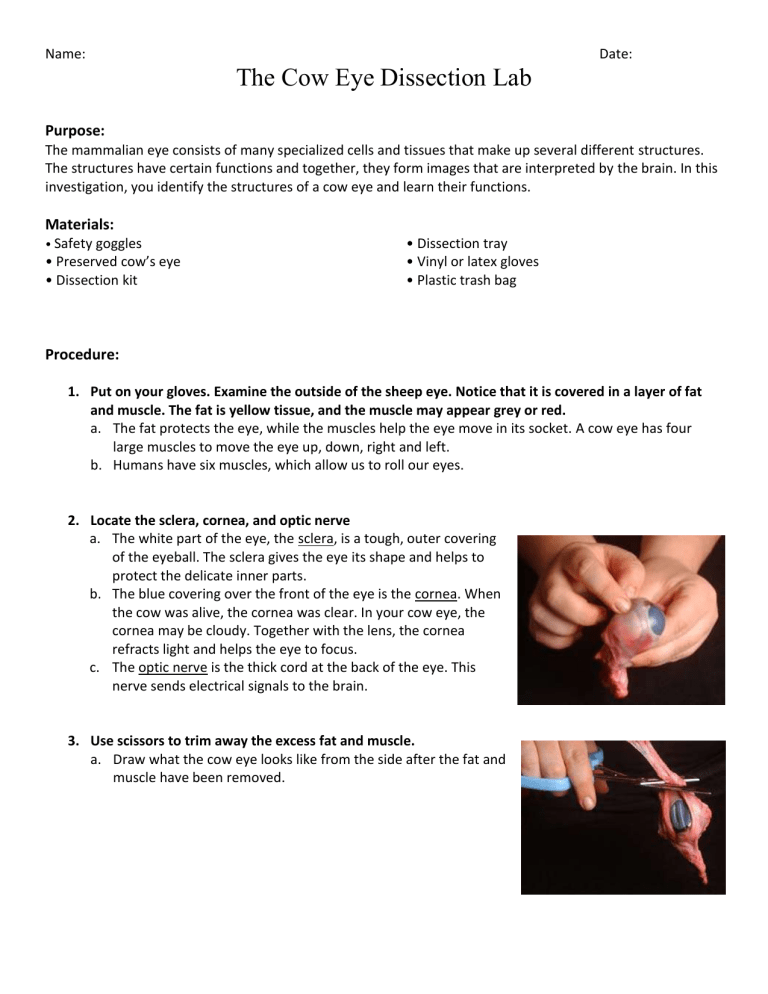
Name: Date: The Cow Eye Dissection Lab Purpose: The mammalian eye consists of many specialized cells and tissues that make up several different structures. The structures have certain functions and together, they form images that are interpreted by the brain. In this investigation, you identify the structures of a cow eye and learn their functions. Materials: • Safety goggles • Preserved cow’s eye • Dissection kit • Dissection tray • Vinyl or latex gloves • Plastic trash bag Procedure: 1. Put on your gloves. Examine the outside of the sheep eye. Notice that it is covered in a layer of fat and muscle. The fat is yellow tissue, and the muscle may appear grey or red. a. The fat protects the eye, while the muscles help the eye move in its socket. A cow eye has four large muscles to move the eye up, down, right and left. b. Humans have six muscles, which allow us to roll our eyes. 2. Locate the sclera, cornea, and optic nerve a. The white part of the eye, the sclera, is a tough, outer covering of the eyeball. The sclera gives the eye its shape and helps to protect the delicate inner parts. b. The blue covering over the front of the eye is the cornea. When the cow was alive, the cornea was clear. In your cow eye, the cornea may be cloudy. Together with the lens, the cornea refracts light and helps the eye to focus. c. The optic nerve is the thick cord at the back of the eye. This nerve sends electrical signals to the brain. 3. Use scissors to trim away the excess fat and muscle. a. Draw what the cow eye looks like from the side after the fat and muscle have been removed. 4. Use the sharp probe to poke a hole in the eye halfway between the cornea and the optic nerve. Some liquid will come out when you do this. Use scissors to cut all the way around the eye, separating it into front and back halves. Try not to disturb the internal parts of the eye as you cut. 5. Separate the eye into two halves. Taking the front half of the eye, which contains the cornea, iris, and lens, try to look through it. It may be possible to see right through the lens and cornea. a. See if you can read text through the eye. b. If you can see an image through the eye, note whether it appears upright or upside down. Write it below. 6. Use the sharp probe to poke a hole at the junction of the cornea and the sclera. Use the scissors to cur a circle around the cornea in order to remove it. a. Under the cornea is the more jelly-like substance that helps support the cornea. 7. Under the cornea is the iris, which is a ring of tissue. Remove it from the cow eye. a. In cows, the iris is an oval shape. In humans it is circular. b. The pupil is seen to be simple a hole in the iris. 8. Remove the lens from the eye and, using a paper towel, wipe all the jelly form it. The lens will probably be yellow, but it is colourless and clear in the living eye. a. See if you can see an image through the lens. The lens will be hard but in the living eye it is soft and can change shape in order to focus. b. Does the object appear bigger or smaller through the lens? Write it below. 9. Empty the eye if there is still liquid present. On the back half of the eyeball, you can see some blood vessels that are part of a thin film. That film is the retina. a. If the retina has come off it will likely be attached to the eye at a single spot, the blind spot. b. Try to push the retina around and find the blind spot. c. There are no photoreceptors at the blind spot, you can’t see anything that lands in that place on the retina. 10. Take a look at the tapetum. Under the retina, there is an iridescent layer. Blue and green. a. This layer helps the cow to see under low-light conditions. b. Humans do not have this layer. 11. Make sure you know how to identify the cornea, retina, sclera, iris, lens, optic nerve, external muscle. Call over Ms. Qiu and point out each part. 12. Clean up!! Discussion: 1. Find your blind spot a. To find your blind spot, use the two dots below. Hold one hand over your left eye and look directly at the cross. At first, you can see both dots even though you're looking directly at only one. As you slowly move the page closer to your eyes, the right-hand dot disappears! If you move your eye, the dot will reappear, but as long as you focus on the first dot, the second will be invisible. Move even closer and the missing dot reappears. You’ve found your blind spot! b. Why does the optic nerve cause a blind spot? 2. a. b. c. d. e. f. g. Match the term to the definition ______ External muscles ______ Retina ______ Cornea ______ Lens ______ Optic nerve ______ Iris ______ Sclera A. A clear structure that refracts light and can change in curvature B. A tiny ring of muscles that change the shape of the lens C. The pigmented ring of muscles that change the size of the pupil D. A thin layer of cells that convert light into nerve signals E. Move the eye around F. Transmits signals from the eye to the brain G. Gives the eye its shape and protects the inner parts 3. What is the purpose of the layer of fat? 4. List three differences between the anatomy of a cow eye and a human eye. 5. Label the eye diagram below with all of the structures you identified. 6. What would happen to a person’s vision if the eye’s lens was unable to change shape? Conclusion 1. Describe how different parts of the eye adapt to bright light and darkness.
