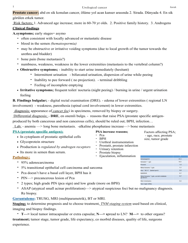
!1 Urological cancer faruk Prostate cancer; abd en sik konulan cancer, ölüme yol acan kanser arasında 2. Sirada. Dünyada 4. En sik görülen erkek tumor. Risk factors; 1. Advanced age increase; more in 60-70 yr olds. 2. Positive family history. 3. Androgens Clinical findings A.symptoms; early stages= asymo - often consistent with locally advanced or metastatic disease - blood in the semen (hematospermia) - may be obstructive or irritative voiding symptoms (due to local growth of the tumor towards the urethra and bladder) - bone pain (bone metastasis?) - numbness, weakness, weakness in the lower extremities (metastasis to the vertebral column?) • Obstructive symptoms; - inability to start urine immediately (hesitant) - Intermittent urination - bifurcated urination, dispersion of urine while peeing - Inability to pee forward ( no projection). - terminal dribbling - Feeling of incomplete emptying • Irritative symptoms; frequent toilet/ nocturia (night peeing) / burning in urine / urgent urination feeling B. Findings bulgular; - digital rectal examination (DRE). - edema of lower extremities ( regional LN involvement) - weakness, paresthesia (spinal cord involvement) in lower extremities. diagnosis; appearance of cancer foci in specimens, removed by biopsy or surgery. Differential diagnosis; - DRE, en onemli bulgu. - reasons that raise PSA (prostate specific antigenproduced by both cancerous and non cancerous cells), should be ruled out; BPH, infection… Lab; - anemia —> long bone metastasis. -alkaline phosphatase increase—> bone metastasis PSA (prostate specific antigen); PSA increase reasons; Factors affecting PSA; - Pca - age, race, prostate • In cytoplasm of prostatic epithelial cells - BPH size, tumor grade - Urethral instrumentation • Glycoprotein structure prostate infarction • Production is regulated by androgen receptors - Prostatit, Urinary retention - Prostate biopsy • Its more in semen than serum. - Ejaculation, inflammation Pathology; - 95% adenocarcinoma - 5% transitional epithelial cell carcinoma and sarcoma - Pca doesn’t have a basal cell layer, BPH has it - PIN—> precancerous lesion of Pca - 2 types; high grade PIN (pca sign) and low grade (more on BPH) - ASAP (atypical small acinar proliferation)—> atypical suspicious foci but no malignancy diagnosis. Re biopsy. Goruntuleme; TRUSG, MRI (multiparametrik), BT or MRI. Staging; to determine prognosis and to choose treatment, TNM staging system used based on clinical, imaging and biopsy findings. - T—> local tumor intracapsular or extra capsular, N—> spread to LN? M—> to other organs? treatment; tumor stage, tumor grade, life expectancy, co morbid diseases, quality of life, surgeons experience. !2 Urological cancer faruk Pca tedavi 3 ana başlık atlında yapılmakta; A. Localized disease treatment; 1. Radical prostatectomy/ radiotherapy (external/ trans perineal brachytherapy), 3. Active izlem 4. Cryotherapy 5. HIFU ; high intensity focused ultrasound B. Treatment in Locally advanced disease; 1. Radiotherapy 2. Hormonal therapy C. Metastatic diseases; hormontherapy—> ablation of androgen source, inhibition of androgen production, anti-androgens LNRH or LH inhibition. 1. Surgical castration 2.medical castration 3. Anti-androgens 4. LHRH antagonists 5. Biphosphanate 6. Bone lesions= palliative radiotherapy Bladder cancer epidemiology; men/woman ratio; 3/1. Woman 8th most seen cancer, 2.5% of women’s cancer. Genellikle ileri yaslarin hastaligi. Etiology and risk factors; smoking, aromatic amines, analgesic, chronic infection, pelvic radiation. Chemical carcinogens. Pathology; bladder tumors 98% from epithelial origin. Epithelial tumors (90% transitional epi ca, 5-7% scc) Squamous cell carcinoma; 5-7%, shistosmiasis, chronic irritation and inflammation are important, bad prognosis. Adenocancer; 1-2%, generally chronic irritation and inflammation related (metaplasmic, urachal, metastatic). Muscle invasion when diagnosed. Secrete mucus. Prognosis worse than others. Transitional epithelial cancer (pathogenesis); 75-85% initially superficial tumors—> become invasive 20-30%. Epithelial hyperplasia, metaplastic changes, dysplasia, cancer development Staging; Primary tumor; T0: no tumor Ta: mucosal involvement Tis; carcinoma in situ T1: lamina propria involved. T2; muscular involvement, T3: perivesical involvement, T4: spread to surrounding organs & tissues Diagnosis; - symptoms seen; painless clotted hematuria en sik. Bladder irritation symptoms, tumor localization in upper and lower urinary system. —>hematuria; microscopic or macro. Hematuria grade. Secondary anemia symptoms. —> bladder irritation symptoms; burning when urinating, frequent urination, frequent urge to urinate, passing urine at night. —> obstructive symptoms; flank pain due to hydronephrosis secondary to ureter obstruction. - sensation of fullness may develop= result of intravesical obstruction and increase in residual urine after voiding. —> metastasis related symp; weight loss, loss of appetite, weakness, anemia… Systoscopy; bladder tumor diagnosis and follow up = gold standard method Testis tumors; 1. Germ cell tumors—> a. Classic, anaplastic, spermatocystic. b) embryonal carcinoma (adult/ juvenile) c. Teratocarcinoma d. Teratoma e. Choriocarcinoma. 2. Gonadostromal tumors; leydig, sertoli 3. Metastatic tumors; lymphoma, prostate, melanoma, AC etiology; gonadal dysgenesis (20-30% cancer), gonadoblastoma !3 Urological cancer faruk Risk factors; etiological; cryptorchidism, klinefelter syndrome, family. History, contralateral tumor, testicular intraepithelial neoplasia (TIN), infertility. pathological; histological type, tumor size, vascular lymphatic peri-humoral invasion Clinical (metastatic diseases); primary localization, non pulmonary visceral metastasis. symptoms; painless mass (hydrocele), gynecomastia (germ cell, 50% sertoli/ leydig), right testis more often Diagnosis physical examination / USG / chest CT (small lymph nodes seen <2cm)/ orchioectomy/ tumor markers (AFP, B-hCG, LDH); after orchioctomy elevations suggest metastasis. Lymph node diagnosis. Differential diagnosis; torsion, epididimitis, hydrocele, hernias. a-fetoprotein; produced in pure embryonal, teratocarcinoma, yolk sac, mixed tumors, high in embryogenesis (increased in liver and GI tract diseases). h-CGT; produced in placenta, produced from syncytiotrophoblastic tissue, all choriocarcinomas (40% embryonal, 5-10% seminoma). Serum tumor markers; Orchioctomy; high inguinal, spermatic cord isolation at external inguinal ring, testicles taken up,. one or both testicles are removed Primary tumor; pT1is; intratubular germ cell neoplasia. pT1; tumor testis and epididymis no nerve, vascular and lymphatic invasion yok. pT2; vascular or lymphatic invasion seen. pT3; spermatic cord involvement. pT4; scrotum involvement. Kidney tumors; • 90% parenchymal (primary kidney tumor), 5-10% calyces and pelvic renal origin. - Primary kidney… from prancheyma divided into benign and malignent. • Adult kidney tumors classification; epithelial tumors—>benign epithelial tumor, cortical papillary adenoma, renal oncocytoma, juxtaglomerular cell tumor, metanephric tumors, renal cell carcinoma. • Mesenchymal tumors—> benign mesenchymal.., angiomyolipoma, medullary fibroma, leimyoma, lipom, hemangiom, malignant mesenchymal tumors, sarcomas, lymphoma. • Blastema kaynakli; mesoblastic nefroma, neuroblastoma • Neuroendocrine tumors; carcinoids, primitive neuroectodermal tumor Renal cell cancer; Urinery sistem kaynaklı maligniteler arasında 3. Sıklıkta. Daha çok 60-70 yaşları. ertiology; smoking (risk increases 30-50%) / extreme obestiy/ hypertension / genetic (major genetic anomaly= loss of chromosome 3p)—> tumor suppressor genes inactivation, oncogenes activation. —> hereditary kidney tumors; Von hippel Lindau disease, familial papillary renal cell carcinoma, BHD syndrome, familial leiyomyomatosis + RCC pathology; conventional RHK, papillary, chromofobe, collecting canal. —>conventional RCC; most commonly seen type (70%), yellow in color due to the abundance of lipids and glycogen in their cytoplasm. -Kökeni proksimal kıvrıntılı tüpler —> papillary RCC; 2nd most common seen type. Cytogenic anomalies. multifocal, type 2 prognosis bad - Kökeni distal kıvrıntılı tüpler —> chromophobe renal cell cancer; 3rd most common. Prognosis very good. unilateral. Oncocytomy diagnosis. Kökeni kortikal toplayıcı kanallar . !4 Urological cancer faruk —> collecting canal RCC; least seen. Kökeni medüller toplayıcı kanal. Very aggressive, prognosis very bad. Symptoms; classic triad = hematuria, side pain, palpable mass RCC paraneoplastic syndroms; hypercalcemia, polisitemi, hypertension, Stauffer syndrome, weight loss, fever, amyloidosis, prolactin and glucagon hormones increase. Physical examination; palpable cysts, vericocele, supraclavicular/ servical adenppathy. Lower extremity edema. Goruntuleme yöntemleri; • USG; non invasive, sensitivity high, kidney cysts. • B; gold standard of kidney tumor diagnosis. Sensitivity and specificity (95%) • MRI; - The most effective radiological imaging method for determining vascular invasion in kidney tumors.. • Differential diagnosis; oncosytoma, angiomyolipoma, abscess, infarcts, vein malformations, pseudotumor, mestasis. Upper urinary system uroepithelial tumors;


