Diabetic Retinopathy Detection: Preprocessing Methods Comparison
advertisement

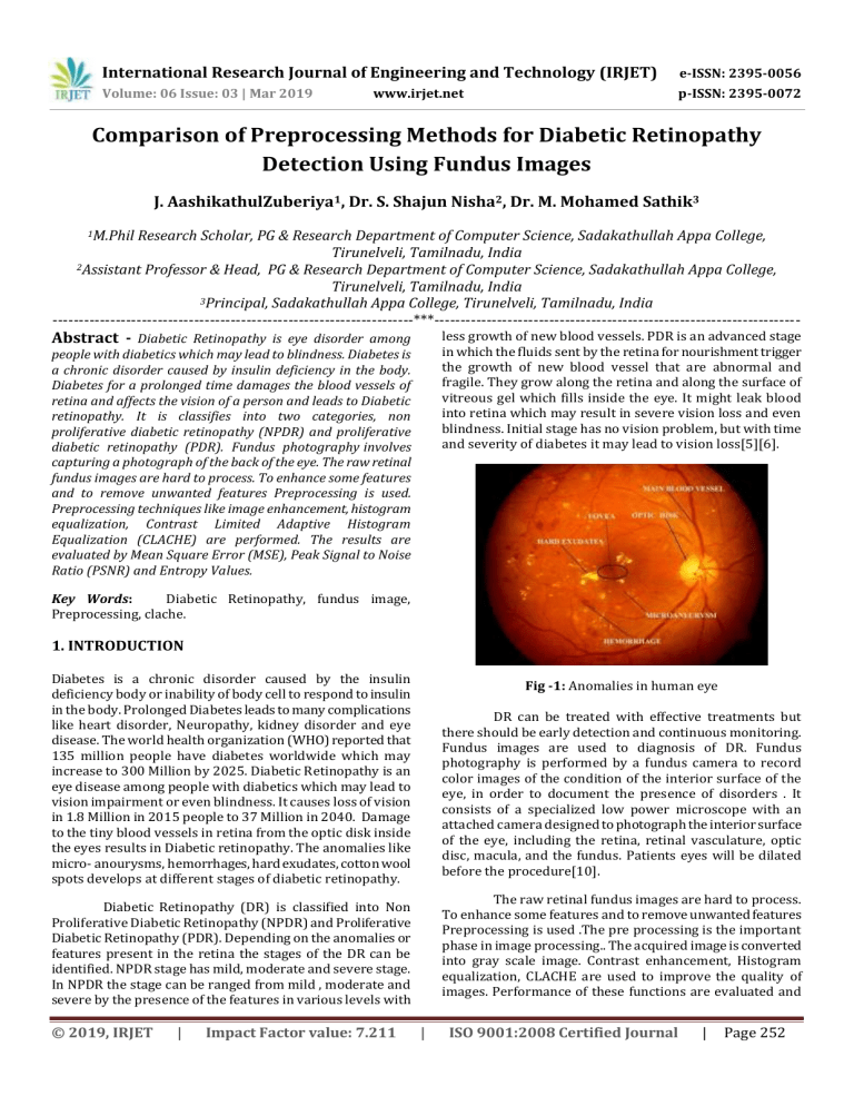
International Research Journal of Engineering and Technology (IRJET) e-ISSN: 2395-0056 Volume: 06 Issue: 03 | Mar 2019 p-ISSN: 2395-0072 www.irjet.net Comparison of Preprocessing Methods for Diabetic Retinopathy Detection Using Fundus Images J. AashikathulZuberiya1, Dr. S. Shajun Nisha2, Dr. M. Mohamed Sathik3 1M.Phil Research Scholar, PG & Research Department of Computer Science, Sadakathullah Appa College, Tirunelveli, Tamilnadu, India 2Assistant Professor & Head, PG & Research Department of Computer Science, Sadakathullah Appa College, Tirunelveli, Tamilnadu, India 3Principal, Sadakathullah Appa College, Tirunelveli, Tamilnadu, India ---------------------------------------------------------------------***---------------------------------------------------------------------- Abstract - Diabetic Retinopathy is eye disorder among less growth of new blood vessels. PDR is an advanced stage in which the fluids sent by the retina for nourishment trigger the growth of new blood vessel that are abnormal and fragile. They grow along the retina and along the surface of vitreous gel which fills inside the eye. It might leak blood into retina which may result in severe vision loss and even blindness. Initial stage has no vision problem, but with time and severity of diabetes it may lead to vision loss[5][6]. people with diabetics which may lead to blindness. Diabetes is a chronic disorder caused by insulin deficiency in the body. Diabetes for a prolonged time damages the blood vessels of retina and affects the vision of a person and leads to Diabetic retinopathy. It is classifies into two categories, non proliferative diabetic retinopathy (NPDR) and proliferative diabetic retinopathy (PDR). Fundus photography involves capturing a photograph of the back of the eye. The raw retinal fundus images are hard to process. To enhance some features and to remove unwanted features Preprocessing is used. Preprocessing techniques like image enhancement, histogram equalization, Contrast Limited Adaptive Histogram Equalization (CLACHE) are performed. The results are evaluated by Mean Square Error (MSE), Peak Signal to Noise Ratio (PSNR) and Entropy Values. Key Words: Diabetic Retinopathy, fundus image, Preprocessing, clache. 1. INTRODUCTION Diabetes is a chronic disorder caused by the insulin deficiency body or inability of body cell to respond to insulin in the body. Prolonged Diabetes leads to many complications like heart disorder, Neuropathy, kidney disorder and eye disease. The world health organization (WHO) reported that 135 million people have diabetes worldwide which may increase to 300 Million by 2025. Diabetic Retinopathy is an eye disease among people with diabetics which may lead to vision impairment or even blindness. It causes loss of vision in 1.8 Million in 2015 people to 37 Million in 2040. Damage to the tiny blood vessels in retina from the optic disk inside the eyes results in Diabetic retinopathy. The anomalies like micro- anourysms, hemorrhages, hard exudates, cotton wool spots develops at different stages of diabetic retinopathy. Fig -1: Anomalies in human eye DR can be treated with effective treatments but there should be early detection and continuous monitoring. Fundus images are used to diagnosis of DR. Fundus photography is performed by a fundus camera to record color images of the condition of the interior surface of the eye, in order to document the presence of disorders . It consists of a specialized low power microscope with an attached camera designed to photograph the interior surface of the eye, including the retina, retinal vasculature, optic disc, macula, and the fundus. Patients eyes will be dilated before the procedure[10]. The raw retinal fundus images are hard to process. To enhance some features and to remove unwanted features Preprocessing is used .The pre processing is the important phase in image processing.. The acquired image is converted into gray scale image. Contrast enhancement, Histogram equalization, CLACHE are used to improve the quality of images. Performance of these functions are evaluated and Diabetic Retinopathy (DR) is classified into Non Proliferative Diabetic Retinopathy (NPDR) and Proliferative Diabetic Retinopathy (PDR). Depending on the anomalies or features present in the retina the stages of the DR can be identified. NPDR stage has mild, moderate and severe stage. In NPDR the stage can be ranged from mild , moderate and severe by the presence of the features in various levels with © 2019, IRJET | Impact Factor value: 7.211 | ISO 9001:2008 Certified Journal | Page 252 International Research Journal of Engineering and Technology (IRJET) e-ISSN: 2395-0056 Volume: 06 Issue: 03 | Mar 2019 p-ISSN: 2395-0072 www.irjet.net compared using metrics to find the results. The outline of the proposed work is shown in Fig 2. were considered[11]. Working with color images makes the task more complex in image processing, so the color images are converted to Gray scale Images(GSI).Then contrast of GSI image is enhanced to boost the high intensity pixel along retinal boundaries. The preprocessed output image is shown in Table 6. 1.1 Motivation Justification As there are various preprocessing methods are available and been introduced, the purpose of using Preprocessing is to enhance some features and to remove unwanted features. To identify the best method, Comparision of different image enhancement are performed. Based on the metrices the result is justified. 3.1 Contrast Enhancement Contrast is an important factor in any subjective evaluation of image quality. Contrast is created by the difference in luminance reflected from two adjacent surfaces. In other words, contrast is the difference in visual properties that makes an object distinguishable from other objects and the background. In visual perception, contrast is determined by the difference in the colour and brightness of the object with other objects.[4] 1.2 Outline of the Paper Low contrast image values concentrated near a narrow range (mostly dark, or mostly bright, or mostly medium values). Contrast enhancement (CE) change the image value change the image value distribution to cover a wide range. Contrast of an image can be revealed by its histogram. imadjust function increases the contrast of the image by mapping the values of the input intensity image to new values such that, by default, 1% of the data is saturated at low and high intensities of the input data. Fig -2: Outline of the proposed work. 3.2 Histogram Equalization 1.3 Organization of the Paper Histogram equalization is used to enhance contrast. It is not necessary that contrast will always be increase in this. There may be some cases were histogram equalization (HE) can be worse. In that cases the contrast is decreased. Histeq performs histogram equalization. It enhances the contrast of images by transforming the values in an intensity image so that the histogram of the output image approximately matches a specified histogram (uniform distribution by default. Paper continuous as follows - Section II consist of the related work that is literature survey , Section III describes methodologies and fundus image enhancement using different techniques, Section IV is about the experimental results and discussion Section V has conclusion. 2. RELATED WORK Swathi.C; Anoop.B.K; D.Anto sahaya dhas; S.Perumal sanker [1] used different types of preprocessing techniques. They used adaptive histogram equalization, Weiner filter , Median filter and adaptive mean filter. PSNR and MSE values are used for measurement of efficiency. 3.3 Contrast Limited Adaptive Histogram Equalization Contrast-Limited Adaptive Histogram (CLACHE) uses adapthisteq function to performs equalization. It enhances the contrast of the grayscale image by transforming the values using contrast-limited adaptive histogram equalization. Unlike histogram equalization, it operates on small data regions (tiles) rather than the entire image. Each tile's contrast is enhanced so that the histogram of each output region approximately matches the specified histogram (uniform distribution by default). The contrast enhancement can be limited in order to avoid amplifying the noise which might be present in the image[7] Sumathy, B.; Poornachandra, S [2] extracted the features of retinal images using the new adaptive histogram. The optical disc is difficult to analyze because of its brightness. The author used the method which gives good results. Chen Hee Ooi; Isa, N.A.M [3] used two different methods for adaptive contrast enhancement and brightness presevation. Using this method they divide the histogram on the basis of median and uses the advancement of bi- histogram equalization. 3. METHODOLOGY Fundus images are used to diagnosis of DR. It classifies the image into normal, NPDR and PDR. For evaluation five images © 2019, IRJET | Impact Factor value: 7.211 | ISO 9001:2008 Certified Journal | Page 253 International Research Journal of Engineering and Technology (IRJET) e-ISSN: 2395-0056 Volume: 06 Issue: 03 | Mar 2019 p-ISSN: 2395-0072 www.irjet.net 4. EXPERIMENTAL RESULTS Table -3: Image Quality Parameters of Image 3 4.1 Performance Metrics Image 3 The performance metrics such as Peak signal to Noise Ratio (PSNR), Mean Squared Error (MSE), Entropy are calculated. MSE PSNR Entropy CE 9615.47 8.34 5.6421 HE 4306.52 11.82 5.2647 CLACHE 366.68 22.52 6.1634 A. Peak Signal To Noise Ratio (PSNR): Table -4: Image Quality Parameters of Image 4 It is the evaluation standard of the reconstructed image quality, is the most wanted feature. PSNR is measured in the decibels (dB) and it is given by CE 3834.98 12.33 6.2004 PSNR = 10log (255 ∕ 2 MSE) HE 4492.04 11.64 5.6039 CLACHE 680.44 19.84 6.7073 Image 4 Where the value 255 is the maximum possible value that can be attained by the image signal. Mean square error is defined as where M*N is the size of the original image. Higher the PSNR value betters the reconstructed image. MSE PSNR Entropy Table -5: Image Quality Parameters of Image 5 Image 5 MSE PSNR Entropy B. Mean Square Error (MSE): CE 7613.48 9.35 5.9684 The average squared difference between the reference signal and distorted signal is called as the mean square error. It can be easily calculated by adding up the squared difference pixel-by-pixel and dividing by the total pixel count. Let m x n is a noise free monochrome image I, and K is defined as the noisy approximation. Then the mean square error between these two signals is defined as HE 4859.93 11.30 5.5001 CLACHE 708.94 19.66 6.5956 4.2 Performance Evaluation In this proposed work preprocessing techniques for retinal fundus images were applied for five images. Techniques such as gray scale image. Contrast enhancement, Histogram equalization, CLACHE are applied and the quality of images are improved. The results of the images are tabulated in Table 6 and Table 7 . MSE = 1 m × n [ I i, j − K i, j ] n − 1 2 j = 0 m − 1 i = 0 C. Entropy: For a given PDF p, entropy Ent[p] is computed. In general, the entropy is a useful tool to measure the richness of the details in the output image. Table -6: Result Of Images on applying Gray scale and Contrast Enhancement. ImageName Ent[p] = −Σk = 0 (k)log2 p (k) MSE PSNR Entropy CE 6546.24 10.00 5.8396 HE 3662.97 12.53 5.4481 CLACHE 411.15 22.02 6.4827 MSE PSNR Image 3 4780.83 11.37 6.1269 HE 2470.17 14.24 5.6041 CLACHE 437.50 21.75 6.7184 | Image 4 Entropy CE © 2019, IRJET CE Image 2 Table -2: Image Quality Parameters of Image 2 Image 2 GSI Image 1 Table -1: Image Quality Parameters of Image 1 Image 1 Input Image Impact Factor value: 7.211 Image 5 | ISO 9001:2008 Certified Journal | Page 254 International Research Journal of Engineering and Technology (IRJET) e-ISSN: 2395-0056 Volume: 06 Issue: 03 | Mar 2019 p-ISSN: 2395-0072 www.irjet.net Table -6: Result of images on applying Histogram Equalization and CLACHE. [5] [6] ImageName HE CLACHE Image 1 Image 2 [7] Image 3 [8] Image 4 Image 5 [9] 5. CONCLUSIONS Swati gupta and Karandikar,”Diagonosis of diabetic retinopathy using machine learning”, Journal of Research and development, 2015 Sarni Suhaila Rahim, Vasile Palade, Chrisina Jayne, Andreas Holzinger, James Shuttle worth, “Detection of Diabetic Retinopathy and Masculopathy in Eye Fundus Images using Fuzzy Image Processing”, Springer International Publishing Switzerland 2015, Y.Guo et al. (Eds.): BIH 2015 , LNAI 9250, pp.379-388, 2015 Jeline Devadhas and R. Binisha, “Early Diagonosis of Diabetic Retinopathy by the Detection of Microaneurysms in Fundus Images”, ICTACT Journal On Image and Video Processing , ISSN: 0976-9102, Volume:08, Issue: 04 G. Prabavathi, Dr.K.Mahesh, “Automated Analysis of Microaneurysm Detection of Diabetic Retinopathy”, International Journal for Modern Trends in Science and Technology, ISSN: 2455-3778, Volume:03, Issue No: 05, May 2017 Salman Sayed, Dr. Vandana Inamdar, Sangram Kapre, “ Detection of Diabetic Retinopathy usimg Image Processing and Machine Learning”, International Journal of Innovative Research in Science, Engineering and Technology, ISSN: 2319-8753, Vol.6, Issue 1, January 2017 Khan Abdul Mukhtar Khan, “Detection of Digital Image Processing A Survey”, International Journal of Research in Computer Application and Robotics”,ISSN 2320-7345 Vol.6 Issue 1, Pg:13-19, January 2018 Shajun Nisha, Ashika Parvin, “Preservation Of Historical Document Using Enhancement Techniques International Journal of Trend in Research and Development, Vol 4(1), ISSN: 2394-9333 Jan- Feb 2016 In this paper preprocessing techniques for retinal fundus images were applied for five images. The image quality obtained after applying these algorithms is assessed with metrics. These metrics include Peak Signal To Noise Ratio (PSNR), Entropy and Mean Square Error (MSE). From the results Contrast Limited Adaptive Histogram Equalization (CLACHE) succeeds because it has higher PSNR and Lower MSE value. Using CLACHE will results in highly enhanced image. [10] REFERENCES BIOGRAPHIES [11] Swathi C, Anoop B.K, Anto Sahaya Dhas D, Perumal Sanker S, “Comparison Of Different Image Preprocessing Methods Used For Retinal Fundus Images”, Proc.IEEE Conference On Emerging Devices And Smart Systems(ICEDSS 2017) 3-4march2017 Vol.,No.,PP.9781-5090-5555-5/17$31.00 [2] Sumathy B, Poornachandra S, “Feature Extraction In Retinal Fundus Images”, Information Communication And Embedded Systems (ICICES), 2013 International Conference On , vol., no., pp.798-802, 21-22 Feb. 2013 [3] Chen Hee Ooi; Isa, N.A.M., “Adaptive Contrast Enhancement Methods With Brightness Preserving”, Consumer Electronics, IEEE Transactions On , vol.56, no.4, pp.2543-2551, November 2010 [4] Preeti Gupta, “Contrast Enhancemet For Retinal Images Using Multi-Objective Genetic Algorithm”, International Journal Of Emerging Trends In Engineering And Development, Issue 6, Vol. 1 (January 2016) ISSN 22496149. J.AashikathulZuberiya, M.Phil Research scholar, currently pursuing at sadakathullah appa college, Tirunelveli. I had completed my UG & PG degree in Computer science at Sadakathullah Appa College, I had certification of NPTEL courses. My research area is in Image processing. [1] © 2019, IRJET | Impact Factor value: 7.211 Dr.S.Shajun Nisha, Assistant Professor and Head of the PG & Research Department of ComputerScience, Sadakathullah Appa College, Tirunelveli. She has completed M.Phil. (Computer Science) M.Tech (Computer and Information Technology) in Manonmaniam Sundaranar University, Tirunelveli and completed Ph.D (Computer Science) in Bharathiyar university,Coimbatore. She has involved in various academic activities and attended so many national and international seminars, conferences and presented | ISO 9001:2008 Certified Journal | Page 255 International Research Journal of Engineering and Technology (IRJET) e-ISSN: 2395-0056 Volume: 06 Issue: 03 | Mar 2019 p-ISSN: 2395-0072 www.irjet.net numerous research papers. She is a member of ISTE and IEANG and her specialization is Image processing and neural network. Dr.M.Mohamed Sathik, Principal Sadakathullah Appa College, Tirunelveli. He has completed Ph.D(Computer science & engineering) Ph.D(Computer science), M.Phil. (Computer Science), M.Tech(Computer Science and Information Technology) in Manonmaniam Sundaranar University, Tirunelveli. He has so far guided more than 35 research scholars. He has published more than 100 papers in International Journals and also two books. He is a member of curriculum development committee of various universities and autonomous colleges of Tamil Nadu. He is a syndicate member Manonmaniam Sundaranar University, Tirunelveli. His specializations are VRML, Image Processing and Sensor Networks. © 2019, IRJET | Impact Factor value: 7.211 | ISO 9001:2008 Certified Journal | Page 256


