Chapter 1
Term
The cell theory
Cell reproduction
Cell differentiation
Stem cells
7 functions of life
(organisms)
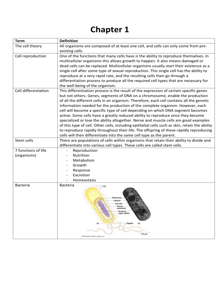
Bacteria
Definition
All organisms are composed of at least one cell, and cells can only come from preexisting cells.
One of the functions that many cells have is the ability to reproduce themselves. In
multicellular organisms this allows growth to happen. It also means damaged or
dead cells can be replaced. Multicellular organisms usually start their existence as a
single cell after some type of sexual reproduction. This single cell has the ability to
reproduce at a very rapid rate, and the resulting cells then go through a
differentiation process to produce all the required cell types that are necessary for
the well-being of the organism.
This differentiation process is the result of the expression of certain specific genes
but not others. Genes, segments of DNA on a chromosome, enable the production
of all the different cells in an organism. Therefore, each cell contains all the genetic
information needed for the production of the complete organism. However, each
cell will become a specific type of cell depending on which DNA segment becomes
active. Some cells have a greatly reduced ability to reproduce once they become
specialized or lose the ability altogether. Nerve and muscle cells are good examples
of this type of cell. Other cells, including epithelial cells such as skin, retain the ability
to reproduce rapidly throughout their life. The offspring of these rapidly reproducing
cells will then differentiate into the same cell type as the parent.
There are populations of cells within organisms that retain their ability to divide and
differentiate into various cell types. These cells are called stem cells.
- Reproduction
- Nutrition
- Metabolism
- Growth
- Response
- Excretion
- Homeostasis
Bacteria
Eukaryotes
Mitochondrion
Chloroplast
Plasmodesmata
Ribosomes
Golgi apparatus
Central vacuole
Plasma membrane
Cell wall
Autotrophs
Smooth endoplasmic
Mitochondria are organelles that carry out respiration. Mitochondria (singular
mitochondrion) are rod-shaped organelles that appear throughout the cytoplasm.
They are close in size to a bacterial cell. Mitochondria have their own DNA, a circular
chromosome similar to that in bacterial cells, allowing them some independence
within a cell. They have a double membrane: the outer membrane is smooth, but
the inner membrane is folded into cristae (singular crista). Inside the inner
membrane is a semiuid substance called the matrix. An area called the inner membrane space lies
between the two membranes. The cristae provide a huge surface area within which
the chemical reactions characteristic of the mitochondria occur. Most mitochondrial
reactions involve the production of usable cellular energy called adenosine
triphosphate (ATP). Because of this, the mitochondria are often called the
powerhouse of a cell. This organelle also produces and contains its own ribosomes;
these ribosomes are of the 70S type. Cells that have high energy requirements, such
as muscle cells, have large numbers of mitochondria.
Chloroplasts are specialized plastids containing the green pigment chlorophyll. They
consist of grana within the colourless stroma. They are the sites for photosynthesis.
Chloroplasts occur only in algae and plant cells. The chloroplast contains a double
membrane and is about the same size as a bacterial cell. Like the mitochondrion, a
chloroplast contains its own DNA and 70S ribosomes. The DNA of a chloroplast takes
the form of a ring.
Occur in all prokaryotic cells and function as sites of protein synthesis.
Golgi apparatus stores, modifies, and packages proteins
Central vacuole has storage and hydrolytic functions
The plasma membrane is found just inside the cell wall and is similar in composition
to the membranes of eukaryotic cells. To a large extent the plasma membrane
controls the movement of materials into and out of the cell, and it plays a role in
binary fission of the prokaryotic cell
The prokaryotic cell wall protects and maintains the shape of the cell. In most
prokaryotic cells this wall is composed of a carbohydrate–protein complex called
peptidoglycan. Some bacteria have an additional layer of a type of polysaccharide
outside the cell wall. This layer makes it possible for some bacteria to adhere to
structures such as teeth, skin, and food
Organisms with the ability to produce energy through photosynthesis
Smooth endoplasmic reticulum is ER without ribosomes. Smooth ER has many
reticulum
Rough endoplasmic
reticulum
Nucleus
unique enzymes embedded on its surface. Its functions are:
• the production of membrane phospholipids and cellular lipids
• the production of sex hormones such as testosterone and oestrogen
• detoxication of drugs in the liver
• the storage of calcium ions in muscle cells, needed for contraction of muscle cells
• transportation of lipid-based compounds
• helping the liver release glucose into the bloodstream when needed.
Rough endoplasmic reticulum is a site of protein synthesis. The endoplasmic
reticulum (ER) is an extensive network of tubules or channels that extends most
everywhere in the cell, from the nucleus to the plasma membrane. Its structure
enables its function, which is the transportation of materials throughout the internal
region of the cell. There are two general types of ER: smooth ER and rough ER.
Smooth ER does not have any of the organelles called ribosomes on its exterior
surface. Rough ER has ribosomes on its exterior.
Nucleus contains most of the cell’s DNA. The nucleus in eukaryotic cells is an isolated
region where the DNA resides. It is bordered by a double membrane referred to as
the nuclear envelope. This membrane allows compartmentalization of the
eukaryotic DNA, thus providing an area where DNA can carry out its functions
without being affected by processes occurring in other parts of the cell. The nuclear
membrane does not provide complete isolation because it has numerous pores that
allow communication with the cell’s cytoplasm
-
Centrosome
Vacuoles
Nucleoid
Nucleolus is a dense, solid structure involved in ribosome synthesis.
Nuclear pore allows communication between the nucleus and the rest of the
cell
The centrosome occurs in all eukaryotic cells. Generally, it consists of a pair of
centrioles at right angles to one another. These centrioles are involved with the
assembly of microtubules, which are important to a cell because they provide
structure and allow movement. Microtubules are also important for cell division.
Cells from higher plants, plants that are thought to have evolved later, produce
microtubules even though they do not have centrioles. The centrosome is located at
one end of the cell close to the nucleus.
Vacuoles are storage organelles that are usually formed from the Golgi apparatus.
They are membrane-bound and have many possible functions. They occupy a very
large space inside the cells of most plants. They may store a number of different
substances, including potential food (to provide nutrition), metabolic waste and
toxins (to be expelled from the cell), and water. Vacuoles enable cells to have higher
surface area to volume ratios even at larger sizes. In plants, they allow the uptake of
water, which provides rigidity to the organism. Vacuoles are smaller in plant cells.
The nucleoid region of a bacterial cell is non-compartmentalized and contains a
single, long, continuous, circular thread of DNA, the bacterial chromosome.
Therefore this region is involved with cell control and reproduction. In addition to
the bacterial chromosome, bacteria may also contain plasmids. These small, circular,
DNA molecules are not connected to the main bacterial chromosome. The plasmids
replicate independently of the chromosomal DNA. Plasmid DNA is not required by
the cell under normal conditions but it may help the cell adapt to unusual
circumstances.
Central vacuole
Animal cells
Centrioles are associated with nuclear division. They are composed of microtubules.
The area in which centrioles are found is called the centrosome. It is present in all
eukaryotic cells, but centrioles are absent from higher plant cells.
Lysosomes are intracellular digestive centres that arise from the Golgi apparatus. A
lysosome does not have any internal structures. Lysosomes are sacs bounded by a
single membrane that contain as many as 40 different enzymes. The enzymes are all
hydrolytic and catalyse the breakdown of proteins, nucleic acids, lipids, and
carbohydrates. Lysosomes fuse with old or damaged organelles from within the cell
to break them down, so that recycling of the components can occur. Lysosomes are
also involved in the breakdown of materials that may be brought into a cell by
phagocytosis. Phagocytosis is a type of endocytosis that is explained on page 37 in
Section 1.4. The interior environment of a functioning lysosome is acidic; this acidic
environment is necessary for the enzymes to hydrolyse large molecules.
Don’t have chloroplast and cell walls, they have small vacuoles. Eukaryotes
Plant cells
Have bigger vacuoles. Eukaryotes.
Lysosome
Hydrophilic
Hydrophobic
Amphipatic
Pili
Lipid
Phospholipids
Binary fission
Cytoplasm
Membrane structure
Phospholipids
Cholesterol
Attracted to water
Not attracted to water
The hydrophilic part is a phosphate group. The hydrophobic part consists of two
hydrocarbon chains.
Transfers genetic material of bacteria. Some bacterial cells contain hair-like growths
on the outside of the cell wall. These structures are called pili and can be used for
attachment. However, their main function is joining bacterial cells in preparation for
the transfer of DNA from one cell to another (sexual reproduction). Some bacteria
have - flagella (plural) or a - flagellum (singular), which are longer than pili. Flagella
allow a cell to move.
Fats
A class of lipids that are a major component of all cell membranes
Prokaryotic cells divide by a very simple process called binary fission. During this
process, the DNA is copied, the two daughter chromosomes become attached to
different regions on the plasma membrane, and the cell divides into two genetically
identical daughter cells. This divisional process includes an elongation of the cell and
a partitioning of the newly produced DNA by microtubule-like fibres called FtsZ.
All eukaryotic cells have a region called the cytoplasm that occurs inside the plasma
membrane or the outer boundary of the cell. It is in this region that the organelles
are found. The fluid portion of the cytoplasm around the organelles is called the
cytosol. The cytoplasm occupies the complete interior of the cell. The most visible
structure with a microscope capable of high magnification is the chromosome or a
molecule of DNA. There is no compartmentalization within the cytoplasm because
there are no internal membranes other than the plasma membrane. Therefore, all
cellular processes within prokaryotic cells occur within the cytoplasm.
• Not all membranes are identical or symmetrical, as the first model implied.
• Membranes with different functions also have a different composition and
different structure, as can be seen with an electron microscope.
• A protein layer is not likely because it is largely non-polar and would not interface
with water, as shown by cell studies
that the ‘backbone’ of the membrane is a bilayer produced from huge numbers of
molecules called phospholipids. Each phospholipid is composed of a three-carbon
compound called glycerol. Two of the glycerol carbons have fatty acids. The third
carbon is attached to a highly polar organic alcohol that includes a bond to a
phosphate group. Fatty acids are not water soluble because they are non-polar. In
contrast, because the organic alcohol with phosphate is highly polar, it is water
soluble. This structure means that membranes have two distinct areas when it
comes to polarity and water solubility. One area is water soluble and polar, and is
referred to as hydrophilic (water-loving). This is the phosphorylated alcohol side. The
other area is not water soluble and is non-polar. It is referred to as hydrophobic
(water-fearing).
These molecules have a role in determining membrane fluidity, which changes with
temperature. The cholesterol molecules allow membranes to function effectively at
a wider range of temperatures than if they were not present. Plant cells do not have
cholesterol molecules; they depend on saturated or unsaturated fatty acids to
maintain proper membrane fluidity.
Proteins
Membrane protein
functions
It is these proteins that create the extreme diversity in membrane function. Proteins
of various types are embedded in the fluid matrix of the phospholipid bilayer. This
creates the mosaic effect referred to in the fluid mosaic model. There are usually
two major types of proteins. One type is referred to as integral proteins and the
other type is referred to as peripheral proteins. Integral proteins show an
amphipathic character, with both hydrophobic and hydrophilic regions within the
same protein. These proteins will have the hydrophobic region in the mid-section of
the phospholipid backbone. Their hydrophilic region will be exposed to the water
solutions on either side of the membrane. Peripheral proteins, on the other hand, do
not protrude into the middle hydrophobic region, but remain bound to the surface
of the membrane. Often these peripheral proteins are anchored to an integral
protein.
•sites for hormone-binding: Proteins that serve as hormone-binding sites have speci
c shapes exposed to the exterior that t the shape of speci c hormones. The
attachment between the protein and the hormone causes a change in the shape of
the protein, which results in a message being relayed to the interior of the cell.
• enzymatic action: Cells have enzymes attached to membranes that catalyse many
chemical reactions. The enzymes may be on the interior or the exterior of the cell.
Often they are grouped so that a sequence of metabolic reactions, called a
metabolic pathway, can occur.
• cell adhesion: Cell adhesion is provided by proteins that can hook together in
various ways to provide permanent or temporary connections. These connections,
referred to as junctions, can include gap junctions and tight junctions.
• cell-to-cell communication: Many of the cell-to-cell communication proteins have
carbohydrate molecules attached. They provide an identity cation label that
represents the cells of different types of species.
• channels for passive transport: Some proteins contain channels that span the
membrane, providing passageways for substances to be transported through. When
this transport is passive, material Make sure you can draw and label all the parts of a
membrane as described in this section for the fluid mosaic model. Follow the
directions given earlier for making a good drawing. In the drawing, the phospholipids
should be shown using the symbol of a circle with two parallel lines attached. It is
also important to show a wide range of proteins with various functions and locations
moves through the channel from an area of high concentration to an area of lower
concentration.
Membrane transport
• pumps for active transport: In active transport, proteins shuttle a substance from
one side of the membrane to another by changing shape. This process requires the
expenditure of energy in the form of ATP. It does not require a difference in
concentration to occur
There are two general types of cellular transport:
• passive transport
• active transport.
transport does not require energy (in the form of ATP), but active transport does.
Diffusion
Facilitated diffusion
Osmosis
Active transport
Endocytosis and
exocytosis
Passive transport occurs in situations where there are areas of different
concentrations of a particular substance. Movement of the substance occurs from
an area of higher concentration to an area of lower concentration. Movement is said
to occur along a concentration gradient. When active transport occurs, the
substance is moved against a concentration gradient, so energy expenditure must
occur.
Diffusion is one type of passive transport. Particles of a certain type move from a
region of higher concentration to a region of lower concentration. However, in a
living system, diffusion often involves a membrane. For example, oxygen gas moves
from outside a cell to inside that cell. Oxygen is used by the cell when its
mitochondria carry out respiration, thus creating a relatively lower oxygen
concentration inside the cell compared with outside the cell. Oxygen diffuses into
the cell as a result. Carbon dioxide diffuses in the opposite direction to the oxygen
because carbon dioxide is produced as a result of mitochondrial respiration
Facilitated diffusion is a particular type of diffusion involving a membrane with speci
c carrier proteins that are capable of combining with the substance to aid its
movement. The carrier protein changes shape to accomplish this task but does not
require energy. It should be evident from this explanation that facilitated diffusion is
very specific depending on the carrier protein. The rate of facilitated diffusion
Osmosis is another type of passive transport: movement occurs along a
concentration gradient. However, osmosis involves only the passive movement of
water across a partially permeable membrane. A partially permeable membrane is
one that only allows certain substances to pass through (a permeable membrane
would allow everything through). A concentration gradient of water that allows the
movement to occur is the result of a difference between solute concentrations on
either side of a partially permeable membrane. A hypertonic (hyperosmotic) solution
has a higher concentration of total solutes than a hypotonic (hypo-osmotic)
solution..
Active transport requires work to be performed. This means energy must be used, so
ATP is required. Active transport involves the movement of substances against a
concentration gradient. This process allows a cell to maintain interior concentrations
of molecules that are different from exterior concentrations. Animal cells have a
much higher concentration of potassium ions than their exterior environment,
whereas sodium ions are more concentrated in the extracellular environment than
in the cells. The cell maintains these conditions by pumping potassium ions into the
cell and pumping sodium ions out of it. Along with energy, a membrane protein
must be involved for this process to occur.
Endocytosis and exocytosis are processes that allow larger molecules to move across
the plasma membrane. Endocytosis allows macromolecules to enter the cell, while
Endocytosis
Exocytosis
Louis Pasteur
Spontaneous
generation
Exceptions to the cell
theory
exocytosis allows molecules to leave. Both processes depend on the - uidity of the
plasma membrane. It is important to recall why the cell membranes are - uid in
consistency: the phospholipid molecules are not closely packed together, largely
because of the rather ‘loose’ connections between the fatty acid tails. It is also
important to remember why the membrane is quite stable: the hydrophilic and
hydrophobic properties of the different regions of the phospholipid molecules cause
them to form a stable bilayer in an aqueous environment.
Endocytosis occurs when a portion of the plasma membrane is pinched off to
enclose macromolecules or particulates. This pinching off involves a change in the
shape of the membrane. The result is the formation of a vesicle that then enters the
cytoplasm of the cell. The ends of the membrane reattach because of the
hydrophobic and hydrophilic properties of the phospholipids and the presence of
water. This could not occur if the plasma membrane did not have a fluid nature.
Exocytosis is essentially the reverse of endocytosis, so the fluidity of the plasma
membrane and the hydrophobic and hydrophilic properties of its molecules are just
as important as in endocytosis. One example of cell exocytosis involves proteins
produced in the cytoplasm of a cell. Protein exocytosis usually begins in the
ribosomes of rough ER and progresses through a series of four steps, outlined below,
until the substance produced is secreted to the environment outside the cell.
1. Protein produced by the ribosomes of the rough ER enters the lumen, inner
space, of the ER.
2. Protein exits the ER and enters the cis side or face of the Golgi apparatus; a
vesicle is involved.
3. As the protein moves through the Golgi apparatus, it is modi ed and exits on
the trans face inside a vesicle.
4. The vesicle with the modi ed protein inside moves to and fuses with the
plasma membrane; this results in the secretion of the contents from the cell.
Louis Pasteur showed that bacteria could not spontaneously appear in sterile
broth.
1. He boiled a nutrient broth.
2. The now sterile nutrient broth was then placed in flasks. Incubation over a
period of time was then allowed.
3. A sample of each - ask was then transferred to a plate containing solid
medium and incubated
The only - ask sample that showed the presence of bacteria was the opened
one. The other two did not show any bacterial growth. This indicated to
Pasteur that the concept of spontaneous generation was wrong.
Living cells occurring from non-living cells
• the multinucleated cells of striated muscle cells, fungal hyphae, and several types
of giant algae
• very large cells with continuous cytoplasm that are not compartmentalized into
separate smaller cells
• viruses
• the problem of explaining the ‘first’ cells without spontaneous generation. These
examples represent exceptions to the ‘normal’ cells that we see in most of the
Theory of
endosymbiosis, origin
of cells
The cell cycle
Interphase
Mitosis
organisms on Earth today. Continued research is needed to see how these
exceptions ‘fit’ in with the current cell theory
• about 2 billion years ago a bacterial cell took up residence inside a eukaryotic cell
• the eukaryotic cell acted as a ‘predator’, bringing the bacterial cell inside
• the eukaryotic cell and the bacterial cell formed a symbiotic relationship, in which
both organisms lived in contact with one another
• the bacterial cell then went through a series of changes to ultimately become a
mitochondrion.
In this process, the eukaryote helped the bacteria by providing protection and
carbon compounds. The bacteria, after a series of changes, became specialized in
providing the eukaryote with ATP. There is a lot of evidence to support this theory.
Mitochondria:
• are about the size of most bacterial cells
• divide by fission, as do most bacterial cells
• divide independently of the host cell
• have their own ribosomes, which allows them to produce their own proteins
• have their own DNA, which more closely resembles the DNA of prokaryotic cells
than of eukaryotic cells
• have two membranes on their exterior, which is consistent with an engulfing
process
The cell cycle describes the behaviour of cells as they grow and divide. In most cases,
the cell produces two cells that are genetically identical to the original. These are
called daughter cells. The cell cycle integrates a growth phase with a divisional
phase. Sometimes, cells multiply so rapidly that they may form a solid mass of cells
called a tumour. We refer to this disease state as cancer. It appears that any cell can
lose its usual orderly pattern of division, because we have found cancer in almost all
tissues and organs.
G1: Growth of cell and increase in number of organelles
S: Replication of chromosomes, with copies remaining attached to one another
G2: Further growth occurs, organelles increase in number, DNA condenses to form
visible chromosomes, microtubules begin to form
One of two main phases in the cell cycle. DNA is replicated in it. 3 phases:
S: the cell replicates the genetic material in the nucleus
G1: some don’t progress past G1 and don’t need to prepare for mitosis and enter
phase G0.
G2: Further growth occurs, organelles increase in number, DNA condenses to form
visible chromosomes, microtubules begin to form through supercoiling – DNA
molecules coil till their supersmall.
Mitosis involves four phases. They are, in sequence:
• prophase:
• metaphase:
• anaphase:
• telophase:
Cytokinesis
Tumour formation
and the cell cycle
Carcinogens
mutagens
The process of division.
Animal: Cell membrane pinches inwards forming cleavage furrows that ultimately
separate the two cells
Plant: Cell plate forms from the inside producing the rigid cell walls that separate the
two cells
Cancer occurs when a cell’s cycle becomes out of control. The result is a mass of
abnormal cells referred to as a tumour.
- A primary tumour is one that occurs at the original site of a cancer.
- A secondary tumour is a metastasis, a cancerous tumour that has spread
from the original location to another part of the organism.
- An example of metastasis is a brain tumour that is in fact composed of breast
cancer cells. In some cases the metastasis of the primary tumour cells is so
extensive that secondary tumours are found in many locations within the
organism.
- Tumours are abnormal cell groups that can develop at any stage of life.
- In cases where they don’t invade nearby tissue, they’re unlikely to damage
and classify as benign
- Malignant tumours = cancer causing
Chemicals and agents causing cancer
Agents causing gene mutations cancer. They’re carcinogens and can be found in
viruses.


