Review
TRENDS in Cell Biology Vol.12 No.2 February 2002
79
Flexible substrata for the detection of
cellular traction forces
Karen A. Beningo and Yu-Li Wang
By modulating adhesion signaling and cytoskeletal organization, mechanical
forces play an important role in various cellular functions, from propelling cell
migration to mediating communication between cells. Recent developments
have resulted in several new approaches for the detection, analysis and
visualization of mechanical forces generated by cultured cells. Combining
these methods with other approaches, such as green-fluorescent protein (GFP)
imaging and gene manipulation, proves to be particularly powerful for
analyzing the interplay between extracellular physical forces and intracellular
chemical events.
Karen A. Beningo
Yu-Li Wang*
Dept of Physiology,
University of
Massachusetts Medical
School, 377 Plantation
Street, Worcester,
MA 01605, USA.
*e-mail: yuli.wang@
umassmed.edu
Cell migration is driven by the physical interactions,
termed ‘traction forces’, that take place between cells
and the surrounding environment [1–3]. Evidence
strongly indicates that these traction forces are
generated by the actin–myosin cytoskeleton and
are coordinated with other events – for example,
adhesion/de-adhesion – to drive directional
movements during such important events as
embryonic development, tissue formation and wound
healing [1–3]. Numerous studies have also indicated
that cells can respond to mechanical signals. Forces
generated by fluid shear [4], sound vibration [5],
physical impact [6,7] or muscle/non-muscle
contractions are transmitted constantly to target
cells. These mechanical signals allow cells to
detect physical changes in a constant chemical
environment and probably have a synergistic role
with chemical signals mediated by chemoattractants
or growth factors.
Over the past two decades, extensive advances
have been made in our understanding of chemical
communications, and of cell–cell and cell–substrate
adhesions. But progress in characterizing the
mechanical interactions has been held back by
technical limitations, in particular the difficulty
in measuring the miniscule physical forces exerted
by single cells over an area no bigger than
50 ·50 microns.
In the past few years, several methods have been
developed to address this problem. Here, we review
the basic principles, strengths and limitations of
these methods and discuss the biological insight
provided by the analysis of traction forces exerted at
the cell–substratum interface that are generated by
migrating cells.
How are cell–substrate mechanical interactions
detected?
Detecting the traction forces generated by single
cells generally involves the use of various forms of
http://tcb.trends.com
transparent, non-toxic, flexible substrata. Mechanical
forces induce deformation (strain) of flexible
substrata, which is detected with a light microscope.
Several studies have used collagen gels for the
detection of traction forces (e.g. Ref. [8]); however, the
poor mechanical characteristics of these gels have
limited their applications.
Silicone substrata
Extensive uses of artificial flexible substrata started
about 20 years ago, when Harris et al. [9] cultured
different cell types on thin films, polymerized with a
flame, on the surface of silicone fluid. Compressive
forces exerted by adherent cells cause the
surface to wrinkle, which is easily visible under
the light microscope. Subsequently, the approach
was optimized by replacing the flame with
ultraviolet light to allow finer control of the flexibility
[10]; this generates softer films with a higher
density of wrinkles for improved sensitivity and
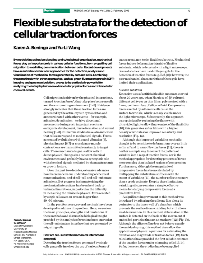
resolution (Fig. 1a).
Although this improved wrinkling method is
thought to be sensitive to deformations over as little
as 1 m m2 and to nano-Newton forces [11], there is
neither a simple way to convert the pattern of
wrinkles into a map of traction forces, nor is the
method appropriate for detecting patterns of forces
more complex than isolated regions of compression.
Furthermore, although the magnitude of
compressive forces has been calculated by
multiplying the substratum stiffness with the
extent of wrinkling [11], the number reflects no more
than a crude estimate. Despite these limitations,
wrinkling silicone remains a simple, effective
means for studying compressive forces at a
qualitative level.
A significant improvement to this method was
introduced by adhering the silicone film along its
perimeter to the inner wall of a chamber, which
prevents the surface from wrinkling but still allows
local deformation. In this method, deformation of the
surface is detected on the basis of the movement of
embedded particles that act as markers ([12]; Fig. 1b).
Although the silicone film does not behave exactly
like an ideal spring, this method does allow the
application of physical equations for estimating the
direction and magnitude of traction forces [12]. Such
calculations have provided the first reliable estimate
of the traction forces under migrating cells [13,14].
So far, however, the studies have been applied
0962-8924/02/$ – see front matter © 2002 Elsevier Science Ltd. All rights reserved. PII: S0962-8924(01)02205-X
80
Fig. 1. Various flexible
substrata used to detect
traction forces. (a) Motile
fish keratocyte on a
wrinkling silicone
substratum. Arrow
indicates direction of
migration. Image kindly
provided by K. Burton.
(b) Motile fish keratocyte
on a non-wrinkling
silicone substratum. Black
tracings indicate the
trajectories of embedded
microbeads; bar, 10 m m.
Reproduced, with
permission, from Ref. [14].
(c) Stationary rat cardiac
fibroblast causing
distortions on a
micropatterned silicone
substratum with regularly
spaced dots. Arrowheads
and magenta dots
underline the pinching
action of the contraction
on the elastomer; bar,
6 m m. Reproduced, with
permission, from Ref.
[15]. (d) Tail region of a
chick embryonic
fibroblast moving across
a detection pad of a
cantilever substratum.
Reproduced, with
permission, from Ref. [20].
(e) Motile NIH 3T3 cell
on a polacrylamide
substratum; bar, 10 m m.
Red arrows indicate local
displacements of beads.
Review
(a)
(b)
(c)
(d)
(e)
http://tcb.trends.com
TRENDS in Cell Biology Vol.12 No.2 February 2002
primarily to fish keratocytes, on a non-physiological
silicone surface tagged with a limited density of
marker particles. Although it should be possible to
optimize the method, the complexity of the
preparation procedure has limited its development
and applications.
A recent development in silicone substrata
involves the preparation of sheets of solid elastomers
using a curing agent ([15]; Fig. 1c). This generates
non-wrinkling substrata with improved mechanical
characteristics. In addition, deformation of the
surface is determined on the basis of micropatterns of
dots or lines, generated by lithography on silicon (Si)
or gallium arsenic (GaAs) molds and imprinted onto
the surface of the substratum. The regular
micropattern has a density of up to 1 dot per 4 m m2,
and allows the direct visualization of strains. But this
approach is currently limited by the availability of
micropatterned molds. Moreover, the micropattern
creates a physically or chemically textured surface,
which might affect cell adhesion and migration
through the contact guidance mechanism [16]. Like
the other types of silicone substrata, a method has yet
to be developed for coating the surface with
extracellular matrix (ECM) proteins to create a more
physiological environment.
Polyacrylamide substrata
As an alternative to silicone, the flexible substratum
can be made from polyacrylamide sheets, which are
easy to prepare and have superior mechanical and
optical properties [17]. The flexibility of the material
is easily controlled by the concentration of acrylamide
and/or bis-acrylamide. Furthermore, the porous
nature of the material provides a more physiological
environment than do solid substrata. Because most
cells show no detectable affinity for polyacrylamide,
several chemical approaches have been developed to
coat the surface with ECM proteins [18], and one can
assume that mechanical interactions with such
substrata are mediated by the coated ECM or
associated proteins.
Deformation is detected by using embedded
fluorescent microbeads as markers [18] (Fig. 1e).
Because the beads are randomly distributed
throughout the substratum and their movements are
dependent on the depth from the surface, the image
must be carefully focused near the surface of the
substratum. In addition, although bead
displacements can be observed directly as the cell
migrates, for stationary or slow-migrating cells the
full extent of deformation must be determined by
comparing images of the stressed substratum with a
null-force image, which must be recorded after
removing the cell by physical or chemical means.
The problem with focusing can be alleviated by the
recently developed technique of stacking a thin layer
of polyacrylamide containing beads on top of a
bead-free substratum; this then confines the beads
to the top surface of the substratum [19].
Review
TRENDS in Cell Biology Vol.12 No.2 February 2002
Micromachined cantilevers
Instead of using uniformly flexible substrata, an
innovative approach has been to use micromachined
cantilevers as force transducers on silicon wafers [20]
(Fig. 1d). Cells adhere and exert forces on
micrometer-sized pads at one end of the flexible
cantilever, causing displacements that are detected
with high precision on a light microscope.
Unlike flexible sheets in which strain propagates
across the surface and requires sophisticated
computational analysis for the calculation of traction
forces (see below), strains are confined to individual
cantilevers and forces can be easily calculated by
multiplying the spring constant of the cantilever with
the distance of movement. Furthermore, this method
can be applied to cells distributed at a high density,
whereas uniformly flexible substrata must be used
with isolated cells. However, the device is difficult to
construct and the surface topology can exert some
effects on cell migration, such as those discussed
above. Moreover, the spatial resolution is limited by
the density of cantilevers and the detection of forces is
limited to one dimension – perpendicular to the axis of
the cantilever.
How are magnitude and direction of traction forces
calculated?
With isolated one-dimensional springs, forces are
easily calculated by the product of displacement and
the spring constant. This simple approach is
applicable to the cantilever method but not to
uniformly flexible substrata, where strains propagate
across the substratum and fall off as a function of
distance from the source of stress. The distribution of
deformation must be mathematically deconvolved –
in a process similar to the deconvolution of optical
images – to obtain the distribution of forces.
Generally, the analysis involves two steps: the
determination of substrate deformation, and the
computation of traction forces.
Originally, substrate deformation was determined
by visually identifying corresponding markers in the
images with and without mechanical stress [14,21].
The coordinates of these markers were then used to
construct a vectorial map. This painstaking process
has since been replaced with automatic computer
programs based on various forms of the optical flow
algorithm [15,22], which searches for the best
regional matches between a pair of images and
generates vectors at a specified density. Under a
correlation-based algorithm, normalized
cross-correlation coefficients are used for identifying
the most likely fit between image subregions [23].
Using interpolation algorithms, deformation at a
given location can then be determined with a
precision of 10–100 nm [23].
Two different approaches have been used to
convert displacement maps acquired from flexible
substrata into maps of traction forces or traction
stress (force per unit area). Both approaches are
http://tcb.trends.com
81
based on the elasticity theory for the semi-infinite
space and can be applied to either micropatterned
or bead-labeled substrata comprising different
elastic materials.
The method developed by Dembo and colleagues
[24,25] makes no a priori assumption of the
distribution of forces other than that forces must be
confined within the boundary of the cell and that net
forces and torques equal zero (given the small mass
and acceleration, the net forces and torques involved
in cell migration are negligible). Because the number
of deformation vectors, superimposed with noise, is
generally insufficient to provide an unambiguous
answer, a probability-based algorithm that favors
minimal complexity (i.e. smooth transitions in forces)
is used to generate a ‘most likely’ map of traction
stress. This approach, although used widely
in signal deconvolution, can limit the precision in
regions where sharp transitions do exist.
Mathematical simulations, using pre-assigned
patterns of point forces [26], have placed the current
resolution at roughly 2 m m (W.A. Marganski and
M. Dembo, unpublished; [27]).
By contrast, the method described by Balaban
et al. [15] for the conversion of marker displacement
to force requires the assumption that forces are
exerted only at focal adhesions, which significantly
reduces the number of possible answers and possibly
allows a more definitive determination of forces at
these sites. It is unclear, however, whether forces are
indeed exerted only at focal adhesions because many
adherent cells show no detectable focal adhesions
[26]. Furthermore, the detection of focal adhesions
requires additional steps of immunofluorescence or
imaging with green-fluorescent protein (GFP) and is
subject to uncertainties – particularly for small focal
adhesions, which might exert stronger forces than
those of large focal adhesions (see below). Such
uncertainties might lead to systematic errors in the
calculated traction forces.
Calculated force or stress distribution can be
visualized as a map of vectorial arrows (Fig. 2a) or
rendered as color images after converting the stress
magnitude into different colors ([27]; Fig. 2b).
In essence, the latter approach functions as a new
form of microscopy and has been referred to as
‘traction force microscopy’. It can be used to generate
a series of force images during cell migration that can
be played back as motion pictures depicting the
dynamics of cell–substrate interactions.
What has been learned about cellular mechanical
interactions?
The above methods have been applied to study forces
exerted during processes such as cell migration
[9,11,13,14,20,27,28], growth cone extension [19] and
cytokinesis [10]. For migrating fibroblasts, strong
traction forces pointing towards the center of the cell
have been localized at the anterior and posterior
regions [2,24]. Such compressive action is consistent
82
Review
(a)
TRENDS in Cell Biology Vol.12 No.2 February 2002
New protrusion
(b)
Integrin
engagement
Nascent focal adhesions
Generation of transient
propelling forces
Mature focal adhesions
Anchorage and
stabilization of protrusion
Migration of
cell body
TRENDS in Cell Biology
Fig. 2. (a) Vector plot of traction stress generated by a fish fin fibroblast
on a polyacrylamide substratum. Arrowheads indicate direction of
forces. (b) Color rendering of the magnitude of traction forces, ‘hot’
colors highlight areas of strongest force and ‘cool’ colors indicate
regions of weaker force.
with the original observations using wrinkling
substrata [9]. Recent studies with myosin inhibitors
and regional detachment of cells with integrinbinding RGD peptides have indicated further that
forces at the front can be generated by active
actin–myosin contractions [21,28], whereas
those in the rear serve as passive anchors [28].
The distribution of forces supports a frontal towing
model of cell migration, in which the frontal regions
serve as the ‘engine’ that tows an adhesive cargo
consisting of the cell body and the tail [22].
Although early studies suggested that frontal
traction forces are generally localized near focal
adhesions [9,21], subsequent scrutiny indicates that
not all adhesions produce detectable traction forces.
Recent systematic analyses using a combination of
traction force microscopy and GFP imaging has led to
the surprising finding that, in the frontal region of
migrating fibroblasts, it is the small, nascent focal
adhesions (sometimes referred to as focal complexes)
that generate the strongest traction stress [27].
The magnitude of forces decreases as focal adhesions
mature and grow in size. Once the focal adhesions
mature, they seem to maintain a constant stress that
is independent of their size [15]. These observations
corroborate the heterogeneity of size, morphology,
protein composition and tyrosine phosphorylation of
focal adhesions [29]. In addition, the transient
propulsion at nascent focal adhesions provides an
elegant, responsive strategy for the cell to coordinate
contractility with migration and adhesion. Together,
these results suggest that different focal contacts
have different mechanical functions depending on
their age and the state of cellular motility (Fig. 3).
Studies with fish keratocytes, which have a
distinct half-moon morphology in which the long axis
lies perpendicular to the direction of cell migration,
have produced results with both similarities and
http://tcb.trends.com
Fig. 3. Relationship between focal adhesions and mechanical forces
during fibroblast migration. The formation of focal adhesions in the
lamellipodium, accompanied by the generation of a pulse of propulsive
forces, drives the forward movement. Cell migration is sustained by
repeated formation of nascent focal adhesions, and thus repeated
pulses of propulsive forces. Mature focal adhesions, such as those
located in the tail, play only a passive role in anchoring cells to the
substrate. Adapted, with permission, from Ref. [27].
differences compared with those obtained from
fibroblasts. Similar to fibroblasts, strong traction
forces lie along the long axis of the cell [30], in a region
where new adhesion sites are forming [31].
In keratocytes, however, this region is located near
the lateral extremities of the cell, and forces are
directed primarily perpendicular to the direction of
cell migration. Although a small propulsive
component has been identified in a recent analysis
[32,33], the results suggest that traction forces are
involved not only in cell migration but possibly in
other functions such as cell–cell communication
and/or mechanosensing (see below).
Flexible substrates have also been used as a means
to apply mechanical stimulations to adherent cells, to
test the ability of cells to sense mechanical changes in
the environment [6,7]. Mechanical forces are exerted
by pushing or pulling on the substrate near the cell
using a blunt microneedle [34,35]. Cells have been
found to reorient towards pulling forces [34],
accompanied by an increase in the number and/or size
of focal adhesions [35,36]. Conversely, pushing
forces cause approaching cells to turn around and
move away.
Cells have been also challenged with substrata
containing a gradient of stiffness and have been found
to move preferentially towards the rigid side [34].
These observations show that cells not only respond
to forces exerted through their adhesion sites,
but also actively probe mechanical properties
of the substratum – a phenomenon termed
‘mechanosensing’.
What is likely to be learned from future investigations?
Clearly, cell–cell and cell–substrate adhesions
represent both a mechanism for passive anchorage
and a mechanism for active physical communications
with the environment. These interactions are likely to
involve transient, localized activities of the
Review
Acknowledgements
We thank Micah Dembo
for helpful discussion.
This study was supported
by NIH NRSA grant
GM-20578 to K.A.B., and
NIH grant GM-32476 and
NASA grant NAG2-1197
to Y-L.W.
TRENDS in Cell Biology Vol.12 No.2 February 2002
actin–myosin cytoskeleton and signal-transduction
enzymes and cannot be investigated without
subcellular characterization of the traction forces,
protein interactions and structural organization.
Traction force microscopy represents a powerful
tool that can be easily combined with other
light-microscopy techniques, such as GFP imaging,
ion imaging, photobleaching, photoactivation, local
drug delivery and micromanipulation, to allow
experimentation at a high spatial and temporal
resolution. The approach has already been combined
with genetic engineering to address the functions
of specific proteins [36]. The simplicity of recently
developed substrata makes qualitative studies of
traction forces feasible for most laboratories.
Although quantitative analyses were initially
performed with supercomputers, a combination
of hardware/software improvements and
availability has enabled personal computers to
handle the task.
Many important issues need to be resolved. For
example, given the differences between nascent focal
complexes and focal adhesions in mechanical output,
it is important to identify the mechanisms that
regulate the production and transduction of
contractile forces during the maturation of focal
adhesions. The process is likely to involve
profound changes in protein–protein interactions.
An intriguing observation is that stationary
fibroblasts appear to maintain an overall magnitude
of traction force similar to that of migrating cells [15],
even though they presumably contain only mature
focal adhesions. During the transition from migrating
to stationary state, therefore, a separate process
might cause the traction forces to stay on focal
adhesions or to transfer the mechanical load from
nascent focal contacts to existing focal adhesions.
References
1 Lauffenburger, D.A. and Horwitz, A.F. (1996)
Cell migration: a physically integrated molecular
process. Cell 84, 359–369
2 Sheetz, M.P. et al. (1998) Cell migration:
regulation of force on extracellularmatrix–integrin complexes. Trends Cell Biol.
8, 51–54
3 Elson, E.L. et al. (1999) Forces in cell locomotion.
Biochem. Soc. Symp. 65, 299–314
4 Davies, P.F. et al. (1997) Spatial relationships
in early signaling events of flow-mediated
endothelial mechanotransduction. Annu. Rev.
Physiol. 59, 527–549
5 Gillespie, P.G. and Walker, R.G. (2001) Molecular
basis of mechanosensory transduction. Nature
413, 194–202
6 Liu, N. et al. (1999) Mechanical force-induced
signal transduction in lung cells. Am. J. Physiol.
277, L667–L683
7 Brown, T.D. (2000) Techniques for mechanical
stimulation of cells in vitro: a review.
J. Biomechanics 33, 3–14
8 Roy, P. et al. (1997) An in vitro force measurement
assay to study the early mechanical interactions
of fibroblasts and collagen matrix. Exp. Cell Res.
232, 106–117
http://tcb.trends.com
83
Equally important is the mechanism of
mechanosensing. Although focal adhesion kinase,
microtubules and myosins have been implicated
in this process, an integrated mechanism remains
to be constructed that probably involves intricate
cross-talk between Ca2+, GTP, proteolysis and
phosphorylation.
Besides focal adhesions, attention has to be
diverted to mechanical interactions at other
structures, including close contacts and cell–cell
junctions. Although the current methods are designed
for analyzing traction forces from isolated single cells,
with some modifications they should be able to deal
with forces generated by a small colony of cells.
An interesting challenge would be to extend the
current studies to a three-dimensional setting.
In a multicellular organism, mechanical interactions
for most cells occur around the whole surface.
The current two-dimensional system for studying
traction forces creates a marked asymmetry between
the dorsal and ventral surfaces, and might yield
results that deviate substantially from those in a true
physiological setting.
Finally, there is strong evidence that mechanical
interactions play an important role in a wide
spectrum of specific processes, including embryonic
morphogenesis [37], neuronal guidance [19],
osteoblast maturation [38] and phagocytosis [39].
Although they probably share some common aspects,
the specific functions of mechanical forces in these
processes are just beginning to be unraveled.
Understanding the interplay between extracellular
physical interactions and intracellular chemical
events is likely to exert a strong impact on many
practical applications, including tissue engineering,
stem cell differentiation and treatments of
autoimmune diseases and cancer.
9 Harris, A.K. et al. (1980) Silicone rubber
substrata: a new wrinkle in the study of cell
locomotion. Science 208, 177–179
10 Burton, K. and Taylor, D.L. (1997) Traction forces
of cytokinesis measured with optically modified
substrata. Nature 385, 450–454
11 Burton, K. et al. (1999) Keratocytes generate
traction forces in two phases. Mol. Biol. Cell 10,
3745–3769
12 Oliver, T. et al. (1998) Design and use of substrata
to measure traction forces exerted by cultured
cells. Methods Enzymol. 298, 497–521
13 Lee, J. et al. (1994) Traction forces generated by
locomoting keratocytes. J. Cell Biol. 127,
1957–1964
14 Oliver, T. et al. (1995) Traction forces in locomoting
cells. Cell Motil. Cytoskeleton 331, 225–240
15 Balaban, N.Q. et al. (2001) Force and focal
adhesion assembly: a close relationship studied
using elastic micropatterned substrates. Nat. Cell
Biol. 3, 466–472
16 Weiss, P. (1958) Cell contact. Int. Rev. Cytol. 7,
391–423
17 Pelham, R.J. and Wang, Y-L. (1997) Cell
locomotion and focal adhesions are regulated by
substrate flexibility. Proc. Natl. Acad. Sci. U. S. A.
94, 13661–13665
18 Beningo, K.A. and Wang, Y-L. Flexible
polyacrylamide substrates for the analysis of
mechanical interactions at cell-substrate
adhesions. Methods Cell Biol. (in press)
19 Bridgman, P.C. et al. (2001) Myosin IIB is required
for growth cone motility. J. Neurosci. 21, 6159–6169
20 Galbraith, C.B. and Sheetz, M.P. (1997) A
micromachined device provides a new bend on
fibroblast traction forces. Proc. Natl. Acad. Sci.
U. S. A. 94, 9114–9118
21 Pelham, R.J. and Wang, Y-L. (1999) High
resolution detection of mechanical forces exerted
by locomoting fibroblasts on the substrate. Mol.
Biol. Cell 10, 935–945
22 Munevar, S. et al. (2001) Traction force microscopy
of migrating normal and H-ras transformed 3T3
fibroblasts. Biophys. J. 80, 1744–1757
23 Marganski, W.A. et al. Measurement of
cell–substrate deformations on flexible substrata
using correlation based optical flow. Methods
Enzymol. (in press)
24 Dembo, M. and Wang, Y-L. (1999) Stresses at the
cell-to-substrate interface during locomotion of
fibroblasts. Biophys. J. 76, 2307–2316
25 Wang, N. et al. (2001) Mechanical behavior in
living cells consistent with the tensegrity model.
Proc. Natl. Acad. Sci. U. S. A. 98, 7765–7770
84
Review
TRENDS in Cell Biology Vol.12 No.2 February 2002
26 Bray, D. (2001) Cell Movement: From Molecule to
Motility, Garland Publishing
27 Beningo, K.A. et al. (2001) Nascent focal
adhesions are responsible for the generation of
strong propulsive forces in migrating fibroblasts.
J. Cell Biol. 153, 881–887
28 Munevar, S. et al. (2001) Distinct roles of frontal
and rear cell-substrate adhesions in fibroblast
migration. Mol. Biol. Cell 12, 3947–3954
29 Zamir, E. et al. (1999) Molecular diversity of
cell–matrix adhesions. J. Cell Sci. 112, 1655–1669
30 Dembo, M. et al. (1996) Imaging the traction
stresses exerted by locomoting cells with the
elastic substratum method. Biophys. J. 70,
2008–2022
31 Small, J.V. et al. (1998) Assembling an actin
cytoskeleton for attachment and movement.
Biochim. Biophys. Acta 1440, 271–281
32 Oliver, T. et al. (1999) Separation of propulsive
and adhesive traction stresses in locomoting
keratocytes. J. Cell Biol. 145, 589–604
33 Galbraith, C. and Sheetz, M.P. (1999) Keratocytes
pull with similar forces on their dorsal and
ventral surfaces. J. Cell Biol. 147, 1313–1323
34 Lo, C-M. et al. (2000) Cell movement is guided by
the rigidity of the substrate. Biophys. J. 79,
144–152
35 Riveline, D. et al. (2001) Focal contacts as
mechanosensors: externally applied local
mechanical force induces growth of focal contacts
36
37
38
39
by an mDia1-dependent and ROCK-independent
mechanism. J. Cell Biol. 153, 1175–1186
Wang, H.B. et al. (2001) Focal adhesion kinase is
involved in mechanosensing during fibroblast
migration. Proc. Natl Acad. Sci. U. S. A. 98,
11295–11300
Oster, G.F. et al. (1983) Mechanical aspects of
mesenchymal morphogenesis. J. Embryol. Exp.
Morphol. 78, 83–125
Burger, E.H. and Klein-Nulen, J. (1999)
Responses of bone cells to biomechanical forces
in vitro. Adv. Dent. Res. 13, 93–98
Beningo, K.A. and Wang, Y-L. Fc-receptor
mediated phagocytosis is regulated by mechanical
properties of the target. J. Cell Sci. (in press)
Apoptotic DNA fragmentation and
tissue homeostasis
Jianhua Zhang and Ming Xu
DNA fragmentation is a hallmark of apoptosis. The tightly controlled
activation of the apoptosis-specific endonucleases provides an effective
means to ensure the removal of unwanted DNA and the timely completion of
apoptosis. Over the past several years, crucial progress has been made in
identifying the long-awaited apoptotic endonucleases, and their importance
in tissue homeostasis is beginning to unfold. Here, we focus on the most
recent discoveries about the functions and mechanisms of these
endonucleases in the context of apoptosis. We also discuss consequences
that defective DNA fragmentation might have for tissue homeostasis and
disease development.
Jianhua Zhang
Ming Xu*
Dept of Cell Biology,
Neurobiology and
Anatomy, University of
Cincinnati College of
Medicine, Cincinnati,
OH 45267-0521, USA.
*e-mail: ming.xu@uc.edu
Multicellular organisms have developed an intricate
control system to balance cell proliferation and cell
death to ensure proper development and tissue
homeostasis. Any abnormalities in the cell death
process can potentially lead to severe human
diseases, including neural degeneration,
autoimmunity and cancer [1–4]. Therefore,
identifying the molecules involved in cell death and
understanding the regulation of the death process are
crucial for prevention and management of these
human diseases.
In normal development and tissue homeostasis,
most of the cells die through physiological or
programmed cell death to remove excessive or
damaged cells [4]. The term ‘apoptosis’ was first used
to describe such cell death with preprogrammed
morphological changes including cell and nuclear
shrinkage, chromatin condensation and apoptotic
body formation in vertebrates [1]. Similar
morphological changes were also observed in
programmed cell death in invertebrates [4].
The apoptotic morphological changes exhibited by
the dying cells are followed by phagocytosis by
http://tcb.trends.com
scavenger cells. A biochemical hallmark of apoptosis
is the cleavage of chromosomal DNA into
oligonucleosome-sized fragments, a process called
DNA fragmentation [2]. Apoptosis eliminates
excessive, mutated, infected and damaged cells and is
actively and inherently controlled [1–4].
Because of the fundamental role of apoptosis in
development and tissue homeostasis, a cell-suicide
program utilizing evolutionarily conserved molecules
is dedicated to the process [5,6]. Elegant genetic and
biochemical work has identified several families of
proteins, such as the Bcl-2 family, that regulate
apoptosis, and caspases that mediate apoptosis by
cleaving downstream molecules. The importance of
these regulators and executors of apoptosis is
underscored by the various developmental
deficiencies and tumorigenic phenotypes of mice that
either are deficient in or overexpress the genes
encoding these molecules [7].
Although most research efforts have focused on the
more upstream death program molecules such as the
Bcl-2 family proteins and caspases, the key
molecules involved in DNA fragmentation and the
role of cleavage of chromosomal DNA in apoptosis
and tissue homeostasis remained elusive. Over
the past few years, biochemical and genetic
work has identified several endonucleases that
play crucial roles in apoptosis. This article focuses on
the latest developments in the functions and
mechanisms of these endonucleases in the context
of apoptosis and discusses the consequences that
compromised functions of the endonucleases
might have for tissue homeostasis and disease
development.
0962-8924/02/$ – see front matter © 2002 Elsevier Science Ltd. All rights reserved. PII: S0962-8924(01)02206-1


