History of Infections Associated With Combat
advertisement
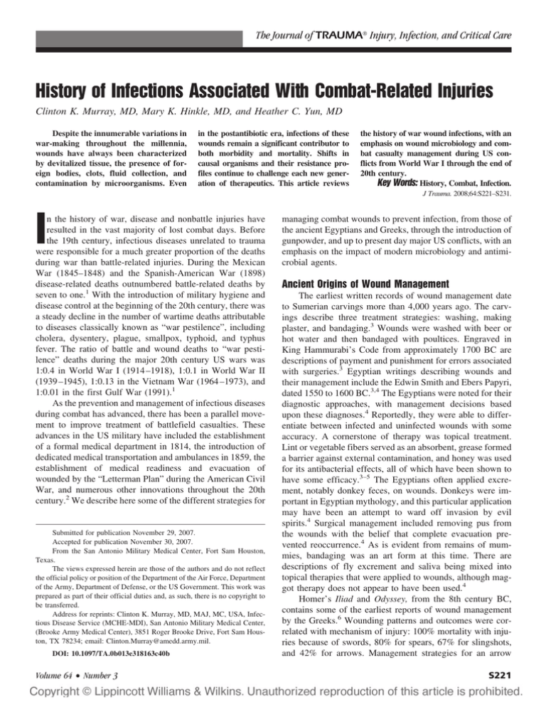
The Journal of TRAUMA威 Injury, Infection, and Critical Care History of Infections Associated With Combat-Related Injuries Clinton K. Murray, MD, Mary K. Hinkle, MD, and Heather C. Yun, MD Despite the innumerable variations in war-making throughout the millennia, wounds have always been characterized by devitalized tissue, the presence of foreign bodies, clots, fluid collection, and contamination by microorganisms. Even in the postantibiotic era, infections of these wounds remain a significant contributor to both morbidity and mortality. Shifts in causal organisms and their resistance profiles continue to challenge each new generation of therapeutics. This article reviews the history of war wound infections, with an emphasis on wound microbiology and combat casualty management during US conflicts from World War I through the end of 20th century. Key Words: History, Combat, Infection. J Trauma. 2008;64:S221–S231. I n the history of war, disease and nonbattle injuries have resulted in the vast majority of lost combat days. Before the 19th century, infectious diseases unrelated to trauma were responsible for a much greater proportion of the deaths during war than battle-related injuries. During the Mexican War (1845–1848) and the Spanish-American War (1898) disease-related deaths outnumbered battle-related deaths by seven to one.1 With the introduction of military hygiene and disease control at the beginning of the 20th century, there was a steady decline in the number of wartime deaths attributable to diseases classically known as “war pestilence”, including cholera, dysentery, plague, smallpox, typhoid, and typhus fever. The ratio of battle and wound deaths to “war pestilence” deaths during the major 20th century US wars was 1:0.4 in World War I (1914 –1918), 1:0.1 in World War II (1939 –1945), 1:0.13 in the Vietnam War (1964 –1973), and 1:0.01 in the first Gulf War (1991).1 As the prevention and management of infectious diseases during combat has advanced, there has been a parallel movement to improve treatment of battlefield casualties. These advances in the US military have included the establishment of a formal medical department in 1814, the introduction of dedicated medical transportation and ambulances in 1859, the establishment of medical readiness and evacuation of wounded by the “Letterman Plan” during the American Civil War, and numerous other innovations throughout the 20th century.2 We describe here some of the different strategies for Submitted for publication November 29, 2007. Accepted for publication November 30, 2007. From the San Antonio Military Medical Center, Fort Sam Houston, Texas. The views expressed herein are those of the authors and do not reflect the official policy or position of the Department of the Air Force, Department of the Army, Department of Defense, or the US Government. This work was prepared as part of their official duties and, as such, there is no copyright to be transferred. Address for reprints: Clinton K. Murray, MD, MAJ, MC, USA, Infectious Disease Service (MCHE-MDI), San Antonio Military Medical Center, (Brooke Army Medical Center), 3851 Roger Brooke Drive, Fort Sam Houston, TX 78234; email: Clinton.Murray@amedd.army.mil. DOI: 10.1097/TA.0b013e318163c40b Volume 64 • Number 3 managing combat wounds to prevent infection, from those of the ancient Egyptians and Greeks, through the introduction of gunpowder, and up to present day major US conflicts, with an emphasis on the impact of modern microbiology and antimicrobial agents. Ancient Origins of Wound Management The earliest written records of wound management date to Sumerian carvings more than 4,000 years ago. The carvings describe three treatment strategies: washing, making plaster, and bandaging.3 Wounds were washed with beer or hot water and then bandaged with poultices. Engraved in King Hammurabi’s Code from approximately 1700 BC are descriptions of payment and punishment for errors associated with surgeries.3 Egyptian writings describing wounds and their management include the Edwin Smith and Ebers Papyri, dated 1550 to 1600 BC.3,4 The Egyptians were noted for their diagnostic approaches, with management decisions based upon these diagnoses.4 Reportedly, they were able to differentiate between infected and uninfected wounds with some accuracy. A cornerstone of therapy was topical treatment. Lint or vegetable fibers served as an absorbent, grease formed a barrier against external contamination, and honey was used for its antibacterial effects, all of which have been shown to have some efficacy.3–5 The Egyptians often applied excrement, notably donkey feces, on wounds. Donkeys were important in Egyptian mythology, and this particular application may have been an attempt to ward off invasion by evil spirits.4 Surgical management included removing pus from the wounds with the belief that complete evacuation prevented reoccurrence.4 As is evident from remains of mummies, bandaging was an art form at this time. There are descriptions of fly excrement and saliva being mixed into topical therapies that were applied to wounds, although maggot therapy does not appear to have been used.4 Homer’s Iliad and Odyssey, from the 8th century BC, contains some of the earliest reports of wound management by the Greeks.6 Wounding patterns and outcomes were correlated with mechanism of injury: 100% mortality with injuries because of swords, 80% for spears, 67% for slingshots, and 42% for arrows. Management strategies for an arrow S221 The Journal of TRAUMA威 Injury, Infection, and Critical Care injury included removal of the arrow, rinsing the wound with warm water, and applying analgesic and styptic herbal medicines to the wound.6 The most well-known figure in Greek medicine was Hippocrates, born around 460 BC.3,6 He produced a vast number of medical texts, whose compilation is referred to as Corpus Hippocraticum.7 He described setting fractures, debridement, bandaging, traction, correction of dislocations, and the use of casts and splints. He recognized the association between fingernail length and disease transmission, which is considered relevant to infection control today. In Hippocrates’ book, On Wounds, he stated that the keys to proper healing include washing the wound in clean water or wine (noted today for antibacterial activity), not allowing the wound to remain moist, and rest and immobility for the afflicted.3,6,8 He also indicated that sutures should be soaked in hot olive oil before use. For compound fractures, he recommended tightly bound bandages to achieve necrosis and autoamputation with subsequent placement of a prosthesis. There are also descriptions of surgical drainage of pus, with a tin pipe known as a “pus-puller” placed into the abscess cavity.3 Fundamental to his teaching was the reduction in inflammation of the wound, although he encouraged the development of pus to meet this goal.7 One of the dominant figures in Roman medicine was Galen of Pergamum (120 –201 AD), a surgeon of Roman gladiators. He was the author of more than 400 works pertaining to medicine. But his overall impact on surgical progress may have been negative as he thought the presence of pus, referred to as “laudable pus”, was beneficial to wounds.7 This premise would persist for centuries. Surgical progress in the Middle Ages was modest for a number of reasons, one of which was the widespread acceptance of Galen’s teaching of the benefit of pus. Equally contributory was the divestment of religious involvement from medical practice, at a time when the majority of physicians were clergymen. It was not until 1267 that the presence of pus was judged to be unnecessary for wound healing, although this idea would not gain mainstream acceptance for almost six more centuries.7 1300 to 1800s The introduction of gunpowder to Europe in the 14th and 15th centuries marked a new era of wound patterns and thereby a shift in wound management. Injuries became more complex, with a rising prevalence of shattered extremities, as well as associated burn injuries further complicating wound management, and ushering in the era of amputations. Surgical techniques of the 14th century focused on the removal of foreign objects, rejoining severed tissue, maintaining tissue continuity, preserving organ substance, and preventing complications.7 Fundamental to the management of injuries at this time was the belief that gunpowder was poisonous and that bullets were contaminated before firing. This prompted therapy to S222 revolve around cautery of the wound with a red hot iron or hot oil.7,9 It was not until the 1500s that this belief would dissipate.7,9 The poisonous nature of gunshot was questioned after Ambrose Paré, a battlefield surgeon, ran out of hot oil and substituted egg yolk, turpentine, and rose oil, and bandaged the wounds.10 He found that wound healing with this method was superior to that obtained with the cautery approach.7,9,10 He also introduced debridement, adopted special tools and new techniques in fracture reduction, and developed a simple tourniquet with vessel ligation.11 Paré also noted the utility of maggot therapy in 1557, which was later supported by Baron D.J. Larrey, Napoleon’s military surgeon in 1829; this therapy is still used today.9,12 Unfortunately, however, Paré still believed in the importance of pus to wound healing. During the Revolutionary War (1775–1781), trauma surgery was greatly influenced by John Hunter, the Surgeon General of the English army. One of his early proposals was that not all wounds need aggressive debridement. Although this application was often used at the time for large wounds, which allowed progression of infection, it is an idea that is supported today for small fragmentation injuries.7,13 John Jones, one of the founders of King’s College Medical School, the precursor of the Columbia University College of Physicians and Surgeons, published a textbook on the management of wounds and injuries for young military surgeons based upon his volunteer surgical experience during the French and Indian Wars (1754 –1763).7,9 The primary emphasis was on removal of bullets within easy reach and avoidance of primary wound closure. If a wound was to be closed, an onion was placed in the wound before closure, and the wound reopened at 24 to 48 hours.7 The wound was expected to develop swelling and pus by the fourth day postinjury, which were thought to be signs of proper wound “digestion” necessary for healing. Amputation continued to be the therapy for compound fractures. Superficial burns were treated with wine and deep full-thickness burns with hog’s lard. Wound care during the War of 1812 (1812–1815) continued to emphasize early amputations to shorten hospital stays, reduce the risk of infection, and to reduce the trauma caused by transportation on horse-drawn vehicles. Management continued to rely upon incision and removal of foreign bodies, with fasciotomy to prevent further tissue damage. During the Napoleonic Wars (1803–1815), amputations were also the standard of care. It was reported that Napoleon’s surgeon, Dominique-Jean Larrey, could perform 200 surgeries a day, or one every 7.2 minutes. Hip and shoulder joint amputations apparently took 15 seconds and 11 seconds, respectively.7 Larrey thought early amputation created a clean viable wound, and was reported to have had a 75% success rate in preventing infection. During the American Civil War (1861–1865) some of the key components of wound care included general anesthesia, delay of primary amputation to reduce the effect of wound shock, bromine to prevent hospital gangrene, use of March Supplement 2008 Infections of Combat Casualties—History well-trained physicians, and the development of paviliontype hospitals.7 More than 50,000 amputations were performed during this conflict.11 In one report Confederate Army troops undergoing primary amputation had a 38% mortality rate (among 1,142 patients) in contrast to secondary or delayed amputation, which had 53% mortality (among 546 patients).7 Although carbolic acid and sodium hypochlorite were available, they were used for treatment, and not prevention, of gangrene. Infections included erysipelas, with a mortality rate of 8%, and hospital gangrene, with a mortality rate of 38% to 62% if untreated, but which fell to 2.6% with the use of topical bromine.14 Patients with these types of infections were housed together to prevent disease transmission.15 Tetanus was rare, but had a mortality rate of 89%. Mortality for pyemia was even higher at 97%. Although pyemia, or sepsis, resulted from only 1% of wounds, it caused 6% of deaths. Typically, penetrating abdominal injuries were not operated on because of an 87% overall mortality, ranging from 59% for colon injuries to 100% for small bowel involvement. Maggot therapy was also used during the American Civil War.9,12 During the Russo-Turkish War (1877–1878), Russian military surgeon Carl Reyher emphasized immediate wound debridement. This was supported by Paul Leopold Friedrich, who recognized that surgery within 4 to 6 hours after wounding usually prevented the development of wound infection.14 Data by Reyher revealed immediate wound excision and antiseptic treatment performed on the battlefield had a mortality of 24% (13 deaths in 55 patients), whereas the same techniques applied after delayed evacuation resulted in 54% mortality (42 deaths in 92 patients). If the delay was by days, the mortality was 55% (11 deaths in 20 patients).14 Around the time of the Spanish-American War (1898) there was acceptance of the germ theory, antiseptic techniques, and more effective anesthesia delivery mechanisms on the battlefield. Antiseptics were carried in first aid pouches on each soldier’s cartridge belt.7 It was also noted that septic shock had a major impact on outcome. At the dawn of the modern microbiology era, it was recognized that wounds were infected with the anaerobic bacteria, Bacillus aerogenes capsulatus (other names used in the literature for the next 50 years included Bacillus welchii, Clostridium welchii, and Clostridium perfringens), resulting in gas gangrene.16 Modern Microbiology and Antiseptics Although it had been postulated that infections were associated with contamination of wounds, were contagious, and could be spread by people and instruments, before the germ theory this remained a controversial issue.14 With the introduction of modern microbiology, there was a rapid transition of the care provided to combat casualties. Louis Pasteur in 1861 identified bacteria as the cause of putrefication and toxic effects, and proposed the germ theory of disease. Joseph Lister advanced the care of combat casualties using carbolic Volume 64 • Number 3 acid spray as a method of antisepsis.17 This intervention was reported in 1867 to reduce the amputation mortality rate from 16 of 35 cases to 6 of 40 cases.7,17,18 In 1865 Lister was also disinfecting instruments with carbolic acid and using antiseptics in dressings for wounds.10,17,19 Robert Koch’s confirmation that bacteria caused disease in 1877 further advanced the field. In 1881 Koch was able to grow bacteria on solid media. The Gram’s stain was developed by Christina Gram in 1884. Although van Leeuwenhoek had been able to describe “animalcules” using the microscope in 1716, the importance of this instrument was not realized until the germ theory came into being. Gowns, masks, and gloves were introduced as a means of infection control in the 1880s.20 Florence Nightingale and her cadre of 37 nurses emphasized sanitation and hygiene in the hospital after it was noted in the Crimean War (1853–1856) that two thirds of the original 25,000 man force died of cholera, dysentery, and scurvy within a year.7 World War I (1914 –1918) Numerous advances in surgical techniques occurred during World War I. Appropriate debridement of combat wounds is credited to Belgian Army Surgeon, Antoine Depage, who recognized that wounds needed to have foreign bodies and necrotic tissues removed.9 Use of silver foil dressings for wounds was implemented, reminiscent of the Roman use of silver and nitrate metal filings in wounds.9 Blood transfusions with ABO compatibility testing became available. Laparotomies were performed in World War I for abdominal injuries, although mortality remained high (⬃50%), likely because of delayed evacuation, inadequate resuscitation, and other factors.7 Harvey Cushing was a key physician and researcher during World War I.7 He noted that an increase in trench depth between 1915 and 1917 resulted in more head injuries.7 Cushing found topical antiseptics containing dichloramine-T to be effective in preventing the development of infection.21 The invention of roentgenogram technology by Wilhelm C. Roentgen in 1895 helped in the management of trauma and localization of foreign bodies. The need for rapid surgical care of war wounds was also confirmed. Rapid evacuation was associated with improved outcome; with a mortality rate of 10% if evacuation occurred within 1 hour, and 75% if evacuation occurred after 8 hours.16 Finally, tetanus antitoxin, first described in 1890, had wide-scale distribution and application during World War I, causing tetanus rates to drop from 9 per 1,000 wounded to 1.4 per 1,000.22 Further progress in tetanus antitoxin purification brought about further reductions, with only 12 cases reported among 2,734,819 hospital admissions for wounds and injuries during World War II.23,24 The ability to describe the involved microorganisms was fundamental to developing appropriate wound management strategies. In 1915, Fleming described the bacteriologic history of war wounds (Fig. 1).25 The first phase was a watery, foul-smelling, reddish-brown discharge notable for anaerobes S223 The Journal of TRAUMA威 Injury, Infection, and Critical Care 100 Percentage of wounds with bacteria 90 80 70 Clostridium perfringens 60 Clostridium tetani 50 Streptococci 40 Coliform bacilli 30 Staphylococci 20 10 0 1-7 days (127 cases) 8-20 days (56 cases) over 20 days (27 cases) Time cultures were obtained Fig. 1. Bacteria recovered from combat-related injuries during World War I. Adapted from Lancet. 1915;2:638 – 643. but also including fecal pathogens and streptococci. After approximately 7 days, these were largely replaced by pyogenic streptococci, although anaerobes were still present.26 The wound drainage lost its bloody character and became more purulent with less foul odor during the next 2 weeks. The third phase, beginning at about 20 days, was characterized by the proliferation of pyogenic bacteria, predominantly streptococcci and staphylcococci. It was notable that about a quarter of the patients whose blood was cultured by Fleming were bacteremic. Fleming noted C. welchii (now C. perfringens) in 81% of wounds from 1 to 9 days after injury, 34% from 8 to 20 days, and 18% at 20 days or more after injury. Pettit noted that the interval from injury to surgical intervention had a substantial impact on the incidence of gas gangrene.27 The presence of gas gangrene among 137 patients was 2.9% after treatment at a casualty clearing station and evaluation at a base hospital.26 Fleming also reported on the use of topical antiseptic therapy of wounds, finding an early benefit on infection rates but a negative overall impact on healing.28 He concluded that topical therapy should not be used and that the surgeon should rely “on his skill alone”. He said, “. . . it seems a pity that the surgeon should wish to share his glory with a chemical antiseptic of more than doubtful utility”. Despite this, Dakin’s solution (hypochlorite) remained a common therapy during World War I.29 One of the primary articles detailing gunshot wound management in World War I described the outcomes of patients treated at a casualty clearing station.30 Observations were made regarding wound bacteriology in relation to primary suturing results. Cultures were obtained from 215 of the 224 wounds that were primarily sutured. Twenty-one of these cultures (9.7%) were negative, all of which healed completely S224 by primary intention. The remaining 194 (90.3%) contained anaerobes (61.3%), hemolytic streptococci (10.3%), nonhemolytic streptococci (32.4%) or “other organisms” (48.9%). Those with hemolytic streptococci typically became infected and resulted in failed primary closure (95% of 20 wounds), whereas failure was rare among wounds containing anaerobes alone. Wounds infected with hemolytic streptococci were described as “violently” suppurating, and accompanied by “severe constitutional disturbances”. In contrast, wounds with nonhemolytic streptococci were, in general, found to be less virulent with minimal complications; 71% still healed with primary closure, and those that did not heal showed milder suppuration than wounds infected with hemolytic streptococci. The average interval between wounding and intervention was 11.7 hours. This interval was shorter among the successful cases of primary closure than those failing primary closure (10.7 hours vs. 12.1 hours), though no statistical comparisons were made. In the study of 106 gunshot wounds to the head, infections were serious complications of injury and had a great impact on outcomes.31 There were 43 infected with Staphylococcus aureus, of whom nine died. Sixteen cases of streptococci recovered from wounds were reported, of whom 11 died. Overall, streptococci were associated with a higher mortality rate regardless of the severity of the injury. Introduction of Antimicrobial Therapy The first commercially available antibacterial was the sulfonamide prontosil with accounts of its efficacy published in 1935. This agent became widely used but was mass produced under uncontrolled standards. Its use resulted in numerous deaths from poisoning because of the diethylene glycol in March Supplement 2008 Infections of Combat Casualties—History the elixir. These deaths led to the enactment of the Federal Food, Drug, and Cosmetic Act. Sulfonamide was widely available during the early years of World War II and was often used as a topical wound agent. Other more active antimicrobials, notably penicillin, were developed, which quickly replaced the sulfonamides in therapy of combat injuries. The discovery of a Penicillium mold producing an antimicrobial substance is credited to Alexander Fleming after his accidental recognition of the mold inhibiting the growth of staphylococci.32 However, the use of molds to treat wounds and infections dates back to the Greeks and Romans. Throughout the late 1800s numerous scientists, including Louis Pasteur, William Roberts, John Tyndall, and others recognized that the presence of mold prevented the growth of bacteria.33 Howard Walter Florey, a chemist, was able to isolate and produce the substance in 1939, enabling it to be used in the first patient in 1941 and by the military in Africa in 1942, with remarkable success.34 –36 Because the mass production of penicillin was not available during the initial stages of the war, it was recrystallized from the urine of treated patients for the purposes of reuse.34,37 World War II (1939 –1945) Chemotherapeutics and introduction of antibiotics such as sulfanilamide and penicillin distinguished World War II from previous wars. Lessons learned during World War I were applied during World War II, including delayed primary closure, pedicle flaps, and external fixation devices.36 Although external and internal fixation devices were used, the concern for the development of infection prevented their widespread application.38 Mortality from abdominal injuries decreased from 66% in World War I to 24% in World War II, likely related to delayed primary closure and the separate exteriorization of injured large intestines.7 Chest injuries, which had 62% mortality in the Civil War and 25% in World War I, had only 10% mortality in World War II.7 On December 5, 1941, Dr. John J. Moorhead, chief surgeon for the New York subway system, presented a series of lectures on the treatment of trauma to the Honolulu Medical Society.39 As part of that lecture series, he presented techniques on debridement, delayed primary closure, and the use of sulfanilamide powder in wounds.2,39 This lecture was attended by Army and Navy medical personnel stationed in Hawaii. The Japanese attacked Pearl Harbor approximately 36 hours later. Dr. Moorhead, along with military and civilian physicians, used these techniques with remarkably good results in the aftermath of the attack. Topical sulfanilamide became part of the standard of care, and individual soldiers carried sulfanilamide with them. However, this reliance on sulfanilamide powder replaced, rather than augmented, appropriate debridement.2 In addition, under field conditions the powder was dumped, not sprinkled, into the wound as was performed in Hawaii. One of the lessons learned during World War II was the role that the environment and healthcare personnel played in Volume 64 • Number 3 hospital-associated infections and nosocomial transmission.40 Recommendations to prevent transmission in the hospitals promoted masks for patients and healthcare workers, dressings changed with clean dry hands, all material handled with sterile instruments, and thoroughly cleaning baths. Leaders were trained to enforce the rules. These techniques appeared especially effective with the most problematic pathogen, Streptococcus pyogenes.40,41 Researchers confirmed many of the findings of World War I and the role of infections during the care of casualties.42 Wounds treated within 6 hours had substantially lower infection rates than those delayed for 8 hours or more. Hospital-associated infections occurred in 86% of wounds. There was also recognition of the increasing role of staphylococci and the gram-negative bacilli, Proteus and B. pyocyaneus (now Pseudomonas aeruginosa) in wound infections.43 These pathogens appeared to be of low virulence but had significant impact on impairing wound healing. Study of head injuries revealed that Staphylococci were commonly recovered, but S. aureus was not typically present in deep wounds.44,45 As these wounds matured there was more recovery of Streptococcus pyogenes, with occasional recovery of Proteus species and P. aeruginosa. Of 700 consecutive neurosurgical cases in the Italian campaign, 28 became infected.46 The difference in infection rates in patients treated with penicillin and sulfa powder versus sulfa powder alone was remarkable (17 of 184 [9.2%] in the penicillin and sulfa powder group; 10 of 32 [31.2%] in the sulfa alone group). The infectious complications reported included abscess (11), meningitis (8), encephalitis (1), anaerobic infections (2), uncomplicated fungal infections (3), and superficial wound infections (3). The highest mortality rate was in the meningitis group (6 of 8 died), despite routine use of systemic sulfadiazine. A follow-up of 200 patients with cranial injuries in the United States revealed 47 (23%) had evidence of infection.47 Of these, 21 (44.6%) had retained bone or metal fragments in their wounds. Among the remaining 153 men who did not develop infections, 73 (47%) were shown to have retained bone or metal fragments on subsequent roentgenograms. It was thereby assumed that retained material had little effect on rates of subsequent infection in patients with head injuries. The first reported use of penicillin in World War II was in Oran, Africa, in November 1942.36 Patients arriving in Bristol, England, after transport by hospital ships from Africa, had no evidence of infection.36 Initially, the British Army used penicillin in wounds whereas the American policy was to reserve the limited supply for systemic administration.48 This resulted in Americans being more aggressive in surgical techniques and the British focusing on attempts at bacteriologic sterilization of wounds. In the Italian theater, it was shown that surgery was fundamental for wound management and that penicillin was an adjunct for the control of invasive infection. S225 The Journal of TRAUMA威 Injury, Infection, and Critical Care Korean War (1950 –1953) The Korean War introduced Mobile Army Surgical Hospitals and the rapid evacuation of casualties from the battlefield using helicopters. Forward surgical care in combination with helicopter evacuations enabled patients to arrive for surgical care between 2 and 4 hours after injury. Other notable advances were vascular repairs, lower amputation rates, and hemodialysis. Wounded mortality rates improved from 4.5% during World War II to approximately 2.5% during the Korean War.11 Surgical research teams in theater also allowed for more rigorous study of combat casualties.7 Microbiology reports from the Korean War indicate that at the time injured personnel presented to medical care their wounds were already contaminated with bacteria.49There were seasonal differences in bacteria recovered during the Korean War, with staphylococci and streptococci predominating in winter months, replaced with fecal bacteria during the summer. In addition, summer months had more Clostridium species recovered from wounds.50 The rates of clostridial infections during the Korean War continued the downward trajectory begun after World War I. Among 4,900 wounds there was a reported 0.08 incidence of gas gangrene and no gangrene-associated mortality.51 During World War I, by comparison, there had been a 5% incidence of gas gangrene, with 28% mortality; during World War II the incidence ranged between 0.3% and 1.5%, depending upon the theater, with 15% mortality.27,52–54 This decrease was largely attributable to decreasing the time from injury to definitive care. Those developing gas gangrene during World War I waited an average of 42 hours from injury to surgery, in contrast to those who never developed gangrene, who underwent surgery within 25 hours of injury. In a study assessing clostridial infection in the Korean War, the average evacuation time was 3.5 hours and all the patients who developed gas gangrene had inadequate initial debridements.51 Microbiologic evaluation of neurosurgical cases from the Tokyo Army Hospital between 1951 and 1952 revealed gram-positive bacteria, including hemolytic and nonhemolytic streptococci, and gram-negative bacteria in the wounds.55 Standard therapy in Korea before evacuation to Tokyo consisted of surgical debridement and the use of penicillin with streptomycin. Of the isolates recovered in Tokyo, resistance to penicillin was demonstrated in 48 of 58 cases, and to streptomycin in 49 of 58 cases. Seven cases were resistant to all agents tested (penicillin, streptomycin, tetracyclines, and chloramphenicol). Inadequate debridement was the most commonly cited cause of infection in 25 of 58 cases, with 16 reported cases of inadequate closure of the scalp and dura mater. The authors concluded that antibiotics were an adjunct to appropriate surgical care and that prophylactic antibiotics were associated with a high incidence of drugresistant microorganisms. An analysis of air sinus wounds associated with craniocerebral injuries revealed a high infection rate (47 of 163), especially with delayed surgery.56 S226 Prompt and radical surgical debridement of the structures along the missile tract with appropriate dural repair prevented subsequent infections. There was a 5% bacteremia rate in combat casualties (total of 170 casualties evaluated); however, the degree of shock, type of injury, the time lag to care, and previous antibiotic therapy did not appear to impact the incidence of positive blood cultures.57 Vietnam War (1959 –1975) Continued advancement occurred in the management of combat casualties in Vietnam. Routine helicopter evacuations reduced times from injury to surgical care by 1 to 2 hours. In addition, well-trained surgeons were in abundant supply, working in state-of-the-art facilities closely located to the battlefield. A research team was also in theater enabling a more complete and rapid assessment of research findings.58 Rates for wound infection were as low as 2%, although reports of these rates typically only included complications occurring in theater and not after evacuation.59 Injury patterns were consistent with previous wars, with 67% involving the extremities, 13% the thorax, 12% the abdomen, and 8% the head or neck. In 1969, an assessment was undertaken to evaluate the medical care provided in theater. This study included 19 different military hospitals in Vietnam, with a total of 132,996 admissions.60 Surgical patients comprised 46% of the admissions, but accounted for 93% of the deaths. Septic shock was the third leading cause of death (12%), after hemorrhagic shock (24%) and head injuries (43%). Another assessment of mortality among nearly 7,000 casualties between January 1966 and June 1968 revealed that among 121 deaths, sepsis (predominantly gram-negative) was the second most common cause of death after hemorrhage, and was the primary cause of death 24 hours after injury.61 Hardaway’s assessment of 17,726 American soldiers injured in Vietnam between March 1966 and July 1967 provides a comprehensive review of the injuries, practices, and complications seen during the war.62 The in-hospital mortality rate was 1.81% compared with 3.3% in World War II and 2.4% in the Korean War. The mortality from abdominal wounds improved from 21% in World War II to 12% in Korea to 4.5% in the Vietnam War. Overall, 31% of wounded patients arrived at the hospital within 1 hour of injury and 86% were admitted within 4 hours. However, the mortality increased from 10% if patients were admitted within 1 hour, to 12% at 3 hours, 33% at 4 hours, and 75% at 8 hours. There was a 3.9% wound infection rate reported, but this included only patients managed in Vietnam and not the 68% of wounded that were evacuated out of Vietnam. Sharp pungi sticks placed in the ground caused a unique lower extremity injury pattern with a 10% infection rate. The mean duration of hospitalization was 9.6 days; however, 39% were evacuated in less than 5 days and 71% were evacuated in less than 15 days. Abdominal and lower extremity wounds were more March Supplement 2008 Infections of Combat Casualties—History 35 Percent of total bacteria by day of culture 30 25 Bacillus subtilus S. epidermidis S. aureus Enterococci P. aeruginosa Enterobacter group Proteus group M-H-B-A group E. coli Serratia K. pneumoniae 20 15 10 5 0 Injury (188 cultures) 3 days (124 cultures) 5 days (146 cultures) Time cultures were obtained Fig. 2. Bacteria recovered from wounds during the Vietnam War. Adapted from Am J Surg. 1970;119:275–278. S. epidermidis, Staphylococcus epidermidis; P. aeruginosa, Pseudomonas aeruginosa; M-H-B-A group, Mimeae-Herellea-Bacterium-Alcaligenes group; K. pneumoniae, Klebsiella pneumoniae. likely to become infected and there were no reported cases of gas gangrene. Seventy percent of wounded personnel received antibiotics, typically intravenously. The most common agent used was penicillin, with 51% also receiving streptomycin and 27% receiving chloromycetin. Eighty percent of injuries underwent debridement and only 2% were treated with topical antibiotics. There was continued emphasis on characterization of the bacteria infecting wounds, and the impact of new antibiotics on outcomes and subsequent infections. An assessment of the bacteria in the wounds of 30 Marines at the time of injury and during the following 5 days was undertaken in Vietnam (Fig. 2).63 The study evaluated wound cultures at the time of presentation, and again on days 3 and 5 after injury. Therapy included penicillin, typically combined with a second agent; most frequently streptomycin, followed by chloramphenicol and colistimethate. In addition to wound cultures, blood cultures were obtained every 8 hours daily or if temperature was greater than 38.5°C. Eighteen patients required amputation and 10 required laparotomies. The usual flora on day 1 was a mixture of gram-positive and gram-negative bacteria, which became predominantly gram-negative by day 5. Pseudomonas aeruginosa became the most commonly recovered bacteria by day 5. Eight of 12 patients had bacteria recovered in their blood that matched their wound cultures. All bacteria recovered in blood were resistant to penicillin and streptomycin. This article is often cited supporting Acinetobacter as the most common pathogen recovered in wounds from VietVolume 64 • Number 3 Fig. 3. Casualties lying on stretchers in the Southwest Pacific during World War II. Reprinted with permission from http://www.army.mil/ cmh/photos/WWII/ErlyYrs/SC180534.jpg. S227 The Journal of TRAUMA威 Injury, Infection, and Critical Care Fig. 4. Evacuation of a Korean War Casualty on a stretcher. Reprinted with permission from http://www.army.mil/cmh/photos/Korea/ kor1951/SC373303.jpg. Fig. 5. Helicopter landing during Vietnam War. Reprinted with permission from http://www.army.mil/cmh/art/A&I/vietnam/cc44261. jpg. Fig. 6. Evacuation of casualties during Operation Iraqi Freedom. nam. The data in this study and others do not support this proposal, and Tong,64 the author of the original work, does not recall if the taxonomy would be equivalent to Acinetobacter today. Noyes found similar changes in bacteria during S228 8 days of monitoring with increasing rates of Pseudomonas.65 Another study evaluating wounds in Vietnam revealed the most common pathogen of 112 wound cultures were Enterobacter (Aerobacter) aerogenes (33), S. aureus (30), Pseudomonas (14), Proteus spp. (14), and Escherichia coli (11); 34 wounds had no growth in this study.66 There was resistance to broad-spectrum antibiotics (of the day) demonstrated among all gram-positive and gram-negative pathogens recovered. As patients were evacuated out of theater, they often were managed at the General Hospital in Japan. A study performed during 1967 and 1968 of 1,531 wound cultures revealed S. aureus in 29% of the cultures, followed by P. aeruginosa (18%), E. coli (17%), and Proteus spp. (6%); 13% were culture negative.67 There were no Acinetobacter species recovered. There were seasonal differences in flora noted, with S. epidermidis most commonly recovered in January, P. aeruginosa in July, and E. coli in June. Autopsy blood cultures were positive in 19 of 65 patients, with seven P. aeruginosa and seven Klebsiella pneumoniae. The most common wound pathogens were P. aeruginosa and S. aureus followed by Enterobacter spp. There was increased resistance against almost all antibiotics tested. In the United States, the bacteria recovered from wounds were predominantly gram-negative pathogens. An assessment of 100 tissue specimens from casualties evacuated to Brooke General Hospital at Fort Sam Houston, TX, revealed 92 with a single bacteria species on culture; eight wounds were polymicrobial.68 The most common bacteria recovered were P. aeruginosa (43%), S. aureus (18%), Proteus spp. (12%), and Klebsiella-Enterobacter group (11%). A study looking at tibial shaft fractures at Brooke General Hospital between January 1965 and September 1968 revealed that patients arrived on average 3 weeks after injury.69 Of the 84 open tibial fractures, only 1 of 23 patients with high-velocity injuries developed an infection with S. aureus, whereas six lower velocity wounds developed infections with Pseudomonas (3), S. aureus (2), and Enterobacter (Aerobacter) spp. (1), requiring an average of 22 weeks to heal. Patients with maxillofacial injuries treated in Vietnam without evacuation had an infection rate of 7.1% in a review of data from more than 2,000 of these injuries.70,71 Of patients stabilized in Vietnam and evacuated to the Philippines with avulsive mandibular defects, all 31 evacuated patients had infections, of which 10% developed osteomyelitis.72 Bacteria noted upon arrival in every case included Pseudomonas, Aerobacter-Klebsiella, or both. Staphylococcus aureus and E. coli were also recovered. Another study of maxillofacial injuries evaluated 168 patients evacuated from Vietnam to the United States. Forty-two percent developed an infection, typically presenting late in their care. Infections presented in only 13% of patients early in their care in contrast to 25% during an intermediate time of their care and 62% during their late care.73 March Supplement 2008 Infections of Combat Casualties—History Among craniocerebral missile wounds in Vietnam presenting to care within 2 to 4 hours of injury, superficial wound cultures documented bacteria in 44 of 45 wounds, predominantly gram-positive alone (68%) or mixed grampositive and gram-negative bacteria (16%).74 Only 6 of 90 brain cultures were positive. Staphylococcus spp. were the only bacteria recovered from bone driven into the brain, consistent with the presence of Staphylococcus on skin cultures. An analysis of 1,221 personnel with penetrating craniocerebral trauma revealed 3% incidence of brain abscess, with 54% mortality and 82% morbidity.75 The most common pathogens were S. aureus and S. epidermidis followed by Klebsiella spp. and E. coli. Deaths were more commonly associated with gram-negative than gram-positive pathogens (56% vs. 14%). Overall, there was a 1.6% infection rate of neurosurgical wounds cared for while in Vietnam, 14% while in Japan, and 4% while in the United States.76 –78 The burn flight team for evacuation of burn patients from around the world to Brooke General Hospital was established in 1951.79 The 106th General Hospital in Japan was established to manage burn patients because of the number of burns associated with incendiary devices and large total body surface area burns. The first burn patients admitted for care was on January 1, 1966.80 The number of patients increased from 144 in 1966, to 1,639 in 1967, to 1,180 in 1968. During the Vietnam War there was substantial work undertaken to assess the utility of topical antiseptics or antimicrobial agents in the management of wounds.65,81– 84 Despite their historical use during World War I and World War II and animal and human studies, the application of topical therapy never became accepted as standard care and its utility is still debated today. Operation Just Cause (Panama—1989 –1990), Operation Desert Storm/Shield (Iraq—1990 –1991), and Operation Restore Hope (Somalia—1992–1993) In the last decade of the 20th century the US military was involved in three significant conflicts. Data are available from each conflict detailing wounds and their infectious complications. During Operation Just Cause there were 37 open fractures, nine of which became infected.85 The predominant bacteria recovered were coagulase-negative staphylococci, with one patient infected with S. aureus and two infected with P. aeruginosa. Only 12 of the initial 37 injuries underwent debridement in Panama; the others were transported to the United States for debridement. Of the nine type III open fractures debrided in Panama, only two became infected. In contrast, six of the nine type III open fractures first debrided in the United States became infected likely as a result of surgical delay because of evacuation policies. There are limited reports of combat-related infections during Operation Desert Storm/Shield; however, it is notable that the average evacuation time for injured casualties was 1 hour during the prewar period and 4 hours during the actual war.86 An assessment of casualties evacuated to a military Volume 64 • Number 3 treatment facility in the United States revealed that 1 of 10 closed fractures and 5 of 11 open fractures developed infections.87 The majority of the injuries were not combat related. In the aforementioned report, there was no description of the fracture type or bacteria causing the infection. There were a number of infections among casualties of Operation Restore Hope at the Battle of the Black Sea, commonly known as Blackhawk Down.88 This battle offers a classic example of the types of injuries and care provided during urban operations. There were 125 combat casualties, resulting in 49 carded for record only, 18 deaths, and 58 wounded in action, 11 of whom developed an infection. The time from injury to surgery typically ranged from 5 to 22.5 hours, although some did not undergo surgery until evacuation to Germany. One of the 11 casualties who developed an infection was a prisoner of war who underwent surgery 11 days after injury. This soldier developed a P. aeruginosa infection of an open femur fracture. Only one other patient had a specific organism described, which was also P. aeruginosa. One of the proposed lessons learned from this operation was that delayed evacuation may become a common scenario in modern urban combat environments, and that perhaps injured soldiers should self-administer antibiotics in situations where evacuation may be delayed. SUMMARY Historically, infectious diseases have been responsible for the majority of deaths during war; however, numerous medical and military advances have reversed this trend, resulting in more deaths from battle than infectious diseases in the 20th century. In addition, there have been remarkable improvements decreasing the mortality rate from combat wounds during each major US conflict in the 20th century: 8% (total wounded 153,000) in World War I, 4.5% (total wounded 599,724) in World War II, 2.5% (total wounded 77,788) in the Korean War, 3.6% (total wounded 96,811) in Vietnam, 2.1% (total wounded 143) in Desert Storm (7th Corps), and 6.4% (total wounded 62) in Somalia.88 Major advances in clinical microbiology, wound management, and antimicrobial therapy have had a remarkable impact on combat casualty care. However, with each successive conflict, the ability to manage wound infections becomes more challenging as pathogens become more resistant. Many of the lessons learned from previous wars can be applied to the management of combat casualties in the current wars in Iraq and Afghanistan and future conflicts. REFERENCES 1. Smallman-Raynor MR, Cliff AD. Impact of infectious diseases on war. Infect Dis Clin North Am. 2004;18:341–368. 2. Hardaway RM. 200 years of military surgery. Injury. 1999;30:387–397. 3. Majno G. The Healing Hand: Man and Wound in the Ancient World. Cambridge, MA: Harvard University Press; 1991. 4. Sipos P, Gyory H, Hagymasi K, et al. Special wound healing methods used in ancient Egypt and the mythological background. World J Surg. 2004;28:211–216. S229 The Journal of TRAUMA威 Injury, Infection, and Critical Care 5. 6. 7. 8. 9. 10. 11. 12. 13. 14. 15. 16. 17. 18. 19. 20. 21. 22. 23. 24. 25. 26. 27. 28. 29. 30. 31. 32. 33. 34. Molan PC. The evidence supporting the use of honey as a wound dressing. Int J Low Extrem Wounds. 2006;5:40 –54. Pikoulis EA, Petropoulos JC, Tsigris C, et al. Trauma management in ancient Greece: value of surgical principles through the years. World J Surg. 2004;28:425– 430. Pruitt BA Jr. Combat casualty care and surgical progress. Ann Surg. 2006;243:715–729. Dolara P, Arrigucci S, Cassetta MI, et al. Inhibitory activity of diluted wine on bacterial growth: the secret of water purification in antiquity. Int J Antimicrob Agents. 2005;26:338 –340. Broughton G II, Janis JE, Attinger CE. A brief history of wound care. Plast Reconstr Surg. 2006;117:6S–11S. Forrest RD. Development of wound therapy from the Dark Ages to the present. J R Soc Med. 1982;75:268 –273. Noe A. Extremity injury in war: a brief history. J Am Acad Orthop Surg. 2006;14:S1–S6. Whitaker IS, Twine C, Whitaker MJ, et al. Larval therapy from antiquity to the present day: mechanisms of action, clinical applications and future potential. Postgrad Med J. 2007;83:409 – 413. Bowyer GW. Management of small fragment wounds in modern warfare: a return to Hunterian principles? Ann R Coll Surg Engl. 1997;79:175–182. Wangensteen OH, Wangensteen SD. Military surgeons and surgery, old and new: an instructive chapter in management of contaminated wounds. Surgery. 1967;62:1102–1124. Bollet AJ. The major infectious epidemic diseases of Civil War soldiers. Infect Dis Clin North Am. 2004;18:293–309. Hardaway RM. Wound shock: a history of its study and treatment by military surgeons. Mil Med. 2004;169:iv. Lister J. On the effects of the antiseptic system of treatment upon the slubrity of a surgical hospital. Lancet. 1870;1:84 –101. Alexander JW. The contributions of infection control to a century of surgical progress. Ann Surg. 1985;201:423– 428. Keen WW. Before and after Lister. Science. 1915;I:845– 853, 881– 891. Miller JT, Rahimi SY, Lee M. History of infection control and its contributions to the development and success of brain tumor operations. Neurosurg Focus. 2005;18:e4. Cushing H. Notes on penetrating wounds of the brain. Br Med J. 1918;1:221–226. Furste W. A golden opportunity. J Trauma. 1998;44:1110 –1112. Bruce D. Tetanus: analysis of 1458 cases, which occurred in home military hospitals during the years 1914 –1918. J Hygiene. 1920;19:1. Long A, Sartwell. Tetanus in the U. S. Army in World War II. Bull US Army Med Dep. 1947;7:371–385. Fleming A. On the bacteriology of septic wounds. Lancet. 1915; 2:638 – 643. Stoddard JL. The occurrence and significance of B. welchii in certain wounds. JAMA. 1918;71:1400 –1402. Pettit RT. Infections in wounds of war. JAMA. 1919;73:494. Fleming A. The action of chemical and physiological antiseptics in a septic wound. Brit J Surg. 1919;7:99 –129. Haller JS Jr. Treatment of infected wounds during the Great War, 1914 to 1918. South Med J. 1992;85:303–315. Stokes A, Tyler WH. Primary and delayed primary suture of gunshot wounds: report on the bacteriology of wounds. Br J Surg. 1918; 19:92–124. Whitaker R. Gunshot wounds of the cranium: with special reference to those of the brain. Br J Surg. 1916;3. Fleming A. On the antibacterial action of cultures of a penicillium, with special reference to their use in the isolation of B. influenzae. Br J Exp Pathol. 1929;10:226 –236. Florey HW. The use of micro-organisms for therapeutic purposes. Yale J Biol Med. 1946;19:101–117. Fraser I. Penicillin: early trials in war casualties. Br Med J (Clin Res Ed). 1984;289:1723–1725. S230 35. Poole LT. Army progress with penicillin. Brit J Surg. 1944;32:110 –111. 36. Kiehn CL. Progress attained in the search for the primary healing of gunshot wounds of the extremities in the ETO in World War II. Bull NY Acad Med. 1989;65:866 – 878. 37. Richards AN. Production of penicillin in the United States (1941–1946). Nature. 1964;201:441– 445. 38. Coats JB, Cleveland M, McFetridge EM. Surgery in World War II. Orthopedic surgery in the European theater of operations. Med Dep US Army. 1956:109 –118. 39. Moorhead JJ. Surgical experience at Pearl Harbor. JAMA. 1942; 118:712–714. 40. Miles AA, Schwabacher H, Cunliffe AC. Hospital infection of war wounds. Br Med J. 1940;2:855– 859, 895–900. 41. McKissock W, Wright J, Miles AA. The reduction of hospital infection of wounds. A controlled experiment. Br Med J. 1941; 2:375–377. 42. DeWaal HL. Wound infection. A preliminary note on a combined clinical and bacteriological investigation of 708 wounds. Edinburgh Med J. 1943;L:577–589. 43. Roy TE, Hamilton JD, Greenberg L. Wound contamination and wound infection. J R Army Med Corps. 1954;100:276 –295. 44. Ashcroft PB, Pulvertaft RJV. The bacteriology of head wounds. Br J Surg. 1947;1:183–186. 45. Ecker AD. A bacteriologic study of penetrating wounds of the brain, from the surgical point of view. J Neurosurg. 1946;3:1– 6. 46. Slemon HV. Forward neurosurgery in Italy. J Neurosurg. 1945; 2:332–339. 47. Maltby GL. Penetrating craniocerebral injuries: evaluation of the late results in a group of 200 consecutive penetrating cranial war wounds. J Neurosurg. 1946;3:239 –249. 48. Lyons C. Penicillin and its use in the war wounded. Am J Surg. 1946;72:315–318. 49. Lindberg RB, Wetzler TF, Marshall JD, et al. The bacterial flora of battle wounds at the time of primary debridement; a study of the Korean battle casualty. Ann Surg. 1955;141:369 –374. 50. Strawitz JG, Wetzler TF, Marshall JD, et al. The bacterial flora of healing wounds; a study of the Korean battle casualty. Surgery. 1955;37:400 – 408. 51. Howard JM, Inui FK. Clostridial myositis; gas gangrene; observations of battle casualties in Korea. Surgery. 1954;36:1115–1118. 52. North JP. Clostridial wound infection and gas gangrene. Surgery. 1947;21:361–372. 53. MacLennan JD. Anaerobic infections of war wounds in the middle east. Lancet. 1943;1:63– 66, 94 –99, 123–126. 54. Neel HB, Cole JP. Gas gangrene in amphibious warfare in the Pacific area. Am J Surg. 1944;66:290 –299. 55. Wannamaker GT, Pulaski EJ. Pyogenic neurosurgical infections in Korean battle casualties. J Neurosurg. 1958;15:512–518. 56. Arendall RE, Meirowsky AM. Air sinus wounds: an analysis of 163 consecutive cases incurred in the Korean War, 1950 –1952. Neurosurgery. 1983;13:377–380. 57. Lindberg RB, Wetzler TF, Newton A, et al. The bacterial flora of the blood stream in the Korean battle casualty. Ann Surg. 1955; 141:366 –368. 58. Matsumoto T, Wyte SR, Moseley RV. Surgical research in the United States Army. Vietnam-Tokyo-Washington, D.C. (Japan, communication zone). Mil Med. 1969;134:1323–1329. 59. Jones EL, Peters AF, Gasior RM. Early management of battle casualties in Vietnam. An analysis of 1,011 consecutive cases treated at a mobile army surgical hospital. Arch Surg. 1968;97:1–15. 60. Arnold K, Cutting RT. Causes of death in United States Military personnel hospitalized in Vietnam. Mil Med. 1978;143:161–164. 61. Feltis JJ. Surgical experience in a combat zone. Am J Surg. 1970; 119:275–278. March Supplement 2008 Infections of Combat Casualties—History 62. 63. 64. 65. 66. 67. 68. 69. 70. 71. 72. 73. 74. 75. Hardaway RM III. Vietnam wound analysis. J Trauma. 1978; 18:635– 643. Tong MJ. Septic complications of war wounds. JAMA. 1972; 219:1044 –1047. Murray CK, Yun HC, Griffith ME, et al. Acinetobacter infection: what was the true impact during the Vietnam conflict? Clin Infect Dis. 2006;43:383–384. Noyes HE, Chi NH, Linh LT, et al. Delayed topical antimicrobials as adjuncts to systemic antibiotic therapy of war wounds: bacteriologic studies. Mil Med. 1967;132:461– 468. Kovaric JJ, Matsumoto T, Dobek AS, et al. Bacterial flora of one hundred and twelve combat wounds. Mil Med. 1968;133:622– 624. Matsumoto T, Wyte SR, Moseley RV, et al. Combat surgery in communication zone. I. War wound and bacteriology (preliminary report). Mil Med. 1969;134:655– 665. Heggers JP, Barnes ST, Robson MC, et al. Microbial flora of orthopaedic war wounds. Mil Med. 1969;134:602– 603. Witschi TH, Omer GE Jr. The treatment of open tibial shaft fractures from Vietnam War. J Trauma. 1970;10:105–111. Jacob E, Setterstrom JA. Infection in war wounds: experience in recent military conflicts and future considerations. Mil Med. 1989; 154:311–315. Tinder LE, Osbon DB, Lilly GE, et al. Maxillofacial injuries sustained in the Vietnam conflict. Mil Med. 1969;134:668 – 672. Morgan HH, Szmyd L. Maxillofacial war injuries. J Oral Surg. 1968;26:727–730. Kelley JF. Management of War Injuries to the Jaws and Related Structures. Washington DC: US Government Printing Office; 1977. Carey ME, Young H, Mathis JL, et al. A bacteriological study of craniocerebral missile wounds from Vietnam. J Neurosurg. 1971; 34:145–154. Rish BL, Caveness WF, Dillon JD, et al. Analysis of brain abscess after penetrating craniocerebral injuries in Vietnam. Neurosurgery. 1981;9:535–541. Volume 64 • Number 3 76. 77. 78. 79. 80. 81. 82. 83. 84. 85. 86. 87. 88. Kapp JP. An analysis of infections in neurosurgical wounds sustained in Vietnam. Mil Med. 1977;142:763–766. Hagan RE. Early complications following penetrating wounds of the brain. J Neurosurg. 1971;34:132–141. Hammon WM. Analysis of 2187 consecutive penetrating wounds of the brain from Vietnam. J Neurosurg. 1971;34:127–131. Wolf SE, Kauvar DS, Wade CE, et al. Comparison between civilian burns and combat burns from Operation Iraqi Freedom and Operation Enduring Freedom. Ann Surg. 2006;243:786 –792; discussion 792–785. Allen BD, Whitson TC, Henjyoji EY. Treatment of 1,963 burned patients at 106th general hospital, Yokohama, Japan. J Trauma. 1970;10:386 –392. Mendelson JA. Topical therapy as an expedient treatment of massive open wounds. Experimental study. Surgery. 1960;48:1035–1047. Matsumoto T, Hardaway RM 3rd, Dobek AS, et al. Antibiotic topical spray applied in a simulated combat wound. Arch Surg. 1967;95:288 –294. Matsumoto T, Hardaway RM 3rd, Dobek AS, et al. Different soils in simulated combat wound. I. Vietnam. Mil Med. 1967;132:893– 895. Mendelson JA, Lindsey D. Sulfamylon (mafenide) and penicillin as expedient treatment of experimental massive open wounds with C. perfringens infection. J Trauma. 1962;2:239 –261. Jacob E, Erpelding JM, Murphy KP. A retrospective analysis of open fractures sustained by U.S. military personnel during Operation Just Cause. Mil Med. 1992;157:552–556. Leedham CS, Blood CG, Newland C. A descriptive analysis of wounds among U.S. Marines treated at second-echelon facilities in the Kuwaiti theater of operations. Mil Med. 1993;158:508 –512. Travis MT, Cosio MQ. A retrospective review of orthopedic patients returning from Operations Desert Shield and Desert Storm to an Army Medical Center. Mil Med. 1993;158:348 –351. Mabry RL, Holcomb JB, Baker AM, et al. United States Army Rangers in Somalia: an analysis of combat casualties on an urban battlefield. J Trauma. 2000;49:515–528; discussion 528 –519. S231
