B r i e f c o m m u n i c at i o n s
Dopaminergic neurons promote
hippocampal reactivation and
spatial memory persistence
npg
© 2014 Nature America, Inc. All rights reserved.
Colin G McNamara, Álvaro Tejero-Cantero, Stéphanie Trouche,
Natalia Campo-Urriza & David Dupret
We found that optogenetic burst stimulation of hippocampal
dopaminergic fibers from midbrain neurons in mice exploring
novel environments enhanced the reactivation of pyramidal
cell assemblies during subsequent sleep/rest. When applied
during spatial learning of new goal locations, dopaminergic
photostimulation improved the later recall of neural
representations of space and stabilized memory performance.
These findings reveal that midbrain dopaminergic neurons
promote hippocampal network dynamics associated with
memory persistence.
Brain representations of space are encoded by pyramidal cell assemblies
in the hippocampus during active behavior1–3. However new representations, such as those formed during exploration of novel environments or learning of new goal locations, are initially labile and therefore
require stabilization to persist as memories4–6. Pharmacological studies
have implicated the neurotransmitter dopamine in the stability of
hippocampus-dependent memory. Blockade of D1/D5 dopaminergic
receptors during spatial learning impairs memory persistence7, whereas
blockade during exploration of novel environments curtails the stability
of new spatial maps8 and prevents novelty-facilitated synaptic plasticity9.
However the contribution of dopamine to hippocampal neuronal
dynamics associated with memory persistence remains to be identified10.
In the hippocampus, memory stabilization is thought to be supported
by ‘sleep reactivation’, whereby assembly firing patterns expressed during exploration are reactivated in subsequent sleep/rest periods during
sharp wave/ripple (SWR, 135–250 Hz) oscillatory events11,12. Indeed,
electrical disruption of SWRs after learning impairs spatial memory13.
We tested whether activation of midbrain dopaminergic neurons during
spatial exploration and learning enhances reactivation of newly encoded
hippocampal representations and improves memory performance.
To identify and control the activity of midbrain neurons expressing the
dopamine transporter (DAT), we injected a Cre-inducible viral construct
encoding Channelrhodopsin-2 fused to enhanced yellow fluorescent
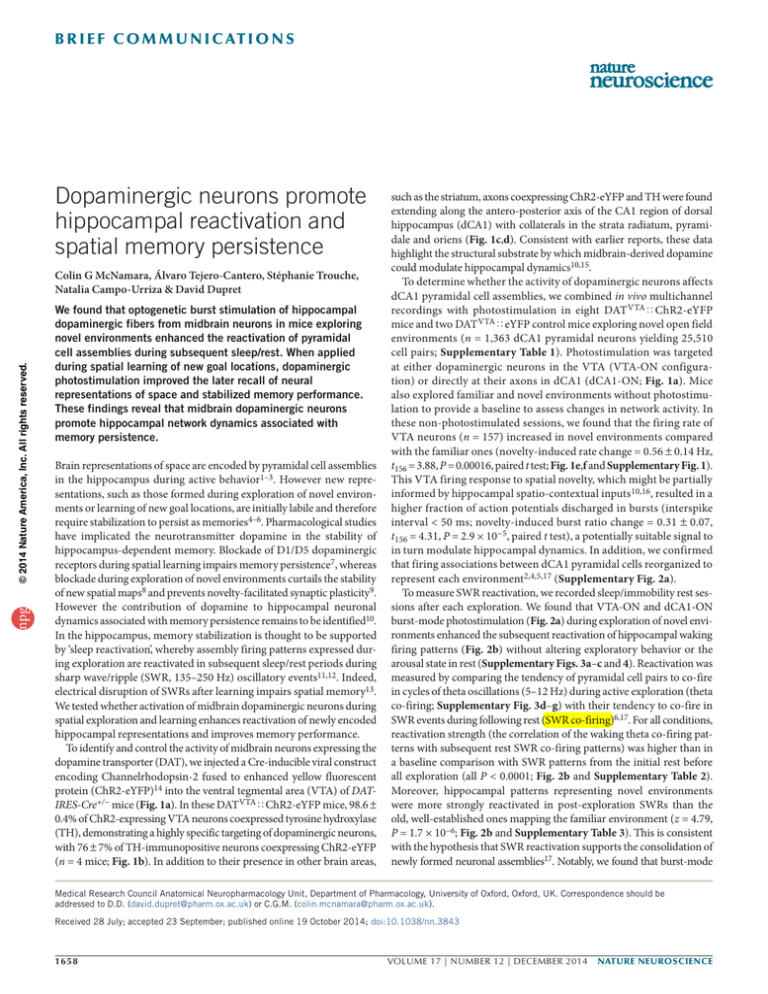
protein (ChR2-eYFP)14 into the ventral tegmental area (VTA) of DATIRES-Cre+/− mice (Fig. 1a). In these DATVTAøChR2-eYFP mice, 98.6 ±
0.4% of ChR2-expressing VTA neurons coexpressed tyrosine hydroxylase
(TH), demonstrating a highly specific targeting of dopaminergic neurons,
with 76 ± 7% of TH-immunopositive neurons coexpressing ChR2-eYFP
(n = 4 mice; Fig. 1b). In addition to their presence in other brain areas,
such as the striatum, axons coexpressing ChR2-eYFP and TH were found
extending along the antero-posterior axis of the CA1 region of dorsal
hippocampus (dCA1) with collaterals in the strata radiatum, pyramidale and oriens (Fig. 1c,d). Consistent with earlier reports, these data
highlight the structural substrate by which midbrain-derived dopamine
could modulate hippocampal dynamics10,15.
To determine whether the activity of dopaminergic neurons affects
dCA1 pyramidal cell assemblies, we combined in vivo multichannel
recordings with photostimulation in eight DAT VTAøChR2-eYFP
mice and two DAT VTAøeYFP control mice exploring novel open field
environments (n = 1,363 dCA1 pyramidal neurons yielding 25,510
cell pairs; Supplementary Table 1). Photostimulation was targeted
at either dopaminergic neurons in the VTA (VTA-ON configuration) or directly at their axons in dCA1 (dCA1-ON; Fig. 1a). Mice
also explored familiar and novel environments without photostimulation to provide a baseline to assess changes in network activity. In
these non-photostimulated sessions, we found that the firing rate of
VTA neurons (n = 157) increased in novel environments compared
with the familiar ones (novelty-induced rate change = 0.56 ± 0.14 Hz,
t156 = 3.88, P = 0.00016, paired t test; Fig. 1e,f and Supplementary Fig. 1).
This VTA firing response to spatial novelty, which might be partially
informed by hippocampal spatio-contextual inputs10,16, resulted in a
higher fraction of action potentials discharged in bursts (interspike
interval < 50 ms; novelty-induced burst ratio change = 0.31 ± 0.07,
t156 = 4.31, P = 2.9 × 10⁻⁵, paired t test), a potentially suitable signal to
in turn modulate hippocampal dynamics. In addition, we confirmed
that firing associations between dCA1 pyramidal cells reorganized to
represent each environment2,4,5,17 (Supplementary Fig. 2a).
To measure SWR reactivation, we recorded sleep/immobility rest sessions after each exploration. We found that VTA-ON and dCA1-ON
burst-mode photostimulation (Fig. 2a) during exploration of novel environments enhanced the subsequent reactivation of hippocampal waking
firing patterns (Fig. 2b) without altering exploratory behavior or the
arousal state in rest (Supplementary Figs. 3a–c and 4). Reactivation was
measured by comparing the tendency of pyramidal cell pairs to co-fire
in cycles of theta oscillations (5–12 Hz) during active exploration (theta
co-firing; Supplementary Fig. 3d–g) with their tendency to co-fire in
SWR events during following rest (SWR co-firing)6,17. For all conditions,
reactivation strength (the correlation of the waking theta co-firing patterns with subsequent rest SWR co-firing patterns) was higher than in
a baseline comparison with SWR patterns from the initial rest before
all exploration (all P < 0.0001; Fig. 2b and Supplementary Table 2).
Moreover, hippocampal patterns representing novel environments
were more strongly reactivated in post-exploration SWRs than the
old, well-established ones mapping the familiar environment (z = 4.79,
P = 1.7 × 10⁻⁶; Fig. 2b and Supplementary Table 3). This is consistent
with the hypothesis that SWR reactivation supports the consolidation of
newly formed neuronal assemblies17. Notably, we found that burst-mode
Medical Research Council Anatomical Neuropharmacology Unit, Department of Pharmacology, University of Oxford, Oxford, UK. Correspondence should be
addressed to D.D. (david.dupret@pharm.ox.ac.uk) or C.G.M. (colin.mcnamara@pharm.ox.ac.uk).
Received 28 July; accepted 23 September; published online 19 October 2014; doi:10.1038/nn.3843
1658
VOLUME 17 | NUMBER 12 | DECEMBER 2014 nature neuroscience
a
Figure 1 Midbrain dopaminergic neurons
increase their discharge in novel environments.
(a) Virus injection and photostimulation
configurations. (b) ChR2-eYFP expression
targeting VTA dopaminergic neurons as
identified by TH immunoreactivity. Cell nuclei
were stained using DAPI. (c) ChR2-expressing
dopaminergic axons in dCA1. Insets show
corresponding TH immunoreactivity (flattened
70-µm z stack; DAPI, 1-µm confocal plane).
(d) Three-dimensional reconstruction of ChR2expressing axons in the dorsal hippocampus
(distances from bregma in mm). (e) Spike
times (top) of an optogenetically identified
dopaminergic neuron in familiar and novel
environments (1-min raster plot, 10-s rows,
ticks represent spikes, bursts shown in black;
Supplementary Fig. 1) with interspike interval
distributions (left), laser-triggered firing
probability histogram (middle) and spontaneous
(VTA-OFF) versus light-evoked (VTA-ON) spike
waveforms (right); photostimulation subsequent
to shown explorations, mean waveforms
superimposed in black. (f) Distribution of firing
rate change in response to spatial novelty for
VTA neurons having a mean rate between 1
and 12 Hz. DG, dentate gyrus; LMol, stratum
lacunosum-moleculare; Or, stratum oriens;
Py, stratum pyramidale; Rad, stratum radiatum;
SNC, substantia nigra pars compacta;
SNR, substantia nigra pars reticulata.
a
dCA1
*
VTA-ON
Familiar
Novel
d
10 µm
Or
Py
Rad
Or
LMol
dCA1
Py
DG
Rad
50 µm
Dorsal
Left
–2.5
Anterior
–0.9
–1.7
Familiar
Firing rate: 2.5 Hz, burst ratio: 0.44
Novel
Firing rate: 11.0 Hz, burst ratio: 2.9
VTA-ON
3
VTA-OFF
Novel
Familiar
2
1
0
0
400 –40
200
Interspike interval (ms)
1 ms
Rest before
Rest after
Novel
Novel
Novel
Novel
dCA1-ON dCA1-ON dCA1-ON dCA1-ON
burst
burst
burst
tonic
eYFP ctrl. SCH23390
nature neuroscience VOLUME 17 | NUMBER 12 | DECEMBER 2014
0
Time (ms)
40
VTA-ON
f
20
VTA cells
(count)
Count ×1,000
e
ChR2-eYFP
DAPI TH
***
Novel
VTA-ON
burst
200 µm
c
0.1
0
TH
DAPI
ChR2-eYFP
DAPI
SNR
dCA1-ON
***
0.2
SNC
DAT-IRES-Cre+/–
473-nm laser
500 ms
0.3
ChR2-eYFP
TH
VTA
VTA
Photostimulation VTA-ON burst
b
b
DIO-ChR2-eYFP
photoactivation of dopaminergic neurons enhanced hippocampal
reactivation further (VTA-ON: z = 2.53, P = 0.011; dCA1-ON: z = 6.52,
P = 7.2 × 10⁻¹¹; Fig. 2b) without altering the mean firing rate, spatial
tuning or cross-environment reorganization of pyramidal assemblies
(Supplementary Fig. 2). Neither the mean firing rate nor the proportion
of spiking pyramidal cells during SWRs were changed (Supplementary
Fig. 5). Enhanced reactivation therefore represents an increased similarity of the SWRs assembly patterns to the previous waking ones. Such
enhanced reactivation was blocked by the D1/D5 antagonist SCH23390
when it was injected before exploration and was not observed in control
mice transfected with a construct coding for eYFP only (Fig. 2b and
Supplementary Table 3). Furthermore, tonic-mode stimulation consisting of the same number of light pulses (now equally spaced) produced
only a weak trend of increased reactivation (z = 1.94, P = 0.052; Fig. 2b).
Collectively these data demonstrate that burst-mode activation of
dopaminergic neurons increases hippocampal reactivation in a D1/D5
receptor–dependent manner.
We next tested whether dopamine-enhanced reactivation was associated with the strengthening of new spatial memories, as expressed by the
reinstatement of newly learned representations alongside stable behavioral
Waking theta co-firing
versus rest SWR co-firing (r)
npg
© 2014 Nature America, Inc. All rights reserved.
b r i e f c o m m u n i c at i o n s
10
0
–4
0
4
8
Firing rate change (Hz)
performance. We performed bilateral dCA1-ON burst-mode photostimulation with dCA1 recording in four DATVTAøChR2-eYFP mice
(560 dCA1 pyramidal cells yielding 12,072 cell pairs; Supplementary
Table 1) trained to a hippocampus-dependent learning task on a
crossword-like maze (Fig. 3a and Supplementary Fig. 6). On each day,
mice first explored the maze without barriers (pre-learning) to provide
a baseline to assess learning-related changes in network activity. Then,
for the ‘learning’ stage, two start boxes were selected and a food reward
was introduced together with a new arrangement of barriers so that
only one path from each start box led to the reward location. Mice
had 20 trials to learn the new configuration and find the reward.
Behavioral performance gradually improved with learning trials, as
measured by a shortening of distance traveled from start box to reward
in each trial (Fig. 3a,b). Notably, photostimulation during learning prevented the degradation of memory performance in a probe test held 1 h
later (t23 = 4.99, P = 4.8 × 10⁻⁵, t test; Fig. 3b), despite not affecting learning performance (F19, 405 = 0.40, P = 0.99, ANOVA; Fig. 3b). The reorganization of hippocampal spatial maps between the pre-learning and the
learning periods was also unaffected by photostimulation (Fig. 3c,d and
Supplementary Fig. 7). However, the reinstatement of the newly learned
spatial maps in the probe test was promoted following activation of dCA1
dopaminergic axons (z = 5.90, P = 3.6 × 10⁻⁹; Fig. 3d), a phenomenon
associated with memory recall6. The persistence of memories was associated with the strength of hippocampal reactivation. Hippocampal firing
patterns expressed during learning were more strongly reactivated in
Figure 2 Burst-mode photostimulation of dopaminergic neurons during
exploration enhances reactivation of new hippocampal assemblies.
(a) High pass–filtered VTA single-channel tetrode recording (left)
showing firing response to burst-mode photostimulation (trains of
20 10-ms pulses at 50-ms intervals) and superimposed waveforms of
all 20 light-driven spikes (right). (b) SWR reactivation of dCA1 co-firing
patterns (Supplementary Tables 2 and 3). r represents Pearson correlation
coefficient. Error bars represent s.e. *P < 0.05, ***P < 0.001.
1659
npg
a
Rest
Pre-learning
or
b
Trial 17
Trial 18
c
dCA1-OFF
dCA1-ON
20
16
Rest
Learning with photostimulation, dCA1-ON
12
Probe
Test 1
Test 4
Trial 2
Trial 3
Trial 19
Trial 20
Pre-learning
Test 2
Test 3
Learning
Probe
7.2
12.2
12.8
10.7
17.0
16.7
Peak
***
8
4
0
dCA1-OFF
dCA1-ON
0.4
***
0.3
0.2
0.1
0
Learning
Pre-learning Pre-learning
versus probe
versus
first versus
learning
second half
Methods
Methods and any associated references are available in the online
version of the paper.
Note: Any Supplementary Information and Source Data files are available in the
online version of the paper.
Acknowledgments
We thank J. Csicsvari, P. Somogyi and P.J. Magill for their comments on a previous
version of the manuscript, C. Etienne for her assistance with axon reconstruction,
S. Cuell for his initial contribution with the crossword maze, S. Threfell and L. Norman
0 (Hz)
e
0.3
0.2
Rest before
Rest after
**
***
dCA1-ON
d
20 Probe
test
dCA1-OFF
4
8
12
16
Learning (trial number)
Waking theta co-firing
versus rest SWR
co-firing (r)
0
subsequent rest than the pre-learning patterns were in the rest after prelearning exploration (z = 4.23, P = 2.3 × 10⁻⁵; Fig. 3e). Photostimulation
during learning further enhanced the reactivation of the concurrently
expressed firing patterns (z = 3.16, P = 0.0016; Fig. 3e), but did not extend
its effect back to the patterns expressed during the pre-learning exploration (z = 0.21, P = 0.84; Fig. 3e). This finding indicates that dopamine
acts specifically on actively expressed neuronal assemblies.
Determining the network-level processes underpinning the specific stabilization of some newly acquired engrams (while others fade) is critical
for a comprehensive understanding of memory. Our findings demonstrate
that dopaminergic activity at the time of formation of new hippocampal
cell assemblies increases the subsequent off-line SWR reactivation of these
assemblies. Such reactivation is thought to enable the incorporation of new
memory engrams, especially those associated with novel and rewarded
outcomes, into stable representations capable of informing future behavior6,11,12,18. Accordingly, we found that dopaminergic-enhanced reactivation prevents the degradation of learned behavioral performance while
concurrently strengthening the reinstatement of the newly acquired spatial
representation. Thus, our results provide insights into the relationship
between midbrain dopaminergic neurons, encoding salient information
about an environment9,10,16,19,20, and hippocampal assemblies, providing
representations of space1–3, in modulating memory according to the value
of the information to be remembered.
1660
Learning without photostimulation, dCA1-OFF
Rest
Trial 1
Trial 4
Path length to reward
(norm)
Figure 3 Burst-mode photostimulation of dCA1
dopaminergic axons during spatial learning on
the crossword maze stabilizes new hippocampal
maps and memory of new goal locations.
(a) Protocol with example single-day singleanimal trajectories. Solid arrows indicate the
reward location for that day; in-use start boxes
are shown in white. (b) Photostimulation did not
alter path shortening to reward during learning
(left), but did stabilize performance in the probe
(right; P = 4.8 × 10⁻⁵, t test, dCA1-ON versus
dCA1-OFF). Error bars represent s.e.m.
(c) Example rate maps for two dCA1 cells (one per
row) establishing a new firing field in learning
that was reinstated for the top cell only during
the probe (Supplementary Fig. 7). Numbers
indicate peak firing rate. (d) Photostimulation
did not affect spatial map reorganization
between pre-learning and learning (left), but
did promote reinstatement of newly established
maps during the probe (right; P = 3.6 × 10⁻⁹).
r represents Pearson correlation coefficient. Error
bars represent s.e. (e) SWR-reactivation after
learning was higher than after pre-learning and
increased further following photostimulation
(left; Supplementary Tables 2 and 3). However,
photostimulation did not extend its effect to
the previously expressed assemblies (right;
see dashed arrow in a). r represents Pearson
correlation coefficient. Error bars represent s.e.
**P < 0.01, ***P < 0.001.
Population map
similarity (r)
© 2014 Nature America, Inc. All rights reserved.
b r i e f c o m m u n i c at i o n s
0.1
0
PreLearning Learning
learning dCA1-OFF dCA1-ON
Prelearning
for their assistance with the DAT-IRES-Cre mice, and K. Deisseroth (Stanford University)
for sharing the DIO-ChR2-eYFP and DIO-eYFP constructs. C.G.M. is funded by a
Medical Research Council Doctoral Training Award. This work was supported by the
Medical Research Council UK (award MC_UU_12020/7) and a Mid-Career Researchers
Equipment Grant from the Medical Research Foundation (award C0443) to D.D.
AUTHOR CONTRIBUTIONS
C.G.M., Á.T.-C. and D.D. designed the experiments. C.G.M., N.C.-U. and
S.T. carried out the experiments. C.G.M. and D.D. analyzed the data and wrote the
manuscript. Á.T.-C. and S.T. edited the manuscript. D.D. conceived and supervised
the project. All of the authors discussed the results and commented on
the manuscript.
COMPETING FINANCIAL INTERESTS
The authors declare no competing financial interests.
Reprints and permissions information is available online at http://www.nature.com/
reprints/index.html.
O’Keefe, J. Exp. Neurol. 51, 78–109 (1976).
Wilson, M.A. & McNaughton, B.L. Science 261, 1055–1058 (1993).
Moser, E.I., Kropff, E. & Moser, M.B. Annu. Rev. Neurosci. 31, 69–89 (2008).
Kentros, C. et al. Science 280, 2121–2126 (1998).
Frank, L.M., Stanley, G.B. & Brown, E.N. J. Neurosci. 24, 7681–7689 (2004).
Dupret, D., O’Neill, J., Pleydell-Bouverie, B. & Csicsvari, J. Nat. Neurosci. 13,
995–1002 (2010).
7. Bethus, I., Tse, D. & Morris, R.G.M. J. Neurosci. 30, 1610–1618 (2010).
8. Kentros, C.G., Agnihotri, N.T., Streater, S., Hawkins, R.D. & Kandel, E.R. Neuron
42, 283–295 (2004).
9. Li, S., Cullen, W.K., Anwyl, R. & Rowan, M.J. Nat. Neurosci. 6, 526–531 (2003).
10.Lisman, J.E. & Grace, A.A. Neuron 46, 703–713 (2005).
11.Buzsáki, G. Neuroscience 31, 551–570 (1989).
12.Wilson, M.A. & McNaughton, B. Science 265, 676–679 (1994).
13.Girardeau, G., Benchenane, K., Wiener, S.I., Buzsáki, G. & Zugaro, M.B.
Nat. Neurosci. 12, 1222–1223 (2009).
14.Tsai, H.-C. et al. Science 324, 1080–1084 (2009).
15.Scatton, B., Simon, H., Le Moal, M. & Bischoff, S. Neurosci. Lett. 18, 125–131
(1980).
16.Luo, A.H., Tahsili-Fahadan, P., Wise, R.A., Lupica, C.R. & Aston-Jones, G.
Science 333, 353–357 (2011).
17.O’Neill, J., Senior, T.J., Allen, K., Huxter, J.R. & Csicsvari, J. Nat. Neurosci. 11,
209–215 (2008).
18.Singer, A.C. & Frank, L.M. Neuron 64, 910–921 (2009).
19.Cohen, J.Y., Haesler, S., Vong, L., Lowell, B.B. & Uchida, N. Nature 482, 85–88
(2012).
20.Schultz, W., Dayan, P. & Montague, P.R. Science 275, 1593–1599 (1997).
1.
2.
3.
4.
5.
6.
VOLUME 17 | NUMBER 12 | DECEMBER 2014 nature neuroscience
ONLINE METHODS
npg
© 2014 Nature America, Inc. All rights reserved.
Approval for experiments with animals. Experiments involving animals
were conducted according to the UK Animals (Scientific Procedures) Act
1986 under personal and project licenses issued by the Home Office following
ethical review.
received either burst-mode photostimulation (trains of 20 light pulses 10 ms in
duration with pulses at 50-ms intervals and a burst train delivered every 40 s)
or tonic-mode photostimulation (single light pulses 10 ms in duration delivered
every 2 s). For the crossword maze experiments additional DAT-IRES-Cre mice
received burst-mode photostimulation with pulse duration of 20 ms and burst
trains delivered every 20 s. For experiments aimed at disrupting hippocampal network activity by local stimulation of PV-expressing interneurons, PV-IRES-Cre
mice received 473-nm photostimulation applied randomly to 80% of the learning
trials using trains of 10-ms pulses every 16 ms. The laser was off during the intertrial intervals. In all of these experiments, each mouse alternated between days
with photostimulation OFF and days with photostimulation ON.
Subjects and surgical procedures. All DAT-IRES-Cre+/− animals used
(Supplementary Table 1) were male adult transgenic heterozygote mice
(3–8 months old) and bred from homozygotes for DAT-internal ribosome
entry site (IRES)-Cre (Jackson Laboratories, B6.SJL-Slc6a3tm1.1(cre)Bkmn/J, stock
number 006660)21. Animals were housed with their littermates until used in
the procedure with free access to food and water in a dedicated housing room
with a 12/12-h light/dark cycle. Surgical procedures were performed under deep
anesthesia using isoflurane (0.5–2%) and oxygen (2 l min−1). Analgesia was
also provided (buprenorphine, 0.1 mg per kg of body weight). To generate expression of ChR2 in dopamine neurons, we used a Cre-loxP approach by injecting a
Cre-inducible recombinant viral vector containing ChR2-eYFP (pAAV2-EF1aDIO-hChR2(H134R)-eYFP-WPRE, 2.5 × 10¹² molecules per µl, Virus Vector
Core) in DAT-IRES-Cre mice at a rate of less than 0.1 µl min−1 using a glass
micropipette as described previously22. Two additional DAT-IRES-Cre+/− animals were injected with a control viral vector coding for the eYFP only (pAAV2EF1a-DIO-eYFP, 2.5 × 10¹² molecules per µl, Virus Vector Core). Viral vector
injections (1 µl each) were performed to the right hemisphere or bilaterally. All
crossword maze animals received a bilateral injection and implant. Each injection was delivered to the VTA using stereotaxic coordinates 3.25 mm posterior,
±0.5 mm lateral from bregma and 3.8–4 mm ventral from the brain surface.
4 weeks were allowed for virus expression before recording commenced. To test
for the hippocampus dependency of the crossword maze task, PV-IRES-Cre+/+
adult male mice (n = 5; Jackson Laboratories, B6;129P2-Pvalbtm1(cre)Arbr/J,
stock number 008069) were used with the same approach, but to transfect
PV-expressing interneurons bilaterally in the dCA1 region with the ChR2-eYFP
viral vector using stereotaxic coordinates 2.00 mm posterior, ±1.6 mm lateral
from bregma and 1.2 mm ventral from the brain surface for injection. Implanted
mice received eight or ten independently movable tetrodes, constructed as described
previously6,17, and one or two optic fibers (Doric lenses, Québec, Canada).
To enable their independent movement, each tetrode was attached to a M1.4 screw
and all tetrodes were assembled in a microdrive containing the optic fibers before
implantation. This also ensured that the optic fiber was completely enclosed to
prevent escaped light illuminating the environment. Optic fibers targeted at dCA1
were positioned 2 mm posterior and 1.6 mm lateral from bregma and lowered
into the brain a distance of 0.9–1 mm. Tetrodes were typically located within
0.5 mm of the optic fiber. For simultaneous midbrain and hippocampal recordings with VTA targeted optic fiber the implant was positioned at a 40 degree angle
from the antero-posterior axis on the horizontal plane and then at an angle of
10 degrees from the dorso-ventral axis. The optic fiber was positioned at
2.7 mm posterior and 1 mm lateral from bregma and lowered into the brain at the
angle described a distance of 3.9–4.2 mm. All tetrodes were initially implanted
slightly above the recording location of interest.
Post-recovery and recording. After initial recovery from implantation, and
before recording commenced, each mouse was connected to the recording
apparatus and familiarized (through spontaneous exploration) with the familiar
open field environment or crossword maze (without barriers) as applicable. This
typically lasted 1–2 h a day for at least a week. During this time, tetrodes were
gradually lowered over days through the short distance remaining to reach the
required brain area. All recorded mice were also familiarized with a 12- × 12-cm
high-walled box containing home cage bedding which was used for rest recordings (sleep and long periods of awake immobility).
At the start of each recording day hippocampal tetrodes were lowered into
stratum pyramidale and midbrain ones were moved slightly in search of action
potentials. The animal was then left in the home cage for at least 1 h before
recording started. All recordings were performed during day time and the
random assignment of each subject to the various experimental conditions is
reported in Supplementary Table 1. At the end of each recording day, hippo­
campal tetrodes were raised slightly to minimize mechanical damage caused to
the stratum pyramidale by tetrodes left in this layer overnight and to maximize
the cell yield and number of recording days per mouse. No further recording
days were performed once the number of cells decreased below the number
of cells obtained in the initial days. Stratum pyramidale was located using the
profile of SWR events and presence of multiunit spiking activity. Recordings
were always performed in the same room under dim lighting with the recording
arena surrounded by a black circular curtain. Rest recordings were approximately 30 min in duration. Investigators were aware as to which experimental
conditions each subject had to be exposed to (for example, familiar versus novel,
photostimulation OFF versus ON, exploration versus sleep). Open field recording arenas consisted of various boxes of different shapes (polygons with some
curved edges surrounded by walls, approximate area generally near 0.25 m2)
constructed from plastic or wood (sometimes lined with brown paper). Open
field exploration recording sessions were approximately 30 min in duration.
To determine whether enhanced SWR reactivation following burst-mode
photostimulation of dopaminergic terminals in dCA1 required activation of
dopaminergic receptors, mice were injected with SCH23390, a selective and
potent antagonist of D1/D5 dopaminergic receptors (Sigma, #D054, 0.1 mg
per kg in 0.9% saline (wt/vol), intraperitoneal) 30 min before the exploration
of a novel environment.
Data acquisition and photostimulation. Each signal was buffered on the head
of the animal using an operational amplifier in a non-inverting unity gain configuration (Axona) and transmitted over a single strand of litz wire to a dual stage
amplifier and band pass filter (gain 1000, pass band 0.1 Hz to 5 kHz, Sensorium).
The amplified and filtered signals along with a synchronization signal from the
position tracking system and the activation signal for the laser were digitized at
a rate of 20 kHz and saved to disk by a PC with 16 bit analog to digital converter
cards (United Electronics Industries). To track the location of the animal three
LED clusters (red, green and blue) were attached to the electrode casing above
the head of the animal and captured at 25 frames per s by an overhead color
camera (Sony). The camera was attached to a PC that extracted the position data
in real time. Photostimulation was generated using a 473-nm diode-pumped
solid-state laser (Laser 2000, Ringstead) and delivered to the brain through two
lengths of optic fiber (with black plastic jackets to prevent illumination of the
environment) and a rotary joint with splitter in the case of bilaterally implanted
animals (Doric Lenses). The final optic fibers (on the animal end) were coupled
to the implanted fiber using an M3 connector. The output intensity delivered to
the implanted fiber was approximately 20 mW. Open field DAT-IRES-Cre mice
Crossword maze. The crossword-like maze (Fig. 3a and Supplementary
Fig. 6a) consisted of four start boxes and eight intersecting open tracks forming
14 intersections inspired by a layout used previously23. The width of each track
was 5 cm with a 1.5-cm-high rim along the edges. The entire maze measured
95 × 95 cm excluding start boxes. The maze was painted black and suspended
5 cm above a black table. Distal cue cards were placed on the curtain surrounding
the maze and some cue objects were placed on the supporting table dispersed
throughout the maze. To promote allocentric spatial navigation by distal cues,
the maze was randomly rotated relative to the cues at the beginning of each day.
Mice performing the crossword maze task were maintained at 85% of their postoperative body weight. The recording protocol used consisted of the following
ordered stages: rest, pre-learning, rest, learning, rest and probe (Fig. 3a). During
the pre-learning stage, the animal explored the maze with the start boxes closed
and in the absence of barriers and rewards for approximately 20 min.
For the learning stage two start boxes and one food reward location (at the
end of one of the five tracks protruding from the maze) were selected as in use
for that day and the maze was configured with a new arrangement of up to
seven barriers (10 cm in height) such that there was only one path from each
doi:10.1038/nn.3843
nature neuroscience
© 2014 Nature America, Inc. All rights reserved.
npg
start box to the reward (Supplementary Fig. 6a). Mice were given up to 20 trials
(mean = 18.7, median = 19.0, quartile range = 18.0–20.0) to learn to find the
reward with the start point randomly switching between the two start boxes. The
per trial reward was 4 µl of condensed milk diluted 30% in water and was placed
on a plastic cap at the goal location. A similar plastic cap (without reward) was
placed in each of the other four tracks protruding from the maze. A glass vial
(with perforated lid) containing an aliquot of the reward yet non accessible was
placed inside the two start boxes to signal the onset of the learning phase to the
animal. The board was cleaned after each learning trial to discourage the use of
an odor guided search strategy.
For the probe stage (conducted 1 h after learning), the maze was maintained
in the same layout as the learning stage, but no reward was present. Mice were
released from one of the in use start boxes and allowed to explore the maze for
3 min. Mice were released a further three times (3 min each, 12-min duration
probe in total) during the probe stage in order to ensure good coverage of the
maze to calculate spatial rate maps. The path length to first reaching of the
reward location was used to evaluate memory performance. When combining across maze configurations, path lengths were normalized by the relevant
shortest possible path (Supplementary Fig. 6a). The number of crossword maze
recording days included in this study is 12 d without photostimulation and 13 d
with photostimulation for experiments involving the DAT-IRES-Cre mice;
13 d (12 probe tests) without photostimulation and 9 d with photostimulation
for experiments involving the PV-IRES-Cre mice.
Spike detection and unit isolation. The recorded signals were digitally band-pass
filtered (800 Hz to 5 kHz) offline for spike detection and unit isolation. Spikes
were detected using a threshold in the r.m.s. power (0.2-ms sliding window) of
the filtered signal. Unit isolation was performed on the first three or four principal
components calculated for each channel recorded by the tetrode in question.
This was achieved by automatic overclustering (KlustaKwik, http://klusta-team.
github.io/) followed by graphically based manual recombination and further
isolation informed by cloud shape in principal component space, crosschannel spike waveforms, auto-correlation histograms and cross-correlation
histograms24,25. To analyze changes in the firing patterns of pyramidal ensembles
over time and reliably monitor cell pairs across recording sessions, we needed
to ensure that our sample of cells was taken from clusters with stable firing. We
therefore clustered together all of the sessions recorded on a given day. Only wellisolated and stable units over the entire recording day were used for further analysis;
namely, units that showed a clear refractory period in their auto-correlations and
well-defined cluster boundaries across all recording sessions of the day. Units
were deemed independent from each other by their distinct spike waveform, the
absence of refractory period in their cross-correlations and separable principal
component boundaries using a measure based on the Mahalanobis distance24,26.
Hippocampal pyramidal cells were identified by their auto-correlogram, firing
rate and spike waveform, as previously described24. The total number of pyramidal cells included in this study is reported in Supplementary Table 1.
Detection of theta oscillations, SWR events and behavioral states. Theta
oscillations and SWR events were detected off-line using digital band-pass filter
methods as previously described6,17,24. First, the theta/delta power ratio was
measured in 1,600-ms segments (800-ms steps between measurement windows),
using Thomson’s multi-taper method24,27,28. The theta-delta power ratio was
used to mark periods of theta activity and individual theta oscillatory waves were
identified within these periods by first applying a conservative band-pass filter
(5–28 Hz) to the wideband signal. This allowed precise detection of the trough
of individual waves from the filtered trace without over smoothing the signal. Next, phase alignment was performed to these minima and theta windows
were demarcated spanning from peak-to-peak. Windows with a corresponding theta frequency of 5–12 Hz were used in further analysis (mean = 7.5 Hz,
median = 7.7 Hz, quartile range = 6.7–8.6). These detected theta windows corresponded to periods when the animals were actively exploring the environment
(Supplementary Fig. 3d). The mean number of theta cycles per waking session
used for spike binning was not different across conditions (open field experiments: 5,699.6 ± 165.5, F3, 143 = 1.41, P = 0.24, ANOVA; crossword maze experiments = 15,038.5 ± 855.6, t23 = 1.31, P = 0.20, t test; mean ± s.e.m.). SWR event
detection was performed in rest epochs when the instantaneous speed of the
animal was less than 2 cm s−1 and the theta-delta ratio was less than 2. Recorded
nature neuroscience
signals were band-pass filtered (135–250 Hz), and the signal from a ripple-free
reference electrode was subtracted to eliminate common-mode noise (such as
muscle artifacts). Next the power (root mean square) of the processed signal
was calculated for each electrode and summed across CA1 pyramidal cell layer
electrodes to reduce variability. The threshold for SWR event detection was set
to 7 s.d. above the background mean power. The proportion of pyramidal cells
active in SWR events was calculated for recordings with at least five pyramidal
cells. The SWR firing rate histograms of pyramidal cells were calculated using
20-ms bins in reference to the SWR peak (that is, peak of ripple-band power).
Novelty-related change in VTA firing activity. Firing response of VTA neurons to spatial novelty was assessed by the change in firing rate and burst ratio
during exploration of familiar versus novel environments in the absence of photostimulation. The burst ratio measure was calculated as the total number of
burst spikes divided by the total number of single spikes. This allowed longer
bursts with many spikes to have a more representative contribution to the ratio
than spike doublets or triplets compared to measures that count all bursts equally
irrespective of length. Bursts were classified as trains of at least two spikes with
interspike intervals of less than 50 ms. This conservative intraburst spike interval
threshold of 50 ms (intraburst frequency of 20 Hz) was chosen over the often used
80-ms (12.5 Hz) start with greater than 160-ms (6.25Hz) end of a burst criteria
established in anesthetized rats29,30 to allow sufficient clearance between the
burst interval threshold frequency and some of the higher mean rates (11 Hz)
seen in our light-identified units from behaving mice. Note that use of a 80-ms
cut-off produced similar findings. The 160-ms end of burst criteria was also not
needed as it was originally included to allow for the observation that there is often
a missed spike in a burst that isolates the later spikes from the first spikes of the
same burst29, but the burst ratio measure used here takes into account all burst
spikes. These measures were generated from the first 10 min of each exploration
to capture best the times of maximal novel experience. Cells included in this
analysis had a mean firing rate of between 1 and 12 Hz in both the familiar and
novel environments calculated separately. These rate boundaries were chosen to
match that reported for VTA dopaminergic neurons in behaving mice19 and the
mean rates of the light-driven units we identified in separate VTA-ON recording
sessions on the same days.
Reactivation. Reactivation strength was measured as the Pearson correlation
coefficient between the tendencies of hippocampal pyramidal cell pairs to fire
together (co-fire) across theta cycles during exploration (theta co-firing) with
their tendency to co-fire in SWR events during rest (SWR co-firing)6,17. To mea­
sure theta co-firing during exploration, we first established for each pyramidal
cell its instantaneous firing rate counts (IFRC) in windows spanning from peakto-peak of detected theta cycles and then we calculated the correlation coefficient between the IFRCs for each cell pair. Likewise, SWR co-firing values were
calculated as the correlation coefficients between IFRCs of pyramidal cell pairs
taken from 100-ms windows centered on the peak power of SWR events. The
reactivation strength was then calculated as the Pearson correlation coefficient
between the theta bin co-firing values and the SWR bin co-firing values. Detected
theta windows corresponded to periods of active exploration and SWR events
were restricted to times of behavioral immobility. Only simultaneously recorded
cells were paired. The total number of pyramidal cell pairs for each condition is
reported in Supplementary Table 1.
Spatial rate maps. The horizontal plane of the recording arena was divided into
bins of approximately 2 × 2 cm to generate spike count maps (number of spikes
fired in each bin) for each unit and an occupancy map (time spent by the animal in each bin). All maps were then smoothed by convolution with a Gaussian
kernel having s.d. equal to one bin width. Finally, spatial rate maps were generated by normalizing (dividing) the smoothed spike count maps by the smoothed
occupancy map.
Population map similarity. The population map similarity measure as applied
previously6,17 compares all the place maps from two different time periods in a
pairwise fashion in the same manner as the co-firing analysis does for cell firing
patterns across LFP events by performing binning over space rather than time. It
represents the degree to which cells which fired in similar regions of space (that is,
overlapping place fields) still fire in similar regions of space later. It is calculated
doi:10.1038/nn.3843
by first computing the place field similarity (PFS) value for each cell pair during
the first time period as the Pearson correlation coefficient from the direct bin wise
comparisons between the spatial rate maps of the two cells limited to valid bins
(occupancy greater than zero). This is repeated for the second time period and
the population map similarity between the two time periods is then calculated
as the Pearson correlation coefficient between the PFS values from each of the
time periods. Hippocampal place cells screened for their spatial tuning using a
sparsity value of no more than 0.3 were included in this analysis6. Note that, in
the case of assessing map reinstatement during the memory probe test, the PFS
values calculated from the second half of the learning trials were compared to
PFS values from the probe tests.
npg
© 2014 Nature America, Inc. All rights reserved.
Spatial tuning measures. Coherence and sparsity were calculated from
the un­smoothed place rate maps as reported before31,32. Coherence reflects
the similarity of the firing rate in adjacent bins, and is the z transform of the
Pearson correlation (across all bins) between the rate in a bin and the average
rate of its eight nearest neighbors31. Sparsity corresponds with the proportion
of the environment in which a cell fires, corrected for dwell time, and is defined
as (ΣPiRi)2/ΣPiRi2, where Pi is the probability of the mouse occupying bin i and
Ri is the firing rate in bin i (ref. 32).
Error intervals and statistical tests. All error intervals quoted are ±s.e.m. or s.e.
of the correlation coefficient calculated as the square root of (1 – r2)/(n – 2), where
r is the Pearson correlation coefficient and n is the number of cell pairs33. All tests
performed were two sided. All P values for the comparison of Pearson correlation coefficients were calculated using Z statistics after application of Fisher’s r
to z transform. Other tests, wherever appropriate, are mentioned along with the
value. Statistical analyses were performed using SciPy (http://www.scipy.org/) and
R (www.r-project.org). Sample sizes in this study are similar to those generally
employed in the field and were not pre-determined by a sample size calculation.
Data distributions for parametric tests were assumed to be normal, but this was
not formally tested.
Tissue processing and immunocytochemistry. Mice were deeply anesthetized
with isoflurane/pentobarbital and transcardially perfused with phosphatebuffered saline (PBS) followed by fixative (paraformaldehyde dissolved in PBS,
4%, wt/vol), and brains were extracted and sectioned into 70-µm-thick coronal
sections. Sections were washed three times in PBS between each of the following
steps. Cell membranes were permeabilized and nonspecific protein binding was
blocked by incubation for 1 h in PBS-triton (0.3%) and normal donkey serum
(NDS, 10%). Next, sections were incubated for 48 h at 4 °C in a solution of primary
antibodies to GFP (used to recognize ChR2-eYFP, 1:500, Aves Labs #GFP-1020,
Nacalai Tesque #GF090R/04404-84)34 and either TH (for the DAT-IRES-Cre
brain sections, 1:500, EDM Millipore #AB152, Abcam #ab76442)35,36 or PV (for
the PV-IRES-Cre brain sections, 1:4,000, EDM Millipore #MAB1572), washed
and then further incubated in a solution with corresponding secondary antibodies (raised in donkey) conjugated with DyLight488 (1:500, Jackson #703-485-155)
or Alexa488 (1:500, Jackson #703-545-155, Invitrogen #A21208) and Cy3 (1:500,
Jackson #711-165-152 and #703-165-155; 1:1000, Jackson #715-165-150). Both
doi:10.1038/nn.3843
antibody solutions were diluted in PBS-triton (0.3%) and NDS (1%). Finally, sections
were counterstained with DAPI (diluted 1:10,000) for 20 min and mounted on
glass slides using VectaShield (Vector Laboratories).
Microscopic analyses and three-dimensional reconstruction. The distribution
of transfected axons across brain areas was consistent with the known distribution
of dopaminergic fibers37. Specificity of virus transfection was quantified from
both hemispheres of four bilaterally injected brains. Three consecutive sections
centered on the injection coordinate (bregma –3.25) were selected38. An exhaustive tiled grid of 20-µm-thick (0.8-µm step) z stacks to span the VTA was acquired
from the top of each section using an epifluorescence microscope (Imager.M2
with filter set 38 and a Plan-Neofluar 40×/1.3 objective, Zeiss). Only grid tiles
which were deemed to be within the VTA regions (A10 cell groups)39 were
included in the counting. Greater than 900 cells were counted per brain. Threedimensional reconstruction of transfected dCA1 axons was completed using the
same microscope and Neurolucida software (MBF Bioscience). Published images
were acquired using a confocal microscope (Imager.Z1 with LSM 710 scan head
and Plan-Neofluar 5×/0.16, Plan-Apochromat 40×/1.3, 63×/1.4 or 100×/1.46
objective, Zeiss) in sequential scanning mode with the following excitation laser
and emission filter wavelengths. DAPI: 405 nm, 409–499 nm; eYFP/DyLight488:
488 nm, 493–571 nm; Cy3: 543 nm, 566–729 nm. Acquisition and analysis were
completed using ZEN 2008, v5.0 (Zeiss) and ImageJ (http://rsb.info.nih.gov/)
software. Z stacks were flattened using maximum intensity projection.
A Supplementary Methods Checklist is available.
21.Bäckman, C.M. et al. Genesis 44, 383–390 (2006).
22.Cetin, A., Komai, S., Eliava, M., Seeburg, P.H. & Osten, P. Nat. Protoc. 1,
3166–3173 (2006).
23.Tolman, E.C. Psychol. Rev. 55, 189–208 (1948).
24.Csicsvari, J., Hirase, H., Czurkó, A., Mamiya, A. & Buzsáki, G. J. Neurosci. 19,
274–287 (1999).
25.Harris, K.D., Henze, D.A., Csicsvari, J., Hirase, H. & Buzsáki, G. J. Neurophysiol.
84, 401–414 (2000).
26.Harris, K.D., Hirase, H., Leinekugel, X., Henze, D.A. & Buzsáki, G. Neuron 32,
141–149 (2001).
27.Thomson, D.J. Proc. IEEE 70, 1055–1096 (1982).
28.Mitra, P.P. & Pesaran, B. Biophys. J. 76, 691–708 (1999).
29.Grace, A.A. & Bunney, B. J. Neurosci. 4, 2877–2890 (1984).
30.Ungless, M.A. & Grace, A.A. Trends Neurosci. 35, 422–430 (2012).
31.Muller, R.U. & Kubie, J.L. J. Neurosci. 9, 4101–4110 (1989).
32.Skaggs, W.E., McNaughton, B.L., Wilson, M.A. & Barnes, C.A. Hippocampus 6,
149–172 (1996).
33.Zar, J.H. Biostatistical Analysis (Prentice Hall, 1999).
34.Tomiyoshi, G., Nakanishi, A., Takenaka, K., Yoshida, K. & Miki, Y. Cancer Sci. 99,
747–754 (2008).
35.Yamamoto, K., Ruuskanen, J.O., Wullimann, M.F. & Vernier, P. Mol. Cell. Neurosci.
43, 394–402 (2010).
36.Samaco, R.C. et al. Proc. Natl. Acad. Sci. USA 106, 21966–21971 (2009).
37.Björklund, A. & Lindvall, O. in Handbook of Chemical Neuroanatomy, Volume 2:
Classical Transmitters in the CNS, Part I (eds. Björklund, A. & Hökfelt, T.) 55–122
(Elsevier, 1984).
38.Franklin, K.B.J. & Paxinos, G. The Mouse Brain in Stereotaxic Coordinates (Academic
Press, 2008).
39.Fu, Y. et al. Brain Struct. Funct. 217, 591–612 (2012).
nature neuroscience

