TECHNICAL ARTICLE
Calibration of ION ABUNDANCE
in a TOF-MS Spectrum
Kevin L. Hunter and Richard W. Stresau
ETP Electron Multipliers, Ermington, Australia
Presented at the 46th ASMS Conference on Mass Spectroscopy and Allied Topics
May 31-June 4, 1998, Orlando, Florida
Ion Signal
Electron
Multiplier
Preamp
Computer
Interface
Storage
Buffer
ADC
Figure 1. Generalized functional layout of the detection system of a Time of Flight Mass Spectrometer.
Detection systems for TOF mass spectrometers in general
make use of an electron-multiplier detector to convert
the ion signal into electrons and then to multiply the
electron current. This signal is then fed into an Analogto-Digital-Converter (ADC) to give a digitized form of the
signal (Figure 1).
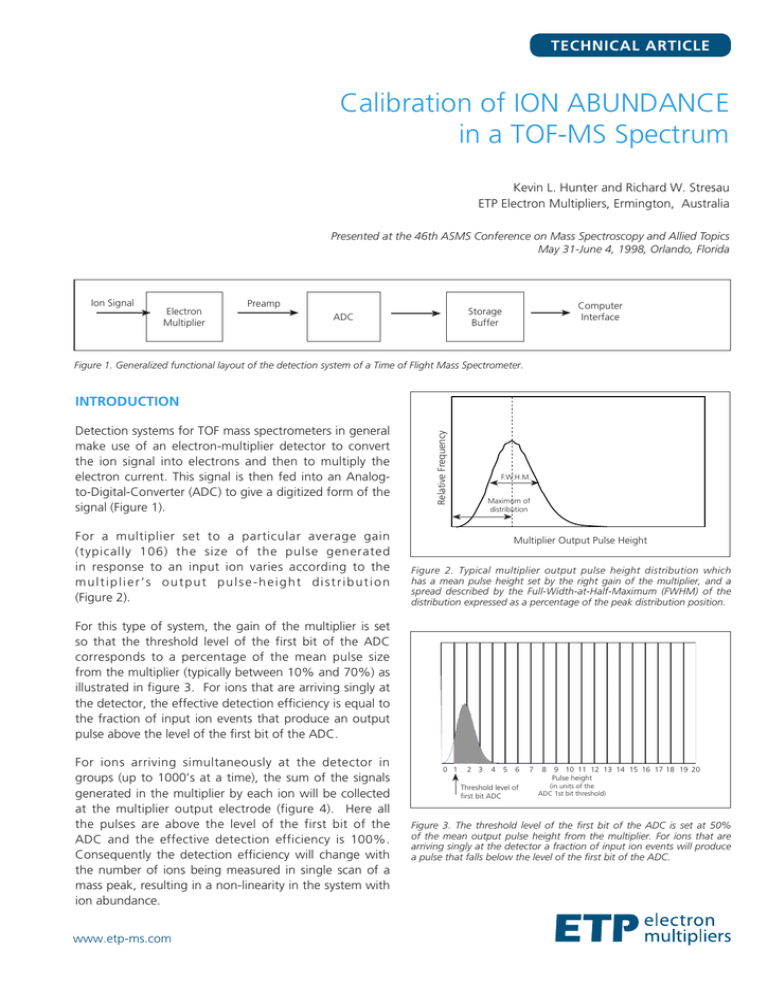
For a multiplier set to a particular average gain
(typically 106) the size of the pulse generated
in response to an input ion varies according to the
multiplier’s output pulse-height distribution
(Figure 2).
Relative Frequency
INTRODUCTION
F.W.H.M.
Maximum of
distribution
Multiplier Output Pulse Height
Figure 2. Typical multiplier output pulse height distribution which
has a mean pulse height set by the right gain of the multiplier, and a
spread described by the Full-Width-at-Half-Maximum (FWHM) of the
distribution expressed as a percentage of the peak distribution position.
For this type of system, the gain of the multiplier is set
so that the threshold level of the first bit of the ADC
corresponds to a percentage of the mean pulse size
from the multiplier (typically between 10% and 70%) as
illustrated in figure 3. For ions that are arriving singly at
the detector, the effective detection efficiency is equal to
the fraction of input ion events that produce an output
pulse above the level of the first bit of the ADC.
For ions arriving simultaneously at the detector in
groups (up to 1000’s at a time), the sum of the signals
generated in the multiplier by each ion will be collected
at the multiplier output electrode (figure 4). Here all
the pulses are above the level of the first bit of the
ADC and the effective detection efficiency is 100%.
Consequently the detection efficiency will change with
the number of ions being measured in single scan of a
mass peak, resulting in a non-linearity in the system with
ion abundance.
www.etp-ms.com
0 1 2 3 4 5 6 7 8 9 1011121314151617181920
Pulse height
(in units of the
Threshold level of
ADC
1st bit threshold)
first bit ADC
Figure 3. The threshold level of the first bit of the ADC is set at 50%
of the mean output pulse height from the multiplier. For ions that are
arriving singly at the detector a fraction of input ion events will produce
a pulse that falls below the level of the first bit of the ADC.
TECHNICAL ARTICLE
5
0
-5
-10
-15
-20
-25
-30
-35
-40
-45
-50
-55
-60
-65
-70
-75
-80
0 1 2 3 4 5 6 7 8 9 1011121314151617181920
Pulse height
(in units of the
Threshold level
ADC 1st bit threshold)
of first bit ADC
Relative Detection Efficiency (%)
REGION II
REGION I
75
70
0.00010.001 0.01
0.1
1
10
100
1000
IONS / Measurement (for a given ion mass)
Figure 5. Detection efficiency curve for the TOF detection system. This
curve has three distinct regions; Region I (single ions), Region III (groups
of many ions), and a transition region, Region II (mix of single ions and
larger groups).
Abundance Linearity of Ion Signal
Pulse height dist = 220%, threshold = 50% on mean pulse height
Deviation from Linearity (%)
10
5
0
Corrected data using standards and equation 2
-5
-10
-15
-20
Raw data
-25
-30
-35
-40
-45
-50
0.00010.001 0.01
0.1
1
0.01
0.1
1
10
100
1000
Figure 7. Theoretical results (using Monte-Carlo simulation) for a
detection system using an electron multiplier with an output pulse
height distribution of FWHM=220%. The threshold level of the 1st bit
of the ADC is set at a range of values from 0.2 to 1.5 (expressed as a
fraction of the mean pulse height).
Method A: Using Selected Standards and the Functional
form of the Calibration Curve.
90
80
0.001
CORRECTING THE ABUNDANCE SCALE
NON-LINEARITY
95
85
1.5
Observed counts per measurement (for a given mass)
REGION III
100
1.0
0.0001
Figure 4. The threshold level of the first bit of the ADC is set to 50% of
the mean output pulse height from the multiplier for a single ion. For
ions that are arriving at the detector in groups of many (up to 1000’s at
a time), essentially all the input ion events are counted.
105
0.2
0.35
0.5
0.0
10
100 1000
Observed counts / measurement
Figure 6. Theoretical results (using Monte-Carlo simulation) for a
detection system using an electron multiplier with an output
height distribution of FWHM=220% (as a percentage of the peak
pulse height) and the threshold level of the 1st bit of the ADC
set at 50% of the mean pulse height. The corrected linearity
curve was obtained using equation 2 and using the values:
A=0.78, B=1.30, C=1.30.
The calibration curve between the measured ion
abundance per measurement, X, and the real number of
ions per measurement reaching the detector, Y, is shown
in figure 5. The mathematical form of this curve is as
follows:
X = Y * K
where K =
1.0-A
2
+
A
2
tanh B log10
(eqn.1)
X
C
(eqn.2)
And the constants A, B and C are:
A - low ion-intensity detection efficiency
B - slope of the inflection region
C - value of X at the inflection point
By using appropriate calibration samples which give ion
peaks on both plateaus and in the transition region of
figure 6, the values of A, B and C can be determined.
Figure 6 shows theoretical results after the correction of
equation 2 was made. The linearity of the system is now
within ±1%.
Method B: Quantization Correction with appropriate
setting of discriminator level
The amount of non-linearity seen in the measured
abundance is strongly dependent on the chosen
setting for the discriminator level (figure 7). Good
linearity can be achieved when the discriminator
www.etp-ms.com
TECHNICAL ARTICLE
10
9
8
Digitized Pulse Size
setting is a small fraction of the mean pulse height.
In practice the minimum discriminator level is
limited by line noise. This situation can be improved
by correcting the signal from the ADC for the error
generated by the digitization of the pulse size
(figure 8). This quantization error can be compensated
for every time a measurement is taken by applying the
expression:
7
6
5
4
After applying equation 1
3
2
Corrected ADC Output = ADC Output + 0.5 (eqn. 3)
1
0
Method C: Quantization Correction and Calibration
Curve
Alternatively the quantization-corrected data in figure 9
could be further corrected by also applying Method A.
In this case the functional form of the corrected data
can be used in conjunction with known ion intensity
standards to provide calibration of the system.
We have also found that for the quantization-corrected
data, the response linearity curves (figure 9) are
not as symmetric as those of the raw data (without
quantization-correction). Better results are obtained if a
different value for the constant B is used in equation 2
for data points with ion intensities greater than the
inflection point of the curve.
01 2345678910
Average Pulse Size
(units of the ADC 1st bit threshold level)
Figure 8. The digitized output of the ADC plotted as a function of the
input pulse size. If a group of pulses with heights evenly distributed
between the thresholds of the third and forth bits of the ADC, then the
average size of these pulses would be 3.5. However the ADC would
read an output of 3 for each pulse.
Deviation from Linearity (%)
Figure 9 shows the same data after using the
quantization correction of equation 3. Good linearity is
achieved when the threshold level is set to 0.35 (fraction
of the mean pulse height). From this result it can be
readily seen that the detection system can be calibrated
by using a set of standards, with a signal intensity that is
on each plateau region, and then adjusting the gain of
the multiplier (or the relative value of the threshold of
the ADC) until the correct ratio of the two standards is
achieved.
5
0.2
0 0.35
0.5
-5 0.0
-10
-15
-20
1.0
-25
-30
-35
-40
-45
-50
1.5
-55
-60
-65
-70
-75
-80
0.00010.001
0.01
0.1
1
10
100
1000
Observed Counts per measurement (for given mass)
Figure 9. The corrected linearity curves were obtained by applying
equation 3 to the data in figure 7. In this case near ideal linearity is
achieved when the threshold level of the 1st bit of the ADC is set at a
value of 0.35 (expressed as a fraction of the mean pulse height).
CONCLUSIONS
Monte-Carlo methods have proven to be a powerful tool
in understanding how ions from a TOF mass spectrometer
interact with the electron multiplier and its associated
electronics. This study has revealed that serious nonlinearities can occur in the measured abundance of ions in
a TOF mass spectrum.
Several methods for correcting the non-linearity in the
measured ion abundance have been determined;
ETP Electron Multipliers
ABN:
35 078 955 521
Address: 31 Hope Street,
Melrose Park NSW 2114
Australia
Tel:
+61 (0)2 8876 0100
Fax:
+61 (0)2 8876 0199
Email:info@etp-ms.com
Web: www.etp-ms.com
©Copyright 2013 ETP Electron Multipliers. All rights reserved. TA-0074-A_RevB
1. Using selected standards and applying the functional
form of the abundance calibration curve determined by
this study (equation 2).
2. Applying a quantization correction to the signal
from the ADC, and then using standards to select a
discriminator level that minimizes the measured
abundance non-linearity.
3. Applying a quantization correction to the signal from
the ADC. Then using standards and the functional form
of the abundance calibration curve (equation 2).


