Introduction to Medical Imaging Lecture 1
advertisement

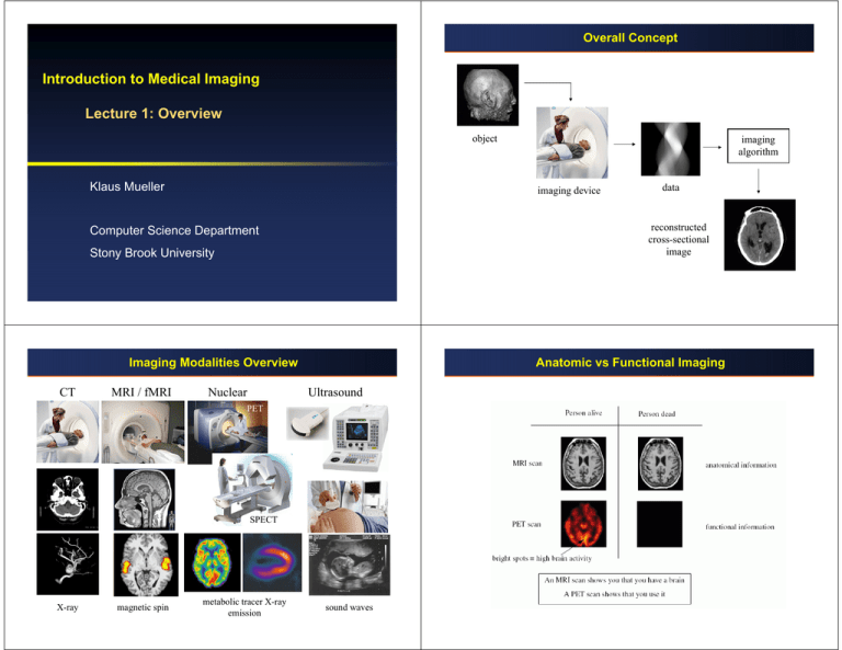
Overall Concept Introduction to Medical Imaging Lecture 1: Overview object Klaus Mueller imaging device Stony Brook University Imaging Modalities Overview MRI / fMRI Nuclear Anatomic vs Functional Imaging Ultrasound PET SPECT X-ray magnetic spin metabolic tracer X-ray emission data reconstructed cross-sectional image Computer Science Department CT imaging algorithm sound waves History: X-Rays Wilhelm Conrad Röntgen • 8 November 1895: discovers X-rays. • 22 November 1895: X-rays Mrs. Röntgen’s hand. • 1901: receives first Nobel Prize in physics An early X-ray imaging system: History: Computed Tomography The breakthrough: • acquiring many projections around the object enables the reconstruction of the 3D object (or a cross-sectional 2D slice) CT reconstruction pioneers: • 1917: Johann Radon establishes the • • • Note: so far all we can see is a projection across the patient: Computed Tomography: Concept mathematical framework for tomography, now called the Radon transform. 1963: Allan Cormack publishes mathematical analysis of tomographic image reconstruction, unaware of Radon’s work. 1972: Godfrey Hounsfield develops first CT system, unaware of either Radon or Cormack’s work, develops his own reconstruction method. 1979 Hounsfield and Cormack receive the Nobel Prize in Physiology or Medicine. Radon Cormack Hounsfield Computed Tomography: Past and Present Image from the Siemens Siretom CT scanner, ca. 1975 • 128x128 matrix. Modern CT image acquired with a Siemens scanner • 512x512 matrix Slice Viewer 3D Visualization Reconstructed object enables: • Enhanced X-ray visualization from novel views: • Maximum Intensity (MIP) visualization: • Shaded object display: More Visualizations Aortic Stent and Arterial Vessels Cartotid Stenosis Virtual Medicine Virtual colonoscopy, endoscopy, arthroscopy Virtual therapy and surgery planning Training platform History: Ultrasound Ultrasound: Present 1942: Dr. Karl Theodore Dussik, • transmission ultrasound investigation of the brain 1955: Holmes and Howry • Subject submerged in water tank to achieve good acoustic coupling image of normal neck 3D Ultrasound 1959: Automatic scanner, Glasgow Intravasular ultrasound twin gestation sacs (s) and bladder (B). Doppler ultrasound History: MRI 1946: Felix Bloch (Stanford) and Edward Purcell (Harvard) demonstrate nuclear magnetic resonance (NMR) MRI Concept MRI measures the effects of magnetic properties of tissue • these effects are tissue-specific • also specific to blood perfusion / oxygenization (functional MRI) MRI is very versatile (but also more expensive than CT) Bloch Purcell Lauterbur 1973: Paul Lauterbur (Stony Brook University) published first MRI (Magnetic Resonance Imaging) image in Nature. • receives the Nobel Prize in Physiology or Medicine in 2003 T1-weighted density-weighted T2-weighted Late 1970’s: First human MRI images conceived Early 1980’s: First commercial MRI systems available slice viewer 1993: Functional MRI in humans demonstrated MRI Applications Cardiac MRI • measures the distortion of “tags” to assess motion of the heart tissue Diffusion Tensor Imaging • measures the diffusion of water • allows the tracking of nerve fibers in the brain (white matter) MRI Applications Functional MRI • allows to assess brain activity during certain tasks • valuable for brain functional studies, but also for surgery planning and diagnosis MRI Applications MR Spectroscopy • measures the distribution of chemicals in each “voxel” of the brain MRI Applications MR Angiography • magnetizes the bolus of blood, enhances vessels • similar effects to X-ray angiography, but non-invasive MR angiography MRI Applications X-ray angiography Credits MR Microscopy • can resolve volumes of down to 50 mm3 (clincial MR does 1mm3) • use for small animal experiments (in place of distructive histology) Most historical data and some images were taken from a similar presentation by Dr. Thomas Liu, UC San Diego Other images are due to (list not complete): • Joe Kniss, U Utah • Gordon Kindlmann, U Utah • Markus Hadwiger, VRVis • Stefan Bruckner, U Vienna • Naeem Shareef, Ohio State U • Viatronix, Inc. • Phillips Medical


