Novel trimethyl lock based enzyme switch for the self
advertisement

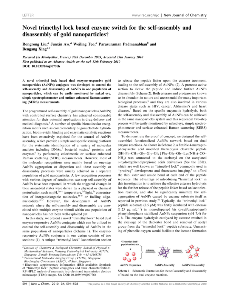
LETTER www.rsc.org/njc | New Journal of Chemistry Novel trimethyl lock based enzyme switch for the self-assembly and disassembly of gold nanoparticlesw Rongrong Liu,a Junxin Aw,a Weiling Teo,a Parasuraman Padmanabhanb and Bengang Xing*a Received (in Montpellier, France) 20th December 2009, Accepted 25th January 2010 First published as an Advance Article on the web 12th February 2010 DOI: 10.1039/b9nj00776h A novel trimethyl lock based dual enzyme-responsive gold nanoparticles (AuNPs) conjugate was developed to control the self-assembly and disassembly of AuNPs in one population of nanoparticles, which can be easily monitored by naked eye, simple spectrophotometer and surface enhanced Raman scattering (SERS) measurements. The programmed self-assembly of gold nanoparticles (AuNPs) with controlled surface chemistry has attracted considerable attention for their potential applications in drug delivery and medical diagnosis.1 A number of specific biomolecular recognition motifs such as complementary oligonucleotide hybridization, biotin-avidin binding and enzymatic catalytic reactions have been extensively exploited for the control of AuNPs assembly, which provide a simple and specific sensing platform for the systematic identification of a variety of molecular analytes including DNAs,2 bacterial toxins,3 proteins and enzymes4 by performing colorimetric or surface enhanced Raman scattering (SERS) measurements. However, most of the molecular recognitions were mainly based on one-step AuNPs aggregation or dispersion and these assembly or disassembly processes were usually achieved in a separate population of gold nanoparticles. A few recognition processes with various degrees of continuous two-step self-assembly of AuNPs have been reported, in which the triggered changes in their assembled states were driven by a physical or chemical perturbation such as pH,5a–c temperature,5d light,5e concentration of inorganic/organic molecules,5f–h or fuelling oligonucleotides.5i–l However, the development of AuNPs network where the self-assembly and disassembly are associated with multiple enzyme stimuli within one population of nanoparticles has not been well-exploited yet. In this study, we present a novel ‘‘trimethyl lock’’ based dual enzyme-responsive AuNPs conjugate which can be utilized to control the self-assembly and disassembly of AuNPs in the same population of nanoparticles (Scheme 1). The enzymeresponsive AuNPs conjugate in our design consists of two sections: (1). A unique ‘‘trimethyl lock’’ lactonization section to release the peptide linker upon the esterase treatment, leading to the self-assembly of AuNPs; (2). A protease active section to cleave the peptide and induce further AuNPs disassembly (Scheme 2). Both esterase and protease are known to be abundant in nature and are essential for many important biological processes,6 and they are also involved in various disease states such as HIV, cancer, Alzheimer’s and heart diseases.7 Based on the specific enzymatic hydrolysis, both the self-assembly and disassembly of AuNPs can be achieved in the same nanoparticles system and this sequential two-step process will be easily monitored by naked eye, simple spectrophotometer and surface enhanced Raman scattering (SERS) measurements. To demonstrate the proof of concept, we designed the selfassembled functionalized AuNPs network based on dual enzyme reactions. As shown in Scheme 2, a flexible 4-mercaptophenylacetic acid modified thermolysin cleavable peptide (SH–Ph–CH2–Gly–Gly–GlykPhe–Gly–Gly–Lys(NH2)–CO– NH2) was connected to the carboxyl on the acetylated o-hydroxyphenolpropionic acids derivatives (See the ESIw), which are well known as ‘‘trimethyl lock’’ commonly used in ‘‘prodrug’’ development and fluorescent imaging,8 to afford the thiol ester and amide bond at each end of the peptide sequence. The advantage of introducing ‘‘trimethyl lock’’ in this investigation is to achieve the effective esterase hydrolysis for the further release of the peptide linker based on lactonization reaction, and also to significantly minimize the selfaggregation of AuNPs caused by enzyme substrate itself as reported in previous study.4d Typically, the ‘‘trimethyl lock’’ peptide substrate (8.5 mM) was firstly incubated with esterase (1.25 mg mL 1) in monodispersed bis (p-sulfonatophenyl) phenylphosphane stabilized AuNPs suspension (pH 7.4) for 2 h. The enzyme hydrolysis catalyzed by esterase resulted in the cleavage of the thiolester bond and removal of acetyl group from the ‘‘trimethyl lock’’ peptide substrate. Unmasking of phenolic oxygen would facilitate the lactone formation a Division of Chemistry & Biological Chemistry, School of Physical & Mathematical Sciences, Nanyang Technological University, 637371, Singapore. E-mail: Bengang@ntu.edu.sg; Tel: +65-63168758 b Translational Molecular Imaging Group (TMIG), Singapore Bio-Imaging Consortium (SBIC), A*Star, Singapore w Electronic supplementary information (ESI) available: Synthesis of ‘‘trimethyl lock’’ peptide conjugates and their characterizations. RP-HPLC analysis of enzymatic hydrolysis and transmission electron microscope (TEM) images. See DOI: 10.1039/b9nj00776h 594 | New J. Chem., 2010, 34, 594–598 This journal is c Scheme 1 Schematic illustration for the self-assembly and disassembly of based on the dual enzyme reactions. The Royal Society of Chemistry and the Centre National de la Recherche Scientifique 2010 Fig. 1 Color image (a) and UV/Vis spectrum (b) for the self-assembly and disassembly of AuNPs induced by the enzyme reactions. State 1: AuNPs with ‘‘trimethyl lock’’ peptide substrate (8.5 mM) only; State 2: AuNPs with esterase (1.25 mg mL 1) treated substrate (8.5 mM); State 3: Aggregated AuNPs in state 2 treated with thermolysin (60 mg mL 1). Scheme 2 Mechanism illustrating the self-assembly and disassembly of AuNPs by ‘‘trimethyl lock’’ peptide substrate, catalyzed by esterase and protease enzymes. with concomitant release of peptide moiety previously attached to the carboxyl groups. The free thiol and positively charged amino group at each end of released peptide initiated the formation of aggregated AuNPs clusters through Au–S bond and electrostatic interactions with the functional groups on the surface of bis (p-sulfonatophenyl) phenylphosphane stabilized AuNPs,9 thus resulting in the significant color change from red to blue. Meanwhile, both an increased absorption band at 600 nm and a decreased absorption at 520 nm were observed in the UV-Vis spectrum (Fig. 1, state 2). As a control, the incubation of AuNPs with only intact ‘‘trimethyl lock’’ peptide substrate could not induce any further color change and spectral shifts (Fig. 1, state 1), indicating the substrate was stable and the self-assembly of AuNPs was mainly from the esterase reaction. The state of AuNPs was observed to be highly aggregated with higher concentration of esterase and the absorbance reached a plateau when the enzyme concentration was higher than 1 mg mL 1 (Fig. 2(a)). The minimum concentration of esterase that could induce the AuNPs self-assembly was found to be 18.5 ng mL 1. In order to examine whether the second enzyme could disassemble the aggregated AuNPs, the aggregated AuNPs were further treated with protease thermolysin (60 mg mL 1) at 37 1C for 4 h. The amide bond between Gly and Phe in the peptide sequence which was connecting to the AuNPs would be selectively recognized and cleaved. The AuNPs aggregation was interrupted and the disassembly of AuNPs resulted in the color change from blue to red and the maximum plasmon resonance peak shifted back from 600 nm to 520 nm (Fig. 1, state 3). The results demonstrated that addition of thermolysin could induce the redispersion of aggregated AuNPs in the solution. This disassembled process could be easily achieved with the thermolysin enzyme concentration as low as 34.1 ng mL 1. The hydrolysis of ‘‘trimethyl lock’’ peptide substrate catalyzed by esterase or thermolysin was monitored by RP-HPLC (see the ESIw). All the results confirmed the active site of This journal is c peptide substrate towards enzyme cleavage and the continuous AuNPs color change in one population of nanoparticles was from the specific enzyme recognitions. This process can be influenced by the concentration of trimethyl lock peptide substrate (see the ESIw). Further evidence of the AuNPs self-assembly and disassembly based on the dual enzyme reactions was obtained from dynamic light scattering (DLS). As shown in Fig. 3, the ‘‘trimethyl lock’’ peptide substrate itself was unable to induce the assembly of the AuNPs and the hydrodynamic size of the monodispersed AuNPs was determined to be 9.6 nm (Fig. 3(a)). Upon the esterase treatment, the specific lactonization of the ‘‘trimethyl lock’’ triggered the release of thiol and amino groups at each end of the peptide, which initiated the distinctive self-assembly of AuNPs. The nanoparticles Fig. 2 (a) Esterase concentration versus absorbance at 600 nm of AuNPs aggregation after addition of esterase treated ‘‘trimethyl lock’’ conjugate. (b) Thermolysin concentration versus absorbance at 520 nm of AuNPs redispersion upon hydrolysis of peptide linker. The Royal Society of Chemistry and the Centre National de la Recherche Scientifique 2010 New J. Chem., 2010, 34, 594–598 | 595 aggregated into larger clusters with the hydrodynamic diameter increasing to 153.2 nm (Fig. 3(b)). Following the addition of thermolysin into the assembled AuNPs for 4 h, DLS showed a population of well-dispersed AuNPs with the average size reducing to 14.6 nm, indicating the disassembly of AuNPs based on the protease cleavage. Compared to the monodispersed AuNPs, the slight size increasing in the disassembled particles revealed the presence of cleaved peptide fragment on the surface of redispersed AuNPs (Fig. 3(c)). Transmission electron microscope (TEM) measurements were also performed to determine the different self-assembly and disassembly states of AuNPs (See the ESIw). The data revealed the dual enzymatic reactions were crucial for the continuous assembly and disassembly of AuNPs, which is consistent with the results in DLS and previous measurements. It has been well known that resonant surface plasmon excitation of the free electrons in metal nanostructures can enhance localized electromagnetic fields around the surface. This enhancement effect becomes particularly strong in the interstitial spaces of aggregated nanoparticles, which is very suitable as platform for surface enhanced Raman scattering (SERS) studies.2b,c In order to monitor the self-assembly and Fig. 3 The hydrodynamic size and size distribution of AuNPs. (a), AuNPs with ‘‘trimethyl lock’’ peptide conjugate (8.5 mM); (b), AuNPs with esterase (1.25 mg mL 1) treated ‘‘trimethyl lock’’ peptide conjugate (8.5 mM); (c), Aggregated AuNPs with thermolysin treatment (60 mg mL 1). 596 | New J. Chem., 2010, 34, 594–598 This journal is c Fig. 4 Raman spectra of (a) 10 1 M dipotassium bis (p-sulfonatophenyl) phenylphosphine dihydrate solution; (b) AuNPs stabilized with dipotassium bis (p-sulfonatophenyl) phenylphosphane; (c) Stabilized AuNPs with esterase treated substrate and (d) Aggregated AuNPs treated with thermolysin, Laser wavelength: 633 nm. disassembly processes of AuNPs induced by the multiple enzyme interactions, the SERS enhancement measurements were performed directly by using stabilizer, dipotassium bis (p-sulfonatophenyl) phenylphosphine dihydrate as molecular reporter. As depicted in Fig. 4(b), the stabilized AuNPs without esterase treatment did not have plasmonic coupling and SERS signals from these dispersed particles were very weak. However, intense signals were detected from AuNPs aggregation upon the addition of ‘‘trimethyl lock’’ peptide conjugate treated with esterase (Fig. 4(c)). The observed SERS signals included the n (CH) at 765 cm 1, d (CH) at 1098 cm 1, and n (CC) at 1603 cm 1 which were in accordance with characteristic bands of stabilizer in aqueous solution (Fig. 4(a)).10 After further treatment with thermolysin, the peptide sequence in the aggregate was cleaved and the aggregated AuNPs went back to the dispersed state and no SERS signals could be observed as shown in Fig. 4(d). In conclusion, we have demonstrated a unique design for the control of self-assembly and disassembly of AuNPs on the basis of dual enzyme reactions in one population of nanoparticles. This process is illustrated by a series of enzymatic reactions in which randomly dispersed AuNPs are first converted into an aggregated structure, and subsequently to a redispersed state upon different enzyme treatments. Based on the consecutive color change from red to blue and finally to red again, visualization of the self-assembly and disassembly of AuNPs in the same nanoparticles system can be observed by the naked eye, simple spectrotrophometer and surface enhanced Raman scattering (SERS) measurements. With more sophisticated design of the substrate or peptide sequence, this strategy could also be readily extended to other enzymatic systems. This sequential two-step assembly and disassembly process initiated by specific enzyme reactions can help us to understand the mechanisms of biomolecular recognitions. It may also have the potential to serve as a valuable platform for multiple enzyme detection and drug screening in biological studies or clinical settings. The authors gratefully acknowledge the start-up, URC (RG56/06), A*Star BMRC (07/1/22/19/534) and SEP (RG139/06) grants in Nanyang Technological University, Singapore. The Royal Society of Chemistry and the Centre National de la Recherche Scientifique 2010 Experimental Synthesis of ‘‘trimethyl lock’’ peptide substrate To a stirred suspension of acetylated ‘‘trimethyl lock’’ compound (15.6 mg, 0.06 mmol) in 200 mL anhydrous dichloromethane, thionyl chloride (43 mL, 0.59 mmol) was added dropwise at 0 1C (ice bath). After addition, the reaction mixture was allowed to warm to room temperature and stirred for 18 h. The solvent was evaporated in vacuum to give the oil product without purification. Then, the purified thermolysin cleavable peptide (SH–Ph–CH2–Gly–Gly–Gly–Phe–Gly–Gly– Lys(NH2)–CO–NH2) (8.6 mg, 0.012 mmol) and triethylamine (13 mL, 0.093 mmol) in 150 mL anhydrous DMF were added into the oil product under nitrogen atmosphere and the reaction mixture was stirred for 12 h. After removing DMF, the crude product was purified by reversed-phase semipreparative HPLC with 20%–80% water–acetonitrile gradient system containing 0.1% TFA to give the final product 7.4 mg (yield 53.1%). ESI-MS Found [M + H+]: 1220.63, [M + Na+]: 1242.85; calculated 1219.6. Colorimetric assay for enzyme hydrolysis of ‘‘trimethyl lock’’ substrate To the solution of 0.4 mL of monodispersed dipotassium bis (p-sulfonatophenyl) phenylphosphane stabilized AuNPs (10 nm) (See the ESIw),9 the peptide substrate and esterase in 0.05 mL of PBS buffer (pH 7.4) were added to afford their final concentrations of 8.5 mM and 1.25 mg mL 1, respectively. The mixture was incubated at 37 1C for enzyme hydrolysis 2 h. Gold nanoparticles were induced to highly aggregated clusters which displayed purple-blue color and surface plasmon resonance shifts from 520 nm to 600 nm. Then, the aggregated gold nanoparticles were further incubated with thermolysin (60 mg ml 1) for 4 h, the aggregated gold clusters were driven disassembly, which displayed red color and surface plasmon resonance shifts back from 600 nm to 520 nm again. This selfassembly and disassembly process were monitored by naked eyes and UV-Vis spectrotrophometer. Dynamic light scattering (DLS) measurements for AuNPs Dynamic light scattering (DLS) measurements were performed using a 90 Plus particle size analyzer (Brookhaven Instruments Corporation). The DLS instrument was operated at 25 1C, 90 degree detector angle with an incident laser wavelength of 660 nm. Size and size distribution of gold nanoparticles were determined in solution. All the samples were measured for 3 min, and the reported values are the average of five repeated consecutive measurements. Surface enhanced Raman Scattering (SERS) measurements Dipotassium bis (p-sulfonatophenyl) phenylphosphane stabilized gold nanoparticles solution dropped on glass slides (approximately 10 mL) was used for SERS measurement. The spectra were excited using 3.5 mW of power at He–Ne 633 nm laser. The laser beam was then focused onto the sample via a dichroic mirror and through an Olympus This journal is c 0.90 NA microscope objective. Raman signals were collected and focused into a 400 mm optical fibre (Ocean Optics, Inc.) which delivered the signals to a single-stage monochromator (DoongWo, Inc.). Spectrum acquisition was started and an integration time is 40 s for all SERS measurements. References 1 (a) D. Peer, J. M. Karp, S. Hong, O. C. Farokhzad and R. Margalit R. Langer, Nat. Nanotechnol., 2007, 2, 751; (b) A. C. Templeton, M. P. Wuelfing and R. W. Murray, Acc. Chem. Res., 2000, 33, 27; (c) Y. C. Cao, R. Jin and C. A. Mirkin, Science, 2002, 297, 1536; (d) E. Katz and I. Willner, Angew. Chem., Int. Ed., 2004, 43, 6042; (e) X. H. Huang, P. K. Jain, I. H. El-Sayed and M. A. El-Sayed, Nanomedicine, 2007, 2, 681; (f) H. W. Gu, P. L. Ho, E. Tong, L. Wang and B. Xu, Nano Lett., 2003, 3, 1261. 2 (a) C. M. Niemeyer, Angew. Chem., Int. Ed., 2001, 40, 4128; (b) X. M. Qian, X. Zhou and S. M. Nie, J. Am. Chem. Soc., 2008, 130, 14934; (c) Y. Lu, J. Liu and J. Liu, Acc. Chem. Res., 2007, 40, 315; (d) J. W. Liu, Z. H. Cao and Y. Lu, Chem. Rev., 2009, 109, 1948; (e) X. Xue, F. Wang and X. G. Liu, J. Am. Chem. Soc., 2008, 130, 3244; (f) S. P. Song, Z. Q. Liang, J. Zhang, L. H. Wang, G. X. Li and C. H. Fan, Angew. Chem., Int. Ed., 2009, 48, 8670; (g) C. D. Medley, J. E. Smith, Z. W. Tang, Y. R. Wu, S. Bamrungsap and W. H. Tan, Anal. Chem., 2008, 80, 1067. 3 C. L. Schofield, R. A. Field and D. A. Russell, Anal. Chem., 2007, 79, 1356. 4 (a) S. Chah, M. R. Hammond and R. N. Zare, Chem. Biol., 2005, 12, 323; (b) J. M. de la Fuente and S. Penadés, Biochim. Biophys. Acta, Gen. Subj., 2006, 1760, 636; (c) Z. Wang, R. Levy, D. G. Fernig and M. Brust, J. Am. Chem. Soc., 2006, 128, 2214; (d) C. Guarise, L. Pasquato, V. De. Filippis and P. Scrimin, Proc. Natl. Acad. Sci. U. S. A., 2006, 103, 3978; (e) A. Laromaine, L. Koh, M. Murugesan, R. V. Ulijn and M. M. Stevens, J. Am. Chem. Soc., 2007, 129, 4156; (f) Y. D. Choi, N. H. Ho and C. H. Tung, Angew. Chem., Int. Ed., 2007, 46, 707; (g) J. Oishi, Y. Asami, T. Mori, J. H. Kang, M. Tanabe, T. Niidome and Y. Katayama, ChemBioChem, 2007, 8, 875; (h) R. R. Liu, R. Liew, J. Zhou and B. G. Xing, Angew. Chem., Int. Ed., 2007, 46, 8799; (i) T. T. Jiang, R. R. Liu, X. F. Huang, H. J. Feng, W. L. Teo and B. G. Xing, Chem. Commun., 2009, 1972; (j) M. Wang, X. G. Gu, G. X. Zhang, D. Q. Zhang and D. B. Zhu, Langmuir, 2009, 25, 2504; (k) Y. L. Wang, D. Li, W. Ren, Z. J. Liu, S. J. Dong and E. K. Wang, Chem. Commun., 2008, 2520. 5 (a) Y. H. Jung, K. B. Lee, Y. G. Kim and I. S. Choi, Angew. Chem., Int. Ed., 2006, 45, 5960; (b) Y. Chen and C. D. Mao, Small, 2008, 4, 2191; (c) J. Sharma, R. Chhabra, H. Yan and Y. Liu, Chem. Commun., 2007, 477; (d) C. A. Mirkin, R. L. Letsinger, R. C. Mucic and J. J. Storhoff, Nature, 1996, 382, 607; (e) H. C. Huang, P. Koria, S. M. Parker, L. Selby, Z. Megeed and K. Rege, Langmuir, 2008, 24, 14139; (f) S. Si, M. Raula, T. K. Paira and T. K. Mandal, ChemPhysChem, 2008, 9, 1578; (g) C. Guarise, L. Pasquato and P. Scrimin, Langmuir, 2005, 21, 5537; (h) H. Otsuka, Y. Akiyama, Y. Nagasaki and K. Kataoka, J. Am. Chem. Soc., 2001, 123, 8226; (i) P. Hazarika, B. Ceyhan and C. M. Niemeyer, Angew. Chem., Int. Ed., 2004, 43, 6469; (j) I. S. Lim, U. Chandrachud, L. Y. Wang, S. Gal and C. J. Zhong, Anal. Chem., 2008, 80, 6038; (k) A. G. Kanaras, Z. X. Wang, A. D. Bates, R. Cosstick and M. Brust, Angew. Chem., Int. Ed., 2003, 42, 191; (l) T. Peng, C. Dohno and K. Nakatani, ChemBioChem, 2007, 8, 483. 6 (a) B. F. Cravatt and E. Sorensen, Curr. Opin. Chem. Biol., 2000, 4, 663; (b) F. Lecaille, J. Kaleta and D. Bromme, Chem. Rev., 2002, 102, 4459. 7 S. Darvesh, D. A. Hopkins and C. Geula, Nat. Rev. Neurosci., 2003, 4, 131. 8 (a) M. G. Nicolaou, C. S. Yuan and R. T. Borchardt, J. Org. Chem., 1996, 61, 8636; (b) R. B. Greenwald, Y. H. Choe, C. D. Conover, K. Shum, D. Wu and M. Royzen, J. Med. Chem., 2000, 43, 475; (c) K. Achilles, Arch. Pharm., 2001, 334, 209; The Royal Society of Chemistry and the Centre National de la Recherche Scientifique 2010 New J. Chem., 2010, 34, 594–598 | 597 (d) S. S. Chandran, K. A. Dickson and R. T. Rains, J. Am. Chem. Soc., 2005, 127, 1652; (e) R. W. Watkins, L. D. Lavis, V. M. Kung, G. V. Los and R. T. Raines, Org. Biomol. Chem., 2009, 7, 3969; (f) B. Wang, S. Gangwar, G. M. Pauletti, T. J. Siahaan and R. T. Borchardt, J. Org. Chem., 1997, 62, 1363; 598 | New J. Chem., 2010, 34, 594–598 This journal is c (g) K. L. Amsberry, A. E. Gerstenberger and R. T. Borchardt, Pharm. Res., 1991, 8, 455. 9 C. J. Loweth, W. B. Caldwell, X. Peng, A. P. Alivisatos and P. G. Schultz, Angew. Chem., Int. Ed., 1999, 38, 1808. 10 F. Zimmermann and A. Wokaun, Mol. Phys., 1991, 73, 959. The Royal Society of Chemistry and the Centre National de la Recherche Scientifique 2010


