Definitions of Arrhythmia’s
Detected by Acuity® Version 7.x
Mortara Instrument’s VERITASTM
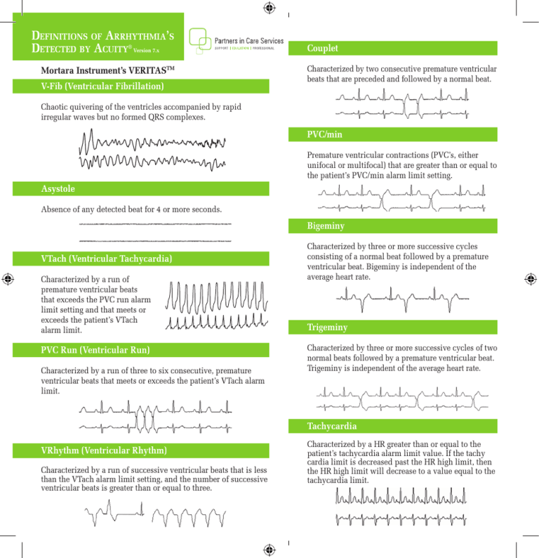
V-Fib (Ventricular Fibrillation)
Couplet
Characterized by two consecutive premature ventricular
beats that are preceded and followed by a normal beat.
Chaotic quivering of the ventricles accompanied by rapid
irregular waves but no formed QRS complexes.
PVC/min
Premature ventricular contractions (PVC’s, either
unifocal or multifocal) that are greater than or equal to
the patient’s PVC/min alarm limit setting.
Asystole
Absence of any detected beat for 4 or more seconds.
Bigeminy
VTach (Ventricular Tachycardia)
Characterized by a run of
premature ventricular beats
that exceeds the PVC run alarm
limit setting and that meets or
exceeds the patient’s VTach
alarm limit.
PVC Run (Ventricular Run)
Characterized by a run of three to six consecutive, premature
ventricular beats that meets or exceeds the patient’s VTach alarm
limit.
Characterized by three or more successive cycles
consisting of a normal beat followed by a premature
ventricular beat. Bigeminy is independent of the
average heart rate.
Trigeminy
Characterized by three or more successive cycles of two
normal beats followed by a premature ventricular beat.
Trigeminy is independent of the average heart rate.
Tachycardia
VRhythm (Ventricular Rhythm)
Characterized by a run of successive ventricular beats that is less
than the VTach alarm limit setting, and the number of successive
ventricular beats is greater than or equal to three.
Characterized by a HR greater than or equal to the
patient’s tachycardia alarm limit value. If the tachy
cardia limit is decreased past the HR high limit, then
the HR high limit will decrease to a value equal to the
tachycardia limit.
Bradycardia
Characterized by a HR less than or equal to the
patient’s bradycardia alarm limit value. If the brady
cardia limit is increased past the HR low limit, then
the HR low limit will increase to a value equal to the
bradycardia limit.
Pause
The R-to-R interval that is greater than or equal to two
times the average R-to-R.
Irregular (Irregular Rhythm)
An irregularity in the R-R interval over a series of at
least 16 non-ventricular beats.
NonCapture (Pacemaker NonCapture)
For pacemaker patients with the Analyze Pacers
option enabled, a beat does not directly follow a
pacer.
Heart Rate and Arrhythmia
For adult and pediatric patients being monitored for arrhythmia, the HR value at the Acuity is
derived from Acuity Arrhythmia Analysis instead of from the patient monitor. Thus for these
patients, the HR value at the monitor may differ from the HR value at the Central Station.
ECG Lead Selection
• In order for Arrhythmia Analysis to take place, an ECG waveform must be displayed in the
ECG 1 location of the Virtual Monitor. If ECG1 is not displayed, arrhythmia analysis will not
be performed. If monitoring with Propaq® LTR or Micropaq®, Acuity will use ECG leads II,
V and III for arrhythmia analysis. If monitoring with Propaq® CS and/or Propaq Encore®,
Acuity uses the leads that the user selects for ECG1 and/or ECG2. Be sure that lead II and
Lead V and if applicable, Lead III has significant amplitude to optimize arrhythmia analysis.
For example, if the Lead II, V or III does not have an average amplitude >200 μV peak-topeak, Acuity may not detect certain arrhythmias, including VFib.
• If false arrhythmia alarms are occurring due to a patient’s unique beat morphology, and if you
are using a 5-lead cable, you can direct Acuity to analyze arrhythmias using one reliable lead
under the Arrhythmia Alarms Setup window.
WARNING: If you turn on Single ECG in response to false lethal arrhythmia alarming (for
example, due to bundle branch block or irregular rate), arrhythmia analysis is limited to one lead.
Typically, 3-lead analysis (via a 5-lead cable) is optimal.
• The ECG cable and electrodes should be checked for damage on a regular basis and replaced
as necessary.
• Lead preparation and placement should be carefully verified
• Consider using stress loops especially for ambulatory patients.
• Tall P and T waves may be incorrectly classified as a QRS complex or PVC and potentially
generate a high heart rate or other alarm condition.
• If biphasic QRS complexes appear, a different monitoring lead should be selected.
• Review the quick reference card: Preparing the Patient for Successful Monitoring.
Paced Patients and Arrhythmia
Warning: Always turn ON Analyze Pacers for paced patients, and always turn OFF Analyze
Pacers for non-paced patients. The default for Analyze Pacers is set to OFF. Therefore, Acuity will
analyze arrhythmias as if the patient is non-paced unless turned ON by the clinician. Once you
have turned the Analyze Pacers Setting ON, this will automatically set the Pacer Display to ON
and will initiate an ST/Arr Relearn
1. In the Arrhythmia Alarms Setup Window, check the Analyze Pacers check box.
2. Set Non Capture to HIGH, MEDIUM, or LOW for Acuity to monitor for a Non Capture
Arrhythmia Event.
Relearn
• The Relearn function enables the clinician to instruct Acuity to relearn a patient’s rhythm
based on the patient’s dominant beat morphology. During a Relearn phase, a Relearn alert will
appear at the device and on the screen at Acuity.
• The Relearn alert presents itself under the following conditions: After a patient connection
or system restart, after some lead changes or lead failures, and/or after ST/Arr Relearn or
Analyze Pacers is clicked in the Arrhythmia Alarms Setup Window.
• During the learning period, Acuity indicates only the VFib and Asystole arrhythmia
conditions. Other vital signs are unaffected.
• Inappropriate use of Relearn can lead to mislabeling of beats and possibly a failure to alarm.
Carefully evaluate the patient’s current rhythm to make sure that you want the Acuity
System to establish it as the patient’s normal sinus rhythm. Periods of noise, artifact, pacer
poison and other alarm conditions may significantly affect the Relearn function. Choose the
best monitoring lead and allow a sufficient time period for stabilization (normally 30 to 40
seconds).
• An appropriate time to use Relearn is when a patient is admitted with a ventricular rhythm
that the Acuity system learns as the patient’s “normal” rhythm. When the patient’s rhythm
converts to the rhythm that is truly normal for that patient, Relearn should be initiated.
In this way, the Acuity system will alarm appropriately if the patient converts back to the
previous ventricular rhythm. Note: The Check Leads alert may appear if this condition
occurs.
8500 SW Creekside Place • Beaverton, OR 97008
1-800-289-2501
©2010, Welch Allyn, Inc. All rights reserved. 08/2010 SM2813 Rev. A

