Recovery of consciousness after brain injury
advertisement
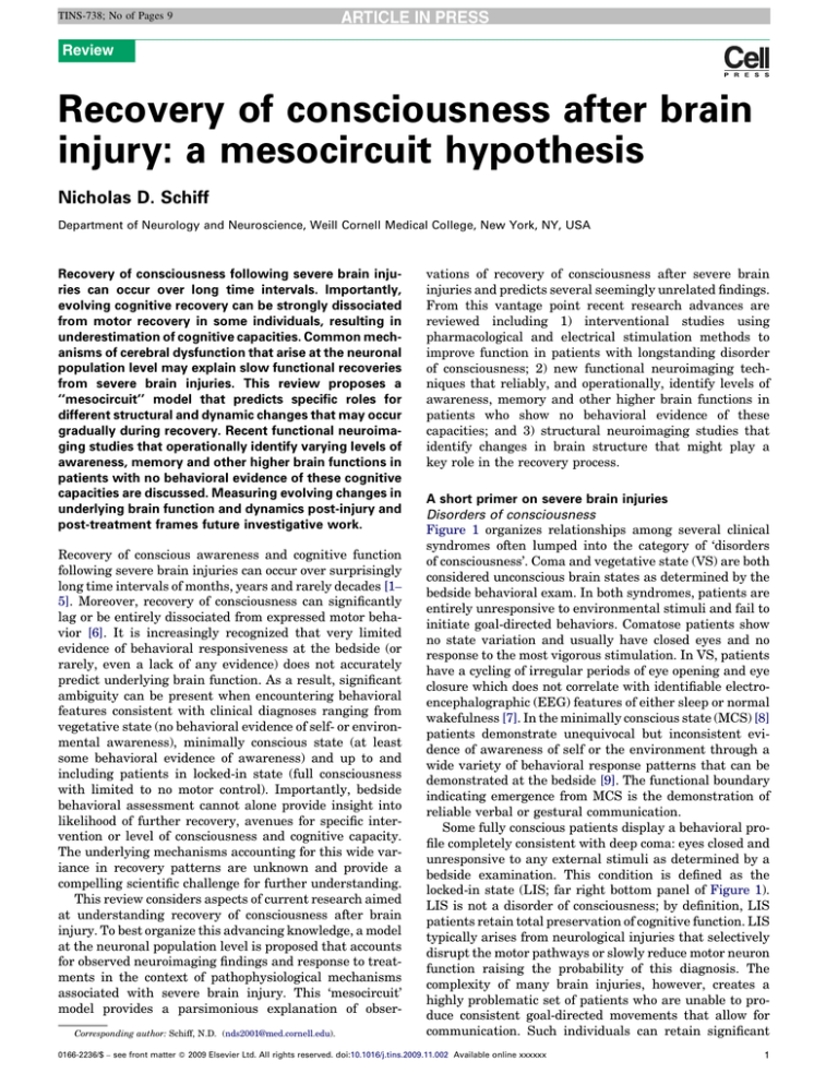
TINS-738; No of Pages 9 Review Recovery of consciousness after brain injury: a mesocircuit hypothesis Nicholas D. Schiff Department of Neurology and Neuroscience, Weill Cornell Medical College, New York, NY, USA Recovery of consciousness following severe brain injuries can occur over long time intervals. Importantly, evolving cognitive recovery can be strongly dissociated from motor recovery in some individuals, resulting in underestimation of cognitive capacities. Common mechanisms of cerebral dysfunction that arise at the neuronal population level may explain slow functional recoveries from severe brain injuries. This review proposes a ‘‘mesocircuit’’ model that predicts specific roles for different structural and dynamic changes that may occur gradually during recovery. Recent functional neuroimaging studies that operationally identify varying levels of awareness, memory and other higher brain functions in patients with no behavioral evidence of these cognitive capacities are discussed. Measuring evolving changes in underlying brain function and dynamics post-injury and post-treatment frames future investigative work. Recovery of conscious awareness and cognitive function following severe brain injuries can occur over surprisingly long time intervals of months, years and rarely decades [1– 5]. Moreover, recovery of consciousness can significantly lag or be entirely dissociated from expressed motor behavior [6]. It is increasingly recognized that very limited evidence of behavioral responsiveness at the bedside (or rarely, even a lack of any evidence) does not accurately predict underlying brain function. As a result, significant ambiguity can be present when encountering behavioral features consistent with clinical diagnoses ranging from vegetative state (no behavioral evidence of self- or environmental awareness), minimally conscious state (at least some behavioral evidence of awareness) and up to and including patients in locked-in state (full consciousness with limited to no motor control). Importantly, bedside behavioral assessment cannot alone provide insight into likelihood of further recovery, avenues for specific intervention or level of consciousness and cognitive capacity. The underlying mechanisms accounting for this wide variance in recovery patterns are unknown and provide a compelling scientific challenge for further understanding. This review considers aspects of current research aimed at understanding recovery of consciousness after brain injury. To best organize this advancing knowledge, a model at the neuronal population level is proposed that accounts for observed neuroimaging findings and response to treatments in the context of pathophysiological mechanisms associated with severe brain injury. This ‘mesocircuit’ model provides a parsimonious explanation of obserCorresponding author: Schiff, N.D. (nds2001@med.cornell.edu). vations of recovery of consciousness after severe brain injuries and predicts several seemingly unrelated findings. From this vantage point recent research advances are reviewed including 1) interventional studies using pharmacological and electrical stimulation methods to improve function in patients with longstanding disorder of consciousness; 2) new functional neuroimaging techniques that reliably, and operationally, identify levels of awareness, memory and other higher brain functions in patients who show no behavioral evidence of these capacities; and 3) structural neuroimaging studies that identify changes in brain structure that might play a key role in the recovery process. A short primer on severe brain injuries Disorders of consciousness Figure 1 organizes relationships among several clinical syndromes often lumped into the category of ‘disorders of consciousness’. Coma and vegetative state (VS) are both considered unconscious brain states as determined by the bedside behavioral exam. In both syndromes, patients are entirely unresponsive to environmental stimuli and fail to initiate goal-directed behaviors. Comatose patients show no state variation and usually have closed eyes and no response to the most vigorous stimulation. In VS, patients have a cycling of irregular periods of eye opening and eye closure which does not correlate with identifiable electroencephalographic (EEG) features of either sleep or normal wakefulness [7]. In the minimally conscious state (MCS) [8] patients demonstrate unequivocal but inconsistent evidence of awareness of self or the environment through a wide variety of behavioral response patterns that can be demonstrated at the bedside [9]. The functional boundary indicating emergence from MCS is the demonstration of reliable verbal or gestural communication. Some fully conscious patients display a behavioral profile completely consistent with deep coma: eyes closed and unresponsive to any external stimuli as determined by a bedside examination. This condition is defined as the locked-in state (LIS; far right bottom panel of Figure 1). LIS is not a disorder of consciousness; by definition, LIS patients retain total preservation of cognitive function. LIS typically arises from neurological injuries that selectively disrupt the motor pathways or slowly reduce motor neuron function raising the probability of this diagnosis. The complexity of many brain injuries, however, creates a highly problematic set of patients who are unable to produce consistent goal-directed movements that allow for communication. Such individuals can retain significant 0166-2236/$ – see front matter ß 2009 Elsevier Ltd. All rights reserved. doi:10.1016/j.tins.2009.11.002 Available online xxxxxx 1 TINS-738; No of Pages 9 Review Trends in Neurosciences Vol.xxx No.x Figure 1. Correspondence of cognitive and motor impairment associated with disorders of consciousness arising following severe brain injuries. The distinctions among clinical disorders of consciousness can be best captured on a two-dimensional axis by comparing degree of impaired cognitive function against degree of motor function. At the bottom left panel, the functional equivalence of coma and vegetative state (VS) as unconscious brain states is indicated by their placement to the left of the vertical dotted line indicating total loss of cognitive function. The large gray box is placed to denote the high degree of uncertainty associated with identifying the cognitive capacities of patients with no controllable motor output channel whose clinical bedside examination can range from minimally conscious state (MCS) to locked-in state (LIS). Note the asterisk (*) indicates that the locked-in state is not a disorder of consciousness and LIS patients retain normal cognitive function by definition. Establishing a true cognitive level for many patients who behaviorally cannot reliably signal through controlled goal-directed movements (dashed horizontal line) is not possible at present. Such patients can retain varying levels of cognitive processing capabilities, awareness, memory and other higher brain functions without detection. Disentangling the potential for cognitive function in setting of severe limitations of motor control and sensorimotor integration mechanisms is among the most important challenges presented by new understanding of the recovery process following severe brain injuries. cognitive capacity near the normal range of cognitive function and yet be indistinguishable from MCS patients. Pathological findings in disorders of consciousness following severe brain injury Anatomic pathologies associated with vegetative state, MCS and severe to moderate cognitive disability following severe injuries have several common features. Autopsy studies of both traumatic and non-traumatic injuries resulting in permanent VS (a prognostic assessment rather than diagnosis, see Ref. [10]) identify widespread neuronal death throughout the thalamus in patients [11]. Importantly, the evident severe bilateral thalamic damage after either trauma or anoxia in permanent VS is not invariably associated with diffuse neocortical neuronal cell death. Moreover, the observation indicates the key functional role for the thalamus for integrative function of the forebrain corticothalamic systems. Recent studies have shown that specific subnuclei of the thalamus demonstrate greater neuronal cell loss as a result of such global and multi-focal cerebral injuries [12]. The nuclei within the central thalamus (the intralaminar nuclei and related paralaminar nuclei) are most involved typically and the degree of neuronal loss observed within these neuronal aggregates grades with outcome [12]. In patients with only moderate disability following severe traumatic brain injury, neuronal loss is primarily identified within the anterior intralaminar nuclei (central lateral nucleus, central medial and paracentralis). Patients with progressively severe disabilities demonstrate neuronal loss involving more ventral and lateral nuclei of the central thalamus (posterior intralaminar 2 group) as illustrated in Figure 2a. These observations are probably a consequence of the unique geometry of connections of the central thalamus. Neurons in these subnuclei have wide point to point connectivity across the cerebral hemisphere and are thus likely to integrate neuronal cell death across these large territories [13,14]. Importantly, the same selected thalamic subpopulations are known to produce global disorders of consciousness (coma, VS and MCS) following bilateral focal injuries [16,17]. Figure 2b illustrates the overlap of the neuronal populations that undergo progressive deafferentation with increasingly severe multi-focal brain injuries and those typically involved in strokes producing initial coma and variable periods of VS and MCS [16,17]. As a consequence of diffuse brain injuries, considerable impact of either focal injury or deafferentation of these central thalamic neurons on forebrain function probably reflects their key contribution to normal mechanisms of arousal regulation [18]. Moreover, deafferentation and dysfunction of these neurons probably plays an important role in producing deficits even when injuries are not severe enough to produce broad neuronal death. Functional specializations of the central thalamus Neuroimaging and electrophysiological studies demonstrate the selective activation of the central thalamus for tasks that require a short-term shift of attention [19–21], sustained cognitive demands of high vigilance [21] or holding information in memory over extended time periods [20,22]. The central thalamus is uniquely situated to support these broad ‘executive’ functions in the forebrain. Central thalamic neurons are strongly innervated TINS-738; No of Pages 9 Review Trends in Neurosciences Vol.xxx No.x Figure 2. Comparison of regions of central thalamus involved in focal and diffuse injuries producing global impairments of consciousness. (a) Regional neuronal cell loss in central thalamus following severe traumatic brain injuries categorized by functional outcomes [12]. Moderately disabled patients have cell loss restricted to the anterior intralaminar regions (red circle). Severely disabled patients have neuronal loss in more caudal and medial components of the central thalamus including the medial aspects of the posterior intralaminar nuclei. Permanent VS is associated with broad loss of central thalamic neurons including the large lateral component of the posterior intralaminar group (the centromedian nucleus) [11,12,16,17]. (b) Focal injury patterns in the central thalamus associated with coma, vegetative state and minimally conscious state (adapted from Ref. [15]). Red circle indicates anterior intralaminar nuclei and surrounding regions, green circle includes area of red circle and more caudal and medial components of the posterior intralaminar region. The figure element of thalamic anatomy is adapted, with permission, from Ref. [15]. by ascending projections from the brainstem/basal forebrain ‘arousal systems’ that control the activity of many cortical and thalamic neurons during the sleep–wake cycle and descending projections from frontal cortical systems that organize goal-directed behaviors and adjust the level of arousal associated with generalized alertness and variations in cognitive effort, stress, sleep deprivation and other variables affecting the wakeful state [14,19,21,23,24] (reviewed in Ref. [18]). The neurons within the central thalamus are further specialized, anatomically and physiologically, by their diffuse projections to supragranular layers of the cerebral cortex [14,25–29] and striatal neurons [30–32]. Both the anterior (CL, Pc) and posterior intralaminar nuclei (centromedian–parafascicular complex, Cm–Pf) and the mesencephalic reticular neurons that monosynaptically project to these neurons [33] activate during the short-term shifting of attention component of a forewarned reaction time task [19]. Activity in the central thalamus covaries with the anterior cingulate cortex (ACC) as well as the pontomesencephalon [21]. The ACC is similarly recruited by a wide range of cognitive demands and shows graded activity with increasing cognitive load, suggesting that this component of the frontal executive systems can drive, or reciprocally increase activity along with, the central thalamus in response to increasing demands of cognitive effort [23,24,34]. The central thalamus and the frontal lobe are closely linked through their direct corticothalamic connections, including supplementary motor, anterior cingular, premotor and prefrontal cortex [35], and indirect links through the frontal cortical–striatopallidal–thalamocortical loop systems [14,25]. Behavioral fluctuations following central thalamic and frontal lobe injuries show strong quantitative and qualitative similarities in experimental behavioral lesion studies in rodents [36]. Similarly, notable fluctuations of behavioral response arise from both direct injuries to the central thalamus (either unilateral [37] or bilateral lesions [38]) and very closely resemble the typical behavioral fluctuations seen in patients and animals with frontal lobe lesions [39]. Linking timeframes of recovery following severe brain injury to underlying pathophysiological mechanisms To date, the majority of longitudinal studies of recovery of consciousness after severe brain injury have focused on metrics that seek to predict the likelihood that a person will not recover past the VS after an initial coma (see Refs [40,41] for reviews of the literature). This focus can be understood in light of important concerns of resource allocation in intensive care units and the high probability of death or permanent VS in coma following cardiac arrest or very severe traumatic brain injuries [41]. However, as immediate in-the-field care for patients with all types of severe brain injury has improved, increasingly large numbers of individuals not only survive their injuries but preserve correspondingly larger numbers of neurons and neuronal connections after the initial injury. Well-established statistics guide the likelihood of permanent VS over time following some patterns of injury [10]. However, similar attempts to link time periods to outcome in patients who demonstrate the limited recovery patterns associated with MCS have shown a poor correlation of long-term outcome with comparable timeframes [1,42]. Most patients who demonstrate evidence of recovery 3 TINS-738; No of Pages 9 Review to MCS within the first 3 months after injuries will recover past MCS by 10 months. Two to five year outcomes can include recovery past the level of severe disability even for patients who remain in MCS for greater than 6 months or a year. Rare cases that demonstrate endpoints of very late recovery from MCS are documented including reemergence of higher functional levels of spoken conversation, autobiographical memory and motor control after years and even decades [2,43]. In part, the differences in timeframes for recovery reflect the differences in underlying pathology present in MCS and related outcomes of severe disability following brain injuries (compared with permanent VS). In these conditions, a mix of effects of neuronal death, deafferentation and dysfunction of remaining neuronal populations play a larger and considerably less well-characterized role. The observations of late recovery from MCS indicate that brain networks can retain functional capacity without expression leaving an important possibility that marked changes in cognitive function can occur without bedside evidence either spontaneously or in response to interventions [2,6]. Role of changes in brain structure in the recovery process Recent studies provide evidence that late recovery of function following severe brain injury can involve structural changes within the brain. Structural magnetic resonance imaging (MRI) studies of a man who at age 40 years spontaneously recovered full expressive and receptive language, after remaining in MCS for 19 years following a severe traumatic brain injury suffered in a motor vehicle accident, revealed evidence of ongoing structural modifications [2]. Using diffusion tensor magnetic resonance imaging (DTI), a technique that quantifies the anisotropy of proton diffusion and thus is a proxy for axonal fiber integrity, extensive white matter injury and cerebral atrophy was noted in brainstem and frontal lobes. Despite evidence of widespread white matter injury, a longitudinal DTI study of the brain identified regions that showed significant change over time. Notably, the midline cerebellar white matter showed increased fractional anisotropy in a second study that correlated with clinical improvements in motor control. In a recent prospective cohort study of severely brain-injured patients followed for a year following initial injury, similar changes in DTI measured fractional anisotropy were identified in the patients who recovered neurological function [44]. In the aggregate, these observations suggest that the normal recovery process also includes a component of structural remodeling that could plausibly relate to reestablishment of goaldirected behaviors and driving of learning and memory mechanisms. However, why such processes might arise at late intervals or not at all requires an examination of potential mechanisms underlying large-scale changes in forebrain dynamics following severe injuries. A ‘mesocircuit’ hypothesis As reviewed above, regularities in the anatomic pathology of different types of severe brain injury suggest that largescale forebrain dysfunction can arise as a result of at least 4 Trends in Neurosciences Vol.xxx No.x three general mechanisms: 1) widespread death of forebrain neurons (i.e. sufficient to produce brain death or permanent VS); 2) widespread deafferentation and disconnection of neurons; and 3) ‘‘circuit’’ level functional disturbances owing to the loss of these neuronal connections [3,18,43,45]. Although the first mechanism is clearly irreversible, some evidence (as reviewed above) suggests that late structural alterations in the brain can arise, altering the effects of the second mechanism. Alterations at the third ‘‘mesocircuit’’ [46] level can arise as a result of global decreases of excitatory neurotransmission producing overall changes in cerebral background activity levels (as produced by anesthesia or direct effects on the function of certain cell types; e.g. hypoxia, see discussion below). Figure 3 illustrates a key vulnerability of the anterior forebrain in the setting of widespread deafferentation and neuronal cell loss that could represent the common denominator in disturbances of consciousness in severe brain injuries. The primary result of disturbances of this network could be to effectively produce a broad decrease in background synaptic activity and excitatory neurotransmission (e.g. diffuse axonal injury, anoxia, hypoxia–ischemia, multi-focal infarction following cerebral vasospasm, encephalitis, etc.; see Ref. [41]). At a neuronal subpopulation level, the medium spiny neurons (MSNs) of the striatum have a key role in maintaining activity in the anterior forebrain through their inhibitory projections to the globus pallidus interna which in turn inhibits the central thalamus [30,47]. Activation of MSN projections, de facto, results in a disinhibition of central thalamic neurons, reestablishes the outflow of thalamocortical transmission and probably promotes a rebound of high frequency thalamocortical activity [48]. The thalamocortical projections from the central thalamus strongly innervate the frontal cortex and have in some instances a joint thalamostriatal projection back to the MSNs [30]; recent studies demonstrate that thalamocortical projections to cortex have a stronger impact on driving excitation within the cortex than cortico–cortical projections [49] and downregulation of thalamic output can be expected to have broad effects across cortical regions. Neurons from the central thalamus (both central lateral nucleus and parafascicular nucleus) strongly project to the MSNs [31] and diffusely innervate the striatum [30]. These thalamostriatal projections use glutamate transmitter proteins with a high probability of synaptic release [32] and could have a strong role in modulating background activity in the striatum. The MSNs have a ‘high threshold’ UP state that keeps them below their firing threshold unless sufficient levels of dopamine neuromodulation are present and there is a high level of spontaneous background synaptic activity arising from excitatory corticostriatal and thalamostriatal inputs [47]. Thus, diffuse brain injuries can lead to a sharp reduction of MSN output as diffuse deafferentation produces withdrawal of both direct excitatory striatal projections from the central thalamus and downregulation of the frontocortical regions that provide the main corticostriatal input. Among frontal cortical regions, the anterior cingulate cortex can play an essential role as it receives strong inputs for the anterior intralaminar nuclei (central lateral nucleus, [35]) and provides a TINS-738; No of Pages 9 Review Trends in Neurosciences Vol.xxx No.x output of the MSNs or direct modulation of mesial frontal cortical neurons would explain the restoration of anterior forebrain activity within the loop connections of the frontal cortex, striatum, pallidum and central thalamus. Figure 3. Proposed ‘‘mesocircuit’’ model underlying forebrain dysfunction and interventions in severe brain injuries. A proposed ‘mesocircuit’ that explains the vulnerability of the anterior forebrain (frontal/prefrontal cortical–striatopallidal– thalamocortical loop systems) following multi-focal brain injuries that produce widespread deafferentation or neuronal cell loss. The thalamocortical projections of the central thalamus are proposed to play an important role in observed reduction of cerebral metabolism in this mesocircuit following different mechanisms of brain injury [43,51]; these projections have a strong activating role driving both cortical and striatal neurons [31,32,49]. The medium spiny neurons (MSNs) of the striatum which send inhibitory projections to the globus pallidus interna (GPi) require high levels of background synaptic activity and dopaminergic neuromodulation to maintain firing rates [47]. Without MSN output the GPi tonically inhibits the central thalamus potentially catalyzing a shutdown of the anterior forebrain. Downregulation of activity within the mesocircuit is predicted to have a broad modulatory impact on the global dynamics of the dominant corticothalamic system [26,27,29,33,48]; specific changes within the cortico–striatopallidal–thalamocortical system identified with alterations of consciousness associated with sleep and anesthesia support this inference [58,61,62,65]. The mesocircuit model also economically accounts for the mix of interventions that have been noted in some patients to restore functions associated with these forebrain systems (e.g. dopaminergic agents, zolpidem and electrical brain stimulation; see text for further discussion). Zolpidem-induced paradoxical arousal in severe brain injury Of particular interest, the mesocircuit model also offers an explanation of a surprising and puzzlingly paradoxical phenomenon recently described that zolpidem (a nonbenzodiazepine hypnotic that potentiates GABAA receptors, also known as ‘Ambien’) can improve alertness and behavioral responsiveness in some severely brain-injured patients [43,55–58]. Brefel-Courbon et al. [43] reported an MCS patient who recovered spoken language, eating and ambulation with zolpidem administration. Figure 4 shows a marked increase in anterior forebrain metabolism associated with zolpidem-administered condition compared with the patient’s off drug state. Similar observations in another zolpidem responsive patient [58] link increases of cerebral metabolism in the frontal cortex, striatum and thalamus to changes in the shape of the spectral content of the EEG (removing abnormal low frequency component) and the coherence architecture (reducing marked low frequency coherence in the off drug state). In accordance with the model in Figure 3, Schiff and Posner [45] proposed the following mechanism for this paradoxical response. Under normal circumstances, the MSNs disinhibit the central thalamus via the globus pallidus interna (GPi; Figure 3). Thus, when MSN activity is reduced as a consequence of brain injury, central thalamic activity is also reduced. Because zolpidem directly inhibits the GPi, it can substitute for the normal inhibition of the GPi from MSNs, and very diffuse regulatory input across large territories of the rostral striatum [50]. Implications of the mesocircuit model for recovery of consciousness after severe brain injury The mesocircuit model presented in Figure 3 organizes and rationalizes recent observations of the response of severely brain-injured subjects to pharmacological and electrophysiological interventions as well as some aspects of normal brain function, as reviewed below. The primary implication of the model in Figure 3 is that frontocortico– striatopallidal–thalamocortical loop frontal systems are selectively vulnerable at the ‘circuit’ level in many types of multi-focal brain injury. This accounts for the observations that selective metabolic depression of the anterior forebrain specifically grades with severity of behavioral impairment following diffuse axonal injury [51]. In addition, the well-known response to dopaminergic agents of severely brain-injured patients with markedly slowed behavioral response following either mesial frontal lobe, basal forebrain or thalamic/midbrain injuries is consistent with the mesocircuit model [52,53]. Behavioral features of these patients range from extreme poverty of movement (‘akinetic mutism’’) to severe disability characterized by very slow but nonetheless accurate responses that allow communication [38,54]. Dopaminergic facilitation of the Figure 4. Changes in cerebral metabolism associated with zolpidem administration in severe brain injury. Fluorodeoxyglucose positron emission tomography studies by Brefel-Courbon et al. [43] of a severe brain-injured patient in minimally conscious state before and after administration of the sedative agent zolpidem (‘Ambien’). In the off drug state (top panels) marked anterior forebrain hypometabolism is noted bilaterally in frontal/prefrontal cortex, thalami and striatum. Following zolpidem administration broad increases of metabolic rates are observed in these regions. Image adapted from [43] with permission. 5 TINS-738; No of Pages 9 Review thus permit a more normal level of central thalamic activity. The GABAA a-1 subunit is expressed in large quantities in the GPi and experimental studies support this mechanism of action [59]. Of note, the MSNs are uniquely vulnerable to cellular dysfunction after hypoxia [60] and several of the reported cases of paradoxical response have followed hypoxic–ischemic injuries [43,55– 58]. In addition to accounting for the paradoxical response to zolpidem, the mesocircuit model provides a plausible framework for related observations in normal subjects. Of note, the model provides an explanation for the observation that the most robust changes in regional cerebral blood flow during the transitions during the sleep–wake cycle are in the striatum [61]. Specifically, increases during the transition from slow wave sleep to rapid eye movement sleep (REM) and decreases in the transition from wakefulness to non-REM sleep. Similar, slightly less significant changes also occur in the ‘centrencephalic’ components of the thalamus and cerebral cortex [61]. The ‘circuit breaker’ effect of withdrawal of cortical and thalamic excitation from the MSN suggests an economical explanation for this otherwise puzzling contribution of the striatum. Similar recovery patterns in metabolic activity of the anterior forebrain are observed during early wakefulness as sleep inertia dissipates [62]. The model suggests reactivation of the frontostriatal systems during sleep states could also provide an explanation for a variety of reports of unusual behaviors (somnambulism, amnestic hyperphagia–nocturnal binge eating without memory trace) arising during sleep specifically associated zolpidem treatment [63,64]. Finally, this mesocircuit model can also account for the common finding of the early selective metabolic downregulation in the mesial frontal and thalamic systems with different anesthetics [65] and variety of specific changes across the induction and recovery from general anesthesia. Electrical stimulation of central thalamus in minimally conscious state Markedly depressed rates of global metabolism are seen in patients with MCS or severe disability [66] and can be produced either by volume loss of neurons and subsequent deafferentation of remaining cells or by neuronal functional impairments and low firing rates. The mesocircuit model shown in Figure 3 predicts that direct activation of excitatory output from the central thalamus in patients with chronically downregulated background synaptic activity following severe brain injury will tend to normalize cortico–striatopallidal–thalamocortical function. Direct activation of central thalamic neurons through electrical stimulation (‘deep brain stimulation’, DBS) has been proposed as an experimental therapeutic strategy that might produce consistent and sustained effects of maintaining the activity within this circuit [67]. A recent single-subject study demonstrated that central thalamic DBS restored arousal regulation and promoted improved behavioral responsiveness in a 38-year-old man after remaining in MCS for 6 years [68]. The patient had remained unable to communicate reliably despite neuroimaging evidence of preservation of large-scale cerebral language networks [69] that suggested a substrate for further recovery. 6 Trends in Neurosciences Vol.xxx No.x Figure 5 illustrates overall design of the study, placement of the electrodes in the central thalamus and main results [68]. Dissociation of expressed motor behavior and integrative cerebral function The need to develop better models for understanding cognitive capacity after severe brain injury is dramatically illustrated by the study of Owen et al. [6]. Via functional MRI (fMRI), these authors demonstrated high-level cognitive function in a patient behaviorally assessed to be in VS for 5 months following a severe traumatic brain injury. When asked to imagine playing tennis the patient exhibited significant fMRI measured brain activation in the supplementary motor areas and when asked to imagine walking through the rooms of her house showed activation in parahippocampal gyrus, posterior parietal cortex and the lateral premotor cortex; both patterns are consistent with those seen in normal control subjects carrying out this task [71]. These observations provide unambiguous demonstrations of command following—a cardinal sign distinguishing VS from MCS [9]. The Owen et al. study raises many important questions. The most critical question is what mechanisms might underlie the failure of the patient to exhibit goal-directed behavior despite the apparent integrity of motor pathways? Owen et al. interrogated the integrity of the patient’s motor pathways using transcranial magnetic stimulation methods and ruled out an interruption of the outflow from the motor cortex to the skeletal muscles accounting for her lack of initiated movements [6]. Collectively, the observations indicate that although this patient could follow commands, she probably remained unable to organize motor responses to carry out goal-directed intentional behaviors because of functional disturbances of forebrain systems associated with motor preparation and action. As noted above, the mesocircuit model provides a parsimonious hypothesis that can be tested to explain these findings. Notably, at the time of study the patient had collapsed regions of skull bilaterally across the frontal lobe visible in the imaging results (see Ref. [6]) demonstrating a marked impact on the frontal systems and suggesting a possible mechanism for persistent dysfunction of the anterior forebrain [72]. Clearly, better understanding of the mechanisms underlying these observations will lead to an improved ability to prospectively identify and riskstratify patients to improve the likelihood of obtaining accurate diagnoses and facilitating recovery of communication. Recent computational modeling studies provide potential insight into the functional role of these long loop frontal–striatopallidal–thalamocortical systems that appear to be selectively vulnerable to shut down after severe brain injury. Goldman [73] has shown that sequentially feedforward circuits (the striatopallidal components add such links to the standard corticothalamic circuit) provide a solution to organizing arbitrary temporal processing demands (such as flexibly reconfiguring sensorimotor contingencies) and holding this information over the long time scales associated with cognition. The feedforward architecture of connections of this model produces response TINS-738; No of Pages 9 Review Trends in Neurosciences Vol.xxx No.x Figure 5. Central thalamic deep brain stimulation (DBS) in the minimally conscious state. (a) Timeline of single-subject study of DBS in the central thalamus in a patient remaining in MCS for 6 years. (b) Location of electrode lead placements within central thalamus of patient’s right (R) and left (L) hemispheres displayed in T1 weighted MRI coronal image and indicated with red arrows. (c). Comparison of presurgical baselines and DBS ON and DBS OFF periods during a 6-month crossover trial. Behavioral baseline evaluations measured using a standardized quantitative assessment tool, the Coma Recovery Scale Revised (CRS-R) were obtained 4 months prior to surgery and for 2 months following surgery prior to titration testing of stimulation parameters (see Ref. [68] for details) showed no change in behavioral responsiveness compared with functional levels measured more than 2 years before the start of the trial. During the early titration testing of electrical stimulation the patient demonstrated immediate and accumulating effects of DBS that included the emergence of consistent and intelligible spoken language, recovery of limb control and the capacity for oral feeding (see Ref. [64] for details). Following a 5-month titration period of testing combinations of stimulation parameters, the patient entered into a blinded 6-month, 30-day alternating ON versus OFF (crossover) study that demonstrated a robust overall effect on behavioral responsiveness measured by CRS-R subscales and supplementary behavioral rating scales. Significant ON versus OFF improvements with electrical stimulation were demonstrated for attentive behavior, oral feeding and limb control. All functional testing showed significant improvements when compared against the 6-month prestimulation baselines. Importantly, observed carryover effects of improvements from the ON to the OFF state were also identified as demonstrated by the high frequency of OFF stimulation ratings compared with prestimulation baselines across all measurements. These effects are comparable to evidence of accumulating behavioral effects of central thalamic electrical stimulation as shown in a rodent model by Shirvalkar et al. [70]. Figure elements have been adapted, with permission, from Ref. [68]. profiles similar to those recorded across frontal cortex [74], striatum [75] and central thalamus [20,22] during performance of ‘executive functions’ such as sustained attention, working memory or motor preparation. This model provides an appealing first-order explanation of the central importance of the anterior forebrain mesocircuit in the global behavioral impairments observed after traumatic brain injury. Graded and variable recovery of function of the entire circuit could be the variable that best categorizes fluctuations in behavioral responsiveness. Reestablishing functional activity across these long loops over times is probably required for the minimal behavioral capacity required to advance motor behaviors beyond the MCS level. Gradually improving the integrity of normal activity patterns within the anterior forebrain could underlie the continuum of outcomes across severe disability whereby the probability of maintaining the anterior forebrain mesocircuit corresponds to different functional outcomes. to guide longitudinal assessments of brain function and 2) the development of novel interventions at the circuit and cellular level to aid recovery. A key overarching goal of these efforts is to identify the potential for communication and support this capacity. Because of the intermittent nature of behaviors in MCS, it is essential to develop tools to more accurately assess patients. Improving the consistency of observed behaviors or providing means for detecting potentially more reliable underlying neurophysiological signals that might enable consistent basic communication is important, even if cognitive capacity remains severely restricted. Fins [76] has recently argued that functional communication represents a major milestone for all patients with severe brain injury across the diagnostic spectrum, their caregivers and family members as it restores a fundamental aspect of the patient’s capacity to reengage the human community and reestablish essential aspects of personhood. Future directions Understanding the circuit mechanisms associated with phases of recovery of consciousness following severe brain injuries will open many directions for future research including 1) the development of new diagnostic tools based on neuroimaging and electrophysiological measurements Acknowledgements This paper was initially presented as a Special Lecture at the Society of Neuroscience 38th Annual Meeting on November 18, 2008. The author thanks Jonathan Victor, Shawniqua Williams, Sudhin Shah and Mary Conte. The support of the James S. McDonnell Foundation and the National Institutes of Health-National Institute of Child Health and Human Development is gratefully acknowledged. 7 TINS-738; No of Pages 9 Review References 1 Lammi, M.H. et al. (2005) The minimally conscious state and recovery potential: a follow-up study 2 to 5 years after traumatic brain injury. Arch. Phys. Med. Rehabil. 86, 746–754 2 Voss, H.U. et al. (2006) Possible axonal regrowth in late recovery from minimally conscious state. J. Clin. Invest. 116, 2005–2011 3 Burruss, J.W. and Chacko, R.C. (1999) Episodically remitting akinetic mutism following subarachnoid hemorrhage. J. Neuropsychiatry Clin. Neurosci. 11, 100–102 4 McMillan, T.M. and Herbert, C.M. (2004) Further recovery in a potential treatment withdrawal case 10 years after brain injury. Brain Inj. 18, 935–940 5 Macniven, J.A. et al. (2003) Emotional adjustment following cognitive recovery from ‘persistent vegetative state’: psychological and personal perspectives. Brain Inj. 17, 525–533 6 Owen, A.M. et al. (2006) Detecting awareness in the vegetative state. Science 313, 1402 7 Kobylarz, E.J. and Schiff, N.D. (2005) Neurophysiological correlates of persistent vegetative and minimally conscious states. Neuropsychol. Rehabil. 15, 323–332 8 Giacino, J.T. et al. (2002) The minimally conscious state: definition and diagnostic criteria. Neurology 58, 349–353 9 Giacino, J.T. and Whyte, J. (2005) The vegetative state and minimally conscious state: current knowledge and remaining questions. J. Head Trauma Rehabil. 20, 30–50 10 Jennett, B. (2002) The Vegetative State, Cambridge University Press 11 Adams, J.H. et al. (2000) The neuropathology of the vegetative state after acute insult. Brain 123, 1327–1338 12 Maxwell, W.L. et al. (2006) Thalamic nuclei after human blunt head injury. J. Neuropathol. Exp. Neurol. 65, 478–488 13 Scannell, J.W. et al. (1999) The connectional organization of the corticothalamic system of the cat. Cereb. Cortex 9, 277–299 14 Van der Werf, Y.D. et al. (2002) The intralaminar and midline nuclei of the thalamus. Anatomical and functional evidence for participation in processes of arousal and awareness. Brain Res. Brain Res. Rev. 39, 107–140 15 Munkle, M.C. et al. (2000) The distribution of calbindin, calretinin and parvalbumin immunoreactivity in the human thalamus. J. Chem. Neuroanat. 19, 155–173 16 Castaigne, P. et al. (1981) Paramedian thalamic and midbrain infarcts: clinical and neuropathological study. Ann. Neurol. 10, 127–148 17 Schiff, N.D. and Plum, F. (2000) The role of arousal and ‘gating’ systems in the neurology of impaired consciousness. J. Clin. Neurophysiol. 17, 438–452 18 Schiff, N.D. (2008) Central thalamic contributions to arousal regulation and neurological disorders of consciousness. Ann. N. Y. Acad. Sci. 1129, 105–118 19 Kinomura, S. et al. (1996) Activation by attention of the human reticular formation and thalamic intralaminar nuclei. Science 271, 512–515 20 Shah, S. et al. Modulation of arousal regulation with central thalamic deep brain stimulation. Conf. Proc. IEEE. (in press) 21 Paus, T. et al. (1997) Time-related changes in neural systems underlying attention and arousal during the performance of an auditory vigilance task. J. Cogn. Neurosci. 9, 392–408 22 Wyder, M.T. et al. (2004) Contextual modulation of central thalamic delay-period activity: representation of visual and saccadic goals. J. Neurophysiol. 91, 2628–2648 23 Paus, T. et al. (1998) Regional differences in the effects of task difficulty and motor output on blood flow response in the human anterior cingulate cortex: a review of 107 PET activation studies. Neuroreport 9, R37–R47 24 Nagai, Y. et al. (2004) Brain activity relating to the contingent negative variation: an fMRI investigation. Neuroimage 21, 1232–1241 25 Gronewegen, H. and Berendse, H. (1994) The specificity of the ‘nonspecific’ midline and intralaminar thalamic nuclei. Trends Neurosci. 17, 52–66 26 Llinas, R.R. et al. (2002) Temporal binding via cortical coincidence detection of specific and nonspecific thalamocortical inputs: a voltagedependent dye-imaging study in mouse brain slices. Proc. Natl. Acad. Sci. U. S. A. 99, 449–454 27 Jones, E.G. (2001) The thalamic matrix and thalamocortical synchrony. Trends Neurosci. 24, 595–601 8 Trends in Neurosciences Vol.xxx No.x 28 Purpura, K.P. and Schiff, N.D. (1997) The thalamic intralaminar nuclei: role in visual awareness. Neuroscientist 3, 8–14 29 Steriade, M. et al. (1993) Electrophysiological properties of intralaminar thalamocortical cells discharging rhythmic (approximately 40 HZ) spike-bursts at approximately 1000 HZ during waking and rapid eye movement sleep. Neuroscience 56, 1–9 30 Deschenes, M. et al. (1996) Striatal and cortical projections of single neurons from the central lateral thalamic nucleus in the rat. Neuroscience 72, 679–687 31 Lacey, C.J. et al. (2007) Novel and distinct operational principles of intralaminar thalamic neurons and their striatal projections. J. Neurosci. 27, 4374–4384 32 Smith, Y. et al. (2009) The thalamostriatal systems: anatomical and functional organization in normal and parkinsonian states. Brain Res. Bull. 78, 60–68 33 Steriade, M. and Glenn, L.L. (1982) Neocortical and caudate projections of intralaminar thalamic neurons and their synaptic excitation from midbrain reticular core. J. Neurophysiol. 48, 352–371 34 Stuss, D.T. and Alexander, M.P. (2007) Is there a dysexecutive syndrome? Philos. Trans. R. Soc. Lond. B Biol. Sci. 362, 901–915 35 Morel, A. et al. (2005) Divergence and convergence of thalamocortical projections to premotor and supplementary motor cortex: a multiple tracing study in the macaque monkey. Eur. J. Neurosci. 21, 1007–1029 36 Mair, R.G. et al. (1998) Lesions of the frontal cortex, hippocampus, and intralaminar thalamic nuclei have distinct effects on remembering in rats. Behav. Neurosci. 112, 772–792 37 Van Der Werf, Y.D. et al. (1999) Neuropsychological correlates of a right unilateral lacunar thalamic infarction. J. Neurol. Neurosurg. Psychiatry 66, 36–42 38 Stuss, D.T. et al. (1988) The neuropsychology of paramedian thalamic infarction. Brain Cogn. 8, 348–378 39 Robertson, I.H. et al. (1997) ‘Oops!’: performance correlates of everyday attentional failures in traumatic brain injured and normal subjects. Neuropsychologia 35, 747–758 40 Young, G.B. (2009) Clinical practice. Neurologic prognosis after cardiac arrest. N. Engl. J. Med 361, 605–611 41 Posner, J. et al. (2007) Plum and Posner’s Diagnosis of Stupor and Coma, (4th edn), Oxford University Press 42 Giacino, J.T. and Kalmar, K. (1997) The vegetative and minimally conscious states: a comparison of clinical features and functional outcome. J. Head Trauma Rehabil. 12, 36–51 43 Brefel-Courbon, C. et al. (2007) Clinical and imaging evidence of zolpidem effect in hypoxic encephalopathy. Ann. Neurol. 62, 102–105 44 Sidaros, A. et al. (2008) Diffusion tensor imaging during recovery from severe traumatic brain injury and relation to clinical outcome: a longitudinal study. Brain 131, 559–572 45 Schiff, N.D. and Posner, J.P. (2007) Another ‘‘Awakenings’’. Ann. Neurol. 62, 5–7 46 Bohland, J.W. et al. (2009) A proposal for a coordinated effort for the determination of brainwide neuroanatomical connectivity in model organisms at a mesoscopic scale. PLoS Comput. Biol. 5, e1000334 47 Grillner, S. et al. (2005) Mechanisms for selection of basic motor programs—roles for the striatum and pallidum. Trends Neurosci. 28, 364–370 48 Llinás, R.R. and Steriade, M. (2006) Bursting of thalamic neurons and states of vigilance. J. Neurophysiol. 95, 3297–3308 49 Rigas, P. and Castro-Alamancos, M.A. (2007) Thalamocortical up states: differential effects of intrinsic and extrinsic cortical inputs on persistent activity. J. Neurosci. 27, 4261–4272 50 Calzavara, R., Mailly, P. and Haber, S.N. (2007) Relationship between the corticostriatal terminals from areas 9 and 46, and those from area 8A, dorsal and rostral premotor cortex and area 24c: an anatomical substrate for cognition to action. Eur. J. Neurosci. 26, 2005–2024 51 Kato, T. et al. (2007) Statistical image analysis of cerebral glucose metabolism in patients with cognitive impairment following diffuse traumatic brain injury. J. Neurotrauma 24, 919–926 52 Matsuda, W. et al. (2003) Awakenings from persistent vegetative state: report of three cases with parkinsonism and brain stem lesions on MRI. J. Neurol. Neurosurg. Psychiatry 74, 1571–1573 53 Meythaler, J.M. et al. (2002) Amantadine to improve neurorecovery in traumatic brain injury-associated diffuse axonal injury: a pilot doubleblind randomized trial. J. Head Trauma Rehabil. 17, 300–313 TINS-738; No of Pages 9 Review 54 Katz, D.I. et al. (1987) Dementia following strokes in the mesencephalon and diencephalon. Arch. Neurol. 44, 1127–1133 55 Whyte, J. and Myers, R. (2009) Incidence of clinically significant responses to zolpidem among patients with disorders of consciousness: a preliminary placebo controlled trial. Am. J. Phys. Med. Rehabil. 88, 410–418 56 Shames, J.L. and Ring, H. (2008) Transient reversal of anoxic brain injury-related minimally conscious state after zolpidem administration: a case report. Arch. Phys. Med. Rehabil. 89, 386–388 57 Cohen, S.I. and Duong, T.T. (2008) Increased arousal in a patient with anoxic brain injury after administration of zolpidem. Am. J. Phys. Med. Rehabil. 87, 229–231 58 Williams, S. et al. (2009) Quantitative neurophysiologic characterization of a paradoxical response to zolpidem in a severely brain-injured human subject. Abstr. Soc. Neurosci. (in press) 59 Chen, L. et al. (2004) Electrophysiological and behavioral effects of zolpidem in rat globus pallidus. Exp. Neurol. 186, 212–220 60 Calabresi, P. et al. (2000) Cellular factors controlling neuronal vulnerability in the brain: a lesson from the striatum. Neurology 55, 1249–1255 61 Braun, A.R. et al. (1997) Regional cerebral blood flow throughout the sleep-wake cycle. An H2(15)O PET study. Brain 120, 1173–1197 62 Balkin, T.J. et al. (2002) The process of awakening: a PET study of regional brain activity patterns mediating the re-establishment of alertness and consciousness. Brain 125, 2308–2319 63 Yang, W. et al. (2005) One rare side effect of zolpidem – sleepwalking: a case report. Arch. Phys. Med. Rehabil. 86, 1265–1266 64 Najjar, M. (2007) Zolpidem and amnestic sleep related eating disorder. Clin. Sleep Med. 3, 637–638 65 Alkire, M.T. et al. (2008) Consciousness and anesthesia. Science 322, 876–880 Trends in Neurosciences Vol.xxx No.x 66 Laureys, S. et al. (2004) Brain function in coma, vegetative state, and related disorders. Lancet Neurol. 3, 537–546 67 Schiff, N.D. and Purpura, K.P. (2002) Towards a neurophysiological basis for cognitive neuromodulation through deep brain stimulation. Thalamus Relat. Syst. 2, 55–69 68 Schiff, N.D. et al. (2007) Behavioral improvements with thalamic stimulation after severe traumatic brain injury. Nature 448, 600–603 69 Schiff, N.D. et al. (2005) fMRI reveals large-scale network activation in minimally conscious patients. Neurology 64, 514–523 70 Shirvalkar, P. et al. (2006) Cognitive enhancement with central thalamic electrical stimulation. Proc. Natl. Acad. Sci. U. S. A. 103, 17007–17012 71 Boly, M. et al. (2007) When thoughts become action: an fMRI paradigm to study volitional brain activity in non-communicative brain injured patients. Neuroimage 36, 979–992 72 Akins, P.T. and Guppy, K.H. (2008) Sinking skin flaps, paradoxical herniation, and external brain tamponade: a review of decompressive craniectomy management. Neurocrit. Care 9, 269– 276 73 Goldman, M. (2009) Memory without feedback in a neural network. Neuron 61, 621–634 74 Messinger, A. et al. (2009) Multitasking of attention and memory functions in the primate prefrontal cortex. J. Neurosci. 29, 5640–5653 75 Hikosaka, O. et al. (1989) Functional properties of monkey caudate neurons. III. Activities related to expectation of target and reward. J. Neurophysiol. 61, 814–832 76 Fins, J.J. (2009) Being conscious of their burden: severe brain injury and the two cultures challenge. Ann. N. Y. Acad. Sci. 1157, 131–147 9
