Photo Quiz
An Abnormal Chest Radiograph
MATTHEW BARNES, CAPT, MC, USAF, Lackland Air Force Base, Texas
DAVID GARCIA, CAPT, MC, USAF, Fort Belvoir Family Medicine Residency, Virginia
genetic condition that causes short stature,
broad thumbs and toes, hirsutism, and often
intellectual disabilities.
On examination, his lungs were clear to
auscultation. He had a regular heart rate
and rhythm with no murmurs. There was
no jugular venous distension. The heart
sounds were shifted to the right, and the
apical impulse was located to the right of
the sternum. There were normal pulses in
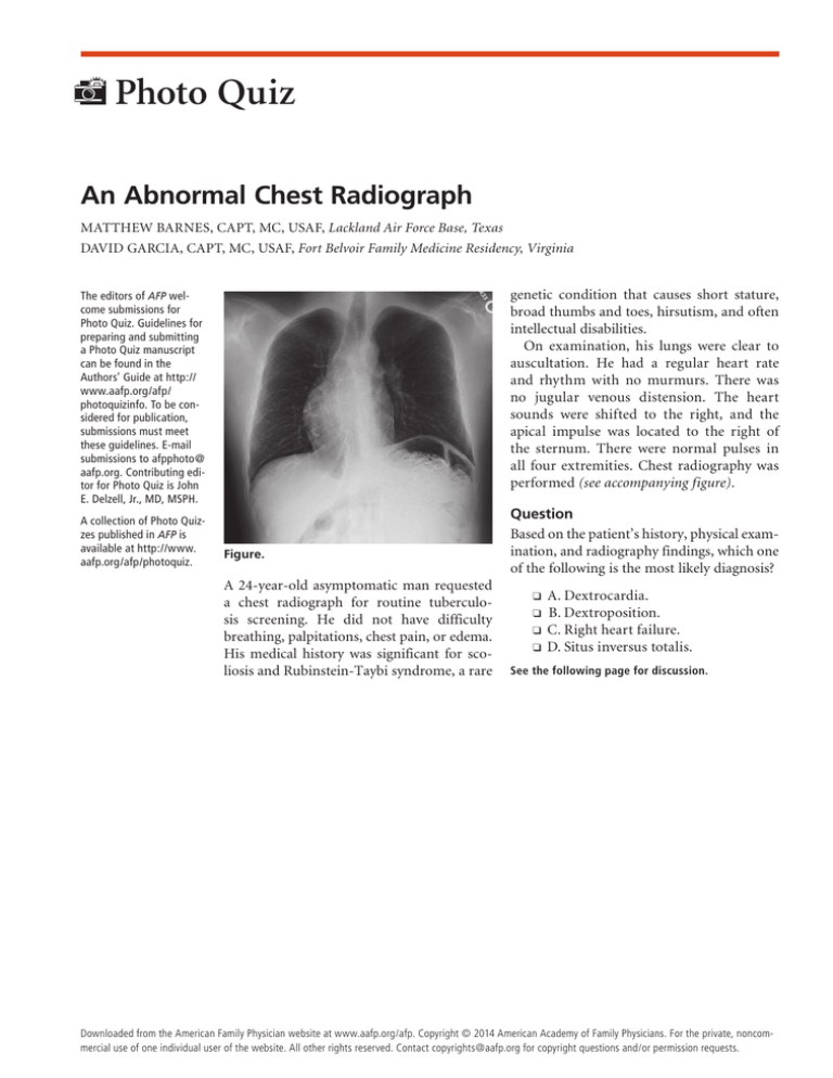
all four extremities. Chest radiography was
performed (see accompanying figure).
The editors of AFP welcome submissions for
Photo Quiz. Guidelines for
preparing and submitting
a Photo Quiz manuscript
can be found in the
Authors’ Guide at http://
www.aafp.org/afp/
photoquizinfo. To be considered for publication,
submissions must meet
these guidelines. E-mail
submissions to afpphoto@
aafp.org. Contributing editor for Photo Quiz is John
E. Delzell, Jr., MD, MSPH.
A collection of Photo Quizzes published in AFP is
available at http://www.
aafp.org/afp/photoquiz.
Figure.
A 24-year-old asymptomatic man requested
a chest radiograph for routine tuberculosis screening. He did not have difficulty
breathing, palpitations, chest pain, or edema.
His medical history was significant for scoliosis and Rubinstein-Taybi syndrome, a rare
Question
Based on the patient’s history, physical examination, and radiography findings, which one
of the following is the most likely diagnosis?
❑
❑
❑
❑
A. Dextrocardia.
B. Dextroposition.
C. Right heart failure.
D. Situs inversus totalis.
See the following page for discussion.
◆ Volume 90, Number 1
July
1, 2014from
www.aafp.org/afp
American Academy of FamilyAmerican
Physician
47
Downloaded
the American Family Physician website at www.aafp.org/afp.
Copyright © 2014
Physicians. Family
For the private,
noncommercial use of one individual user of the website. All other rights reserved. Contact copyrights@aafp.org for copyright questions and/or permission requests.
Photo Quiz
Discussion
The answer is A: dextrocardia. Dextrocardia
is the most common congenital positional
abnormality of the heart, occurring in about
one out of 12,000 persons.1,2 Dextrocardia
refers to the rightward pointing of the cardiac apex.3,4 The malpositioning of the heart
occurs during fetal development. RubinsteinTaybi syndrome is associated with cardiac
abnormalities in 32% of patients.5
Dextrocardia cannot be diagnosed from a
single chest radiograph. Further evaluation
of cardiac position and anatomy with computed tomography and echocardiography
is necessary to confirm the diagnosis. In
this patient, cardiac auscultation and palpation raised initial suspicion about cardiac
placement. In this case, further evaluation
with echocardiography demonstrated dextrocardia. This finding is incidental and not
of clinical concern, unless it is associated
with other anatomic abnormalities that may
compromise functional capability.
Dextroposition refers to a shifting of the
heart into the right hemithorax, often from
mechanical causes.6 Although congenital dextroposition may be associated with
Summary Table
Characteristics
Dextrocardia
Cardiac apex points to the right hemithorax; associated
with severe cardiac malformations
Shifting of the heart into the right hemithorax
Enlargement of the right heart; most commonly caused
by left heart failure
Mirror-image arrangement of normal organs; gastric
bubble will appear on the right side
48 American Family Physician
Address correspondence to David Garcia, CAPT, MC,
USAF, at david.s.garcia.mil@health.mil. Reprints are not
available from the authors.
Author disclosure: No relevant financial affiliations.
The opinions and assertions contained herein are the
private views of the authors and are not to be construed
as official or as reflecting the views of the U.S. Air Force
Medical Department or the U.S. Air Force at large.
REFERENCES
1.Moore KL, Arthur FD, Agur AM. Clinically Oriented
Anatomy. Philadelphia, Pa.: Lippincott Williams &
Wilkins; 2006.
2.Bohun CM, Potts JE, Casey BM, Sandor GG. A
population-based study of cardiac malformations and
outcomes associated with dextrocardia. Am J Cardiol.
2007;100(2):305-309.
3.Longo DL. Harrison’s Principles of Internal Medicine.
New York, NY: McGraw-Hill; 2012.
Condition
Dextroposition
Right heart
failure
Situs inversus
totalis
hypoplastic right lung, it also increasingly
occurs with congenital heart disease from
left to right shunts.6
Right heart failure causing enlargement of
the right heart may present with the cardiac
silhouette in the right thorax. The most
common cause of right heart failure is left
heart failure.6
Situs inversus totalis is a mirror-image
arrangement of normal organs. It is caused
by an abnormal rotation of the organs during fetal development and is associated with
dextrocardia.7 The gastric bubble will appear
on the right side.
www.aafp.org/afp
4.Wolfgang D. Radiology Review Manual. Philadelphia,
Pa.: Wolters Kluwer Health/Lippincott Williams &
Wilkins; 2011.
5.Hennekam RC. Rubinstein-Taybi syndrome. Eur J Hum
Genet. 2006;14(9):981-985.
6.Brant WE, Helms CA. Fundamentals of Diagnostic Radiology. Philadelphia, Pa.: Lippincott Williams & Wilkins;
2007.
7.Carlson BM, Kantaputra PN. Human Embryology and
Developmental Biology. 5th ed. Philadelphia, Pa.: Saunders; 2014. ■
Volume 90, Number 1
◆
July 1, 2014


