An exonic splicing silencer represses spliceosome assembly after
advertisement
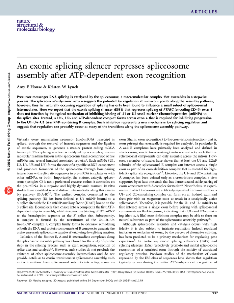
© 2006 Nature Publishing Group http://www.nature.com/nsmb ARTICLES An exonic splicing silencer represses spliceosome assembly after ATP-dependent exon recognition Amy E House & Kristen W Lynch Precursor messenger RNA splicing is catalyzed by the spliceosome, a macromolecular complex that assembles in a stepwise process. The spliceosome’s dynamic nature suggests the potential for regulation at numerous points along the assembly pathway; however, thus far, naturally occurring regulation of splicing has only been found to influence a small subset of spliceosomal intermediates. Here we report that the exonic splicing silencer (ESS1) that represses splicing of PTPRC (encoding CD45) exon 4 does not function by the typical mechanism of inhibiting binding of U1 or U2 small nuclear ribonucleoproteins (snRNPs) to the splice sites. Instead, a U1-, U2- and ATP-dependent complex forms across exon 4 that is required for inhibiting progression to the U4–U6–U5 tri-snRNP–containing B complex. Such inhibition represents a new mechanism for splicing regulation and suggests that regulation can probably occur at many of the transitions along the spliceosome assembly pathway. Virtually every mammalian precursor (pre)-mRNA transcript is spliced, through the removal of intronic sequences and the ligation of exonic sequences, to generate a mature protein-coding mRNA transcript. This splicing reaction is catalyzed by a complex, macromolecular machine known as the spliceosome that is comprised of five snRNAs and several hundred associated proteins1. Each snRNA (U1, U2, U4, U5 and U6) forms the core of a specific snRNP component and promotes formation of the spliceosome through base-pairing interactions with splice site sequences in pre-mRNA templates or with other snRNAs, or both2. Importantly, the mature, catalytic spliceosome (complex C) is not a preformed enzyme; rather, it assembles on the pre-mRNA in a stepwise and highly dynamic manner. In vitro studies have identified several distinct intermediates along this assembly pathway (E-A-B)2,3. The earliest complex committed to the splicing pathway (E) has been defined as U1 snRNP bound to a 5¢ splice site with the U2 snRNP auxiliary factor (U2AF) bound to the 3¢ splice site. E complex is then chased into A complex in the first ATPdependent step in assembly, which involves the binding of U2 snRNP to the branchpoint sequence at the 3¢ splice site. Subsequently, B complex is formed by the recruitment of the U4–U6–U5 tri-snRNP complex. C complex finally forms by extensive remodeling of both the RNA and protein components of B complex to generate the active enzymatic spliceosome capable of catalyzing the splicing reaction. Isolation of the distinct E, A and B intermediate complexes along the spliceosome assembly pathway has allowed for the study of specific steps in the splicing process, such as exon recognition, selection of splice sites and catalysis2,3. However, such studies do not preclude the existence of other spliceosome-assembly intermediates and do not provide details as to crucial transitions in spliceosome assembly, such as the transition from spliceosomal subunits interacting across an exon (that is, exon recognition) to the cross-intron interaction (that is, exon pairing) that eventually is required for catalysis3. In particular, E, A and B complexes have primarily been analyzed and defined in studies using simple two-exon/single-intron constructs, such that the spliceosomal components can only assemble across the intron. However, a number of studies have shown that at least the U1 and U2AF subunits of the spliceosomal E complex can interact across a single exon as part of an exon-definition complex that is essential for highfidelity splice site recognition4,5. Likewise, the U1- and U2-containing A complex has been defined only as a cross-intron complex, a view supported by at least one study that has demonstrated stable pairing of exons concurrent with A complex formation6. Nevertheless, in experiments in which two exons are artificially separated from one another, a U1- and U2-containing complex can form on an isolated exon and then pair with an exogenous exon to result in a catalytically active spliceosome7. Therefore, it is possible for the U1 and U2 snRNPs to first interact across a single exon before pairing with spliceosomal components on flanking exons, indicating that a U1- and U2-containing (that is, A-like) exon-definition complex may be able to form on natural substrates as part of the spliceosome assembly pathway2,3. Although spliceosome assembly and catalysis occurs with high fidelity, it is also subject to intricate regulation. Indeed, regulated inclusion or exclusion of exons, by the process of alternative splicing, has been predicted to be a primary mechanism for regulating gene expression2. In particular, exonic splicing enhancers (ESEs) and splicing silencers (ESSs) respectively promote and inhibit spliceosome recognition of a regulated exon through the activity of associated regulatory proteins. Previous studies of the mechanism of exon repression by the ESS class of sequences have shown that regulation typically occurs during the initial ATP-independent recognition of Department of Biochemistry, University of Texas Southwestern Medical Center, 5323 Harry Hines Boulevard, Dallas, Texas 75390-9038, USA. Correspondence should be addressed to K.W.L. (kristen.lynch@utsouthwestern.edu). Received 13 March; accepted 30 August; published online 24 September 2006; doi:10.1038/nsmb1149 NATURE STRUCTURAL & MOLECULAR BIOLOGY VOLUME 13 NUMBER 10 OCTOBER 2006 937 © 2006 Nature Publishing Group http://www.nature.com/nsmb ARTICLES Figure 1 ESS1 represses splicing of CD45 exon 4 alt-ESS∆E3 in vitro by stalling spliceosome assembly at an WT mESS1 alt-ESS – + – + – + : ESS1 comp unusual step. (a) RNA derived from CD45 mESS1∆E3 minigenes containing regulated exon 4 (with alt∆E3 m∆E3 : ESS1 comp – + – + WT, mESS or alt-ESS sequence) flanked by constitutive exons 3 and 7 was incubated in JSL1 nuclear extract in the absence (–) or 43 39 36 30 : % spliced 21 22 2 2 : % 3-exon 2 27 presence (+) of exogenous competitor (comp) ESS1 RNA for 110 min, and products were WT∆E3 analyzed by low-cycle RT-PCR16. Accuracy of all WT∆E3 alt-ESS∆E3 spliced products was confirmed by cloning and : Comp RNA –ESS1 +ESS1 sequencing. % 3 exon, percentage of three-exon mESS1∆E3 0 5 15 30 60 90 120 0 5 15 30 60 90 120 : Time (min) products. (b) Top, RNA derived from the WTDE3 WT∆E3 alt∆E3 m∆E3 – + – – : ESS1 comp minigene was spliced as in a. For quantification, 0 10 30 0 10 30 0 30 0 30 : Time (min) see Figure 6a. Bottom, 32P-labeled WTDE3 RNA B was incubated in JSL1 nuclear extract under B A A splicing conditions in the absence (–) or presence * * H/E H/E (+) of exogenous ESS1 competitor RNA, and resulting spliceosome assembly complexes were resolved on a nondenaturing polyacrylamide gel. Comigrating H and E complexes (H/E), A and B complexes and the stalled complex (asterisk) are labeled. In b, B complex in the presence of ESS is more diffuse at 15–60 min than in d, because we held these reactions on ice with heparin for the remainder of the extended time course before loading. (c) Splicing of RNA derived from alt-ESSDE3 (altDE3) and mESS1DE3 (mDE3) minigenes. Splicing reactions for these and all subsequent experiments were terminated at 110 minutes. (d) Spliceosome assembly gels as in b, using 32P-labeled versions of the RNAs shown. a c b d splice sites by U2AF or the U1 snRNP3,8–11. However, the inherently dynamic nature of the spliceosome suggests the potential for biologically relevant regulation of splicing at any of the intermediates along the spliceosome assembly pathway. The CD45 gene is an excellent model system for understanding the mechanism and regulation of alternative splicing events. CD45 is a transmembrane protein tyrosine phosphatase whose activity is crucial for T-cell development and signaling12. The gene encoding CD45, PTPRC (hereafter called CD45) contains three variable exons (exons 4, 5 and 6) that are alternatively spliced in human T cells, resulting in distinct isoforms that differ in activity13. Previous analysis of cis-acting elements that govern the regulation of exon 4 splicing has identified an exonic silencer element (ESS1) that is required for exon repression14,15. Moreover, at least three heterogeneous nuclear RNPs (hnRNPs) bind specifically to a core sequence within ESS1, and at least one of these, hnRNP L, functions to repress exon splicing16. However, as the mechanism by which hnRNP L functions to block inclusion of exon 4 has been unknown, we set out to identify the stage of spliceosome assembly blocked by the ESS1-binding proteins. Here we show that a spliceosomal complex that resembles A complex (dependent on U1, U2 and ATP) forms efficiently on ESS1-repressed substrates in extracts from human T cells, but subsequent spliceosome assembly is blocked. Characterization of the sequence requirements for the ESS1-stalled spliceosomal complex show that it is dependent on the integrity of both the 5¢ and 3¢ splice sites at the boundaries of the regulated exon but is not dependent on the presence of upstream or downstream flanking splice sites. Moreover, formation of this specific stalled complex is required for exon regulation. Together these data demonstrate that ESS-mediated exon repression can occur after the ATP-dependent recognition of an exon by U1 and U2, and suggest that the interaction of these two snRNPs across an exon can be regulated to prevent pairing with neighboring exons or stable recruitment of the tri-snRNP. RESULTS ESS1 stalls spliceosome assembly at an unusual step Previously we have demonstrated that a 60-nucleotide (nt) exonic splicing silencer sequence (ESS1) is necessary and sufficient for the 938 VOLUME 13 skipping of the CD45 variable exon 4 both in vivo and in vitro15,16. In vitro splicing reactions using a minigene containing the wild-type exon 4 sequence flanked by the CD45 constitutive exons 3 and 7 (Fig. 1a, WT) show a low level of exon 4 inclusion, or ‘three-exon’ product. However, upon addition of exogenous ESS1 RNA, there is a marked increase in the level of exon inclusion (Fig. 1a, +), consistent with the exogenous ESS1 titrating functionally important ESS1-binding proteins away from the pre-mRNA substrate, thereby alleviating repression. As expected from previous studies16, the ability of exogenous ESS1 to promote exon inclusion is dependent on the regulated exon containing a functional ESS1 element. Splicing of the mESS1 minigene, which contains point mutations in ESS1 that abolish silencing activity15, results in an increased basal level of exon inclusion that cannot be further enhanced by addition of competitor ESS1 RNA (Fig. 1a, mESS1). Similarly, the addition of ESS1 competitor has no effect on the splicing of a minigene in which the entire ESS1 element is removed and replaced with a heterologous sequence from CD45 exon 14 that binds hnRNP A1 and A2 (UTSWMC; A. Melton and K.W.L., unpublished data) and contains unrelated silencing activity in some contexts15,16 (Fig. 1a, alt-ESS). The exon 14 alt-ESS and mESS1 sequences also have no ability to competitively alter the splicing of the WT RNA substrate16. Together, these studies show that when ESS1 is bound by its cognate repressor proteins, it functions to inhibit splicing by preventing use of exon 4 by the spliceosome. As an initial step toward understanding the mechanism of exon repression, we wanted to determine the step in spliceosome assembly that is blocked by ESS1. We first inactivated the 5¢ splice site of the first exon (CD45 exon 3) in the WT minigene so that splicing and spliceosome assembly occurred only on the downstream intron (WTDE3). This construct thus allowed us to observe spliceosome assembly and catalysis at the regulated exon 4 only, without complications from the exon 3–exon 7 splicing pathway that occurs upon exon 4 repression in the WT construct. Splicing of the WTDE3 minigene shows ESS1-dependent regulation, in that exon 4 remains predominantly unspliced (repressed) in the absence of competitor RNA but is efficiently spliced to the downstream exon (derepressed) in the presence of exogenous ESS1 competitor RNA (Fig. 1b, top; see below for quantification). We do note that there was some NUMBER 10 OCTOBER 2006 NATURE STRUCTURAL & MOLECULAR BIOLOGY ARTICLES a b WT∆E3 WT∆E3 Mock 0 30 –U2 0 15 30 –U1 0 15 30 –U6 : snRNA inactivated 0 15 30 : Time (min) –ATP 0 15 30 * * H/E H/E 40 % stalled complex © 2006 Nature Publishing Group http://www.nature.com/nsmb c +ATP 0 15 30 : Time (min) Mock –U6 30 20 3 2 1 0 –U1 –ATP –U2 0 10 20 Figure 2 Stalled complex has the hallmarks of a canonical A complex. (a) The stalled complex is dependent on U1 and U2 snRNAs. Oligonucleotide-directed RNase H–mediated inactivation of snRNAs was used to deplete functional U1, U2 or U6 snRNPs from JSL1 nuclear extract. 32P-labeled WTDE3 RNA was incubated with either mock-treated or snRNP-depleted nuclear extract (–U1, –U2 or –U6) under conditions otherwise favorable to spliceosome assembly, and complexes were resolved as in Figure 1b. (b) The stalled complex is dependent on ATP. 32P-labeled WTDE3 RNA was incubated in JSL1 nuclear extract depleted of ATP under conditions in which both ATP and phosphocreatine were absent from the assembly reaction (–ATP) and compared to reactions containing ATP (+ATP). (c) Summary of results from assembly reactions as in a and b. Percentage of stalled complexed relative to total spliceosome complexes was calculated using densitometry. Each value is an average from four to six independent experiments; error bars indicate s.d. 30 Time (min) degradation of the RNA after 2 h of incubation in extract under both repressed and derepressed conditions. Although this instability is not unusual in extract and is independent of the regulation under study, we routinely terminated the splicing reactions just before 2 h to minimize degradation. To identify the step in the splicing of exon 4 that is blocked by ESS1, we next analyzed spliceosome assembly on WTDE3 in parallel with the in vitro splicing reactions. Resolution of splicing reactions by nondenaturing gel electrophoresis results in a well-defined pattern of shifts corresponding to sequential complexes along the assembly pathway17. Spliceosome assembly on WTDE3 progresses efficiently under derepressed (+ESS1) conditions, as A and B complexes were readily detected after a 15- to 30-min incubation with nuclear extract (Fig. 1b,d). The catalytic C complex cannot be resolved on these gels because of the large size of the template pre-mRNA; however, it must also form efficiently, as up to 50% of WTDE3 pre-mRNA is converted to spliced product under these derepressed conditions. Notably, under repressed conditions, a complex migrating at the same position as the prespliceosomal A complex (marked with asterisk) forms as rapidly and efficiently as in derepressed conditions, but no B complex is evident (Fig. 1b, bottom, –ESS1). Even after a 2-h incubation, at which point the stalled A-like complex had accumulated in large amounts on the WTDE3 substrate, little or no B complex was observed (Fig. 1b). To further determine whether the block we observed in spliceosome assembly on WTDE3 is specifically due to ESS1-mediated repression, we engineered the DE3 mutation into the mESS1 and alt-ESS substrates. The splicing of both of these constructs is not influenced by the addition of exogenous ESS1 RNA, and in this context, the alt-ESS no longer functions as a silencer (Fig. 1c). Notably, for both the alt-ESSDE3 and mESS1DE3 substrates, we observed efficient progression to B complex in the absence of ESS1 RNA (Fig. 1d; see below for confirmation of B complex assignment). Moreover, use of a nonspecific competitor RNA did not allow progression to B complex on the WTDE3 substrate (Supplementary Fig. 1 online). Together, these data demonstrate that the lack of B complex formation on WTDE3 is indeed dependent on the ESS1 repressor activity. Finally, we resolved the spliceosome assembly reactions with the WTDE3 substrate on nondenaturing agarose gels to separate H and E complexes18, and we observed no difference in the efficiency of E complex formation in the absence or presence of ESS1 competitor (data not shown). Although the ESS1-dependent stalled complex migrates at a position consistent with it being an A, or A-like, complex17,19, we set out to more rigorously characterize this complex by determining its dependence on ATP and individual snRNPs (Fig. 2). The canonical A complex is defined as the ATP-dependent association of the U1 snRNP with the 5¢ splice site on the upstream exon and the U2 snRNP bound at the 3¢ splice site branchpoint sequence of the downstream exon, in a cross-intron configuration, whereas a canonical exondefinition complex does not involve the ATP-dependent association of the U2 snRNA2. To determine the dependency of the stalled complex on specific snRNPs, oligonucleotide-directed RNase H cleavage of the U1, U2 or U6 snRNA was used to remove the region of the snRNA required for binding the pre-mRNA20,21. Primer extension of each spliceosomal snRNA showed that cleavage was specific and that greater than 90% of the target snRNA was inactivated, consistent with the diminished ability of the depleted extracts to catalyze pre-mRNA a mESS1∆E3 –U6 Mock : snRNA inactivated 0 15 30 0 15 30 : Time (min) B A H/E b Figure 3 Complex assigned as B complex has hallmarks of tri-snRNP association. (a) Spliceosome assembly reaction as in Figure 1b, using the mESS1DE3 RNA, which efficiently assembles an apparent B complex. Reactions were done in mock-depleted extracts or extracts depleted of U6 snRNA as in Figure 2a. (b) Primer-extension analysis of RNA eluted from complexes assembled either on WTDE3 RNA under derepressed conditions (+ESS1) or on mESS1DE3 RNA. Indicated complexes were excised from nondenaturing gels and RNA was extracted and analyzed with primers specific for U1, U2 and U5 snRNAs (pooled). Assignment of products is based on reactions done with individual primers using total RNA from nuclear extract (NE lanes). NATURE STRUCTURAL & MOLECULAR BIOLOGY VOLUME 13 NUMBER 10 WT∆E3 +ESS1 H/E 'A' mESS1 ∆E3 'B' 'B' NE U1 U2 U5 –U2 –U1 –U5 OCTOBER 2006 939 ARTICLES a b WT∆E7 WT∆E7 – + : ESS1 comp – + : ESS1 comp 0 15 30 0 15 30 : Time (min) B * A 15 55 : % spliced c H/E WT∆E3 was strongly enriched in the ‘B’ complex formed on both mESSDE3 and WTDE3 in the presence of ESS, with a corresponding decrease in U1 signal (Fig. 3b), consistent with our assignment of this complex. Together, the experiments shown in Figures 1–3 strongly support the idea that ESS1 blocks spliceosome assembly in the transition from an A, or A-like, complex to the tri-snRNP–associated B complex. Notably, such a ESS-mediated block is past the stage at which the vast majority of previously observed splicing regulation occurs8–11 and suggests a new mechanism for exon silencing. WT∆E7 */AEC H/E Figure 4 ESS1-dependent repression and formation of the stalled complex does not require either flanking splice site. (a) Splicing of RNA derived from the WTDE7 minigene, in which only the upstream intron is functional. Splicing was done in the absence (–) or presence (+) of exogenous competitor (comp) ESS1 RNA and quantified as in Figure 6. (b) Spliceosome assembly of [32P]WTDE7 RNA in the absence or presence of exogenous ESS1 RNA. (c) Parallel assembly on [32P]RNAs shown. Stalled complex represents an exon-defined A complex As there is little precedent for an ESS functioning to repress exon inclusion after the first ATP-dependent step in splicing, we wanted to better understand the step at which ESS1 stalls spliceosome assembly. Traditionally, A complex has been thought of as being ‘introndefined’—that is, comprising an upstream U1 snRNP interacting with a downstream U2 snRNP across the intervening intron. However, in vitro trans-splicing studies have shown that an A-like complex, containing the U1 and U2 snRNPs, can form across an isolated exon in an exon-defined manner7. To determine whether the ESS1-dependent block in splicing before B complex formation is specific for the intron downstream of exon 4 (the only usable intron in the WTDE3 construct) or is a more generalized block of exon 4 itself, we generated a version of the WT construct in which the 3¢ splice site branchpoint sequence of the downstream exon 7 is inactivated (WTDE7), so that assembly and splicing can occur only on the upstream intron. As observed for the WTDE3 construct (Fig. 1b), splicing of WTDE7 is also inhibited under normal conditions, and this inhibition is alleviated upon addition of competitor ESS1 RNA (Fig. 4a). Notably, the WTDE7 pre-mRNA shows an ESS1-dependent block at the same stage of spliceosome assembly as observed with the WTDE3 minigene (Fig. 4b). As for the WTDE3 minigene, efficient formation of the stalled complex on WTDE7 also requires the presence of ATP, U1 snRNP and U2 snRNP in the assembly reaction (data not shown). The observation that ESS1-mediated repression of spliceosome assembly occurs on both the upstream (WTDE7) and downstream (WTDE3) introns in the same manner suggests that the exon containing the ESS1 sequence is itself the unit of regulation. In other words, these data indicate that the ESS1-dependent stalled complex is an A-like complex splicing (see Supplementary Fig. 2 online). Notably, in the absence of the U1 or U2 snRNP, formation of the stalled complex was greatly reduced relative to mock-depleted extract, whereas inactivation of U6 snRNP had no effect (Fig. 2a,c). Formation of A complex also has a strict requirement for ATP, making this the first ATP-dependent step in the assembly pathway. Depletion of ATP from the assembly reactions completely abolished formation of the stalled complex (Fig. 2b). Together, the requirements of ATP and U1 and U2 snRNAs for the robust formation of the ESS1-stalled complex demonstrate that this complex has the primary hallmarks of an A-like complex. A slowly migrating complex seen under derepressed conditions does not form on the WTDE3 substrate under repressed conditions (Fig. 1b,d). We initially assigned this slowly migrating complex as the tri-snRNP–containing B complex on the basis of its migration, which was consistent with previous observations of this complex17. a ∆E3E7 c To further confirm this assignment of B Mock –U1 –U2 –U6 : snRNA inactivated 20 0 15 30 0 15 30 0 15 30 0 15 30 : Time (min) complex, we first tested whether U6 snRNA ∆E3E7 AEC is required in the formation of this complex. 15 H/E Depletion of the U6 snRNA abolished the ∆5′ slowly migrating complex on the mESSDE3 10 ∆BPS ∆E3E7 b construct, which forms this complex in large ∆5′∆E3E7 5 amounts under mock conditions (Fig. 3a). ∆BPS∆E3E7 –U1 To determine whether U5 snRNA was also ∆E3E7 ∆5′∆E3E7 ∆BPS∆E3E7 –U2 0 5 15 30 0 5 15 30 0 5 15 30 : Time (min) recruited to this slowly migrating complex, 0 10 20 30 AEC Time (min) we eluted complexes formed on mESSDE3 H/E and WTDE3 under derepressed conditions (+ESS) from assembly gels and assayed for the presence of the U1, U2 and U5 snRNAs Figure 5 Stalled complex has the hallmarks of an A-like complex assembled across an exon. 32 by primer extension. In the ‘A’ complex from (a) Assembly on [ P]RNA derived from the DE3E7 minigene, under mock-treated or snRNP-depleted conditions as in Figure 2a. (b) Spliceosome assembly on [32P]RNA derived from DE3E7 minigenes WTDE3, we detected signal for primarily the containing the indicated additional mutations. (c) Summary of efficiency of AEC formation in assembly U1 and U2 snRNAs (Fig. 3b), consistent with reactions lacking U1, U2 or the binding sites for these snRNAs, as in a and b. DE3E7 is a mockthe studies shown in Figure 2. A small treated positive control. Percentage of stalled AEC complex relative to total spliceosome complexes amount of U5 snRNA was detected in the was calculated using densitometry. Each value is an average of four to six independent experiments; ‘A’ complex; by contrast, the U5 snRNA signal error bars indicate s.d. % AEC © 2006 Nature Publishing Group http://www.nature.com/nsmb ∆E3E7 WT∆E3 WT∆E7 ∆E3E7 : pre-mRNA 0 15 30 0 15 30 0 15 30 : Time (min) 940 VOLUME 13 NUMBER 10 OCTOBER 2006 NATURE STRUCTURAL & MOLECULAR BIOLOGY ARTICLES Figure 6 Formation of an AEC is required for ESS1-dependent repression of splicing. (a) RTE34 E47 PCR analysis of in vitro splicing with three-exon WT∆E3 WT∆BPS WT∆5′ – + – + : ESS1 comp – + – + – + : ESS1 comp wild-type (WTDE3) or splice site mutant (WTDBPS, WTD5¢) minigenes. RNA derived from minigenes was incubated under splicing conditions in the absence (–) or presence (+) of exogenous ESS1 competitor (comp) RNA, as in 4 28 11 8 12 11 : % spliced 38 40 26 26 : % spliced Figure 1a. Percentage of spliced product was determined by phosphorimaging and represents the average of at least four independent experiments. s.d. for all splicing quantifications is o10% of average value shown. (b) RT-PCR analysis of in vitro splicing with single-intron constructs. RNA derived from either the E34 or E47 minigene was spliced and analyzed as in a. b AEC should circumvent ESS1-mediated splicing repression. As both the 5¢ and 3¢ splice sites are important for AEC formation (Fig. 5b,c), we wanted to determine the requirement for cross-exon interactions in ESS1-dependent splicing regulation. Quantification of the splicing of WTDE3 (as shown in Fig. 1b) reveals only B4% splicing to exon 4 under repressed conditions, but a six- to eight-fold increase in splicing efficiency upon addition of exogenous ESS1 competitor to derepress exon 4 (Fig. 6a). Notably, parallel reactions with constructs containing mutations in the splice sites surrounding exon 4 (Fig. 6a, WTDBPS or WTD5¢) showed little or no increase in splicing upon addition of exogenous ESS1 RNA, suggesting that ESS1 is not a functional repressor of constructs in which the formation of the AEC complex is inhibited. However, analysis of the WTDBPS or WTD5¢ constructs is complicated by the fact that, in an appreciable portion of the RNA, the flanking exons (gray in Fig. 6) are spliced directly together owing to the weakening of exon 4 between them (data not shown). To circumvent this problem, we also analyzed single-intron exon 4 constructs, in which only intron-defined spliceosome assembly can occur and no alternative splicing pattern is possible (Fig. 6b). Notably, neither of the single-intron E34 or E47 substrates showed any ESS1mediated repression, as addition of exogenous ESS1 competitor had no effect on splicing efficiency (Fig. 6b). Together, the requirements for the 5¢ and 3¢ splice sites of exon 4, both for the efficient formation of the stalled AEC and the regulation of splicing, indicate that the formation of an AEC is essential for ESS1-mediated exon repression. in which the U1 and U2 snRNPs bound to the exon 4 splice sites interact across the repressed exon. As such a complex is distinct from either a canonical E or A complex, we refer to this ESS-dependent stalled complex as an A-like exon-definition complex (AEC). To provide further evidence that the ESS1-stalled complex is an AEC, we wanted to determine whether a complex with the characteristics of the stalled complex can form on exon 4 in the absence of intact introns (that is, with no possibility of intron definition). Therefore, we designed a minigene with both the upstream exon 3 splice site and the downstream exon 7 splice site inactivated (DE3E7) so that assembly can occur only across the regulated exon. Notably, a complex identical in mobility to the ESS1-dependent stalled complex was detected using DE3E7 (Figs. 4 and 5). This complex on DE3E7 requires ATP and U1 and U2 snRNAs for formation, whereas depletion of U6 has little to no effect (Fig. 5a,c and data not shown). To show that recognition of the splice sites by U1 and U2 snRNAs is also required for the DE3E7 complex, as predicted for an AEC, we generated pre-mRNAs corresponding to the DE3E7 substrate with additional modifications to weaken either the U1snRNP-binding site at the 5¢ splice site of exon 4 (D5¢DE3E7) or the U2 snRNP-binding site at the 3¢ splice site branchpoint sequence of exon 4 (DBPSDE3E7). Disruption of either functional splice site markedly inhibited complex formation (Fig. 5b), dropping the efficiency to less than half that of the wild-type site (see Fig. 5c). Together, the characterization of the complex on the DE3E7 minigene and the comparison of this to the ESS1-dependent stalled complex strongly indicate that ESS1 functions by stalling spliceosome assembly at an AEC intermediate. hnRNP L stalls spliceosome assembly at the AEC Previously, we have shown that a complex of hnRNP proteins, including hnRNP L, hnRNP E2 (also called PCBP2) and PTB, bind the ESS1 silencer element16. Of these proteins, hnRNP L is the primary functional component responsible for mediating ESS1-dependent AEC is required for ESS1-dependent exon repression If the formation of a stalled AEC complex is indeed the mechanism by which ESS1 represses exon inclusion, then inhibiting formation of the a b WT∆E3 – + 100 30 30 30 30 30 : L : E2 100 30 10 100 : PTB + + + + + + : ESS1 comp B A/AEC Relative % 3-exon product No protein HnRNP L L + other 100 50 0 100 30 30 + 100 N R B PT L + hn + P N P E2 30 + Protein (ng) 100 L 0 L H/E hn R © 2006 Nature Publishing Group http://www.nature.com/nsmb a NATURE STRUCTURAL & MOLECULAR BIOLOGY VOLUME 13 NUMBER 10 OCTOBER 2006 Figure 7 Proteins that cause ESS1-dependent exon skipping inhibit B complex formation. (a) Spliceosome assembly of [32P]WTDE3 RNA in the absence (–) or presence (+) of 2.5 pmol exogenous competitor (comp) ESS1 RNA and purified recombinant hnRNP proteins as indicated. A/AEC, comigrating A and AEC complexes. (b) Summary of two to four experiments in which purified recombinant hnRNP proteins were added to a derepressed in vitro splicing reaction, in which inclusion of exon 4 had been stimulated by addition of exogenous ESS1 RNA. Error bars indicate s.d. Percent exon 4 inclusion (3-exon product) is normalized to the amount of inclusion observed in the presence of ESS1 competitor but in the absence of additional recombinant protein. 941 ARTICLES a A/AEC © 2006 Nature Publishing Group http://www.nature.com/nsmb B b Figure 8 Model for ESS1 function in hyperstabilizing an AEC. (a,b) Under normal splicing conditions (a), U1 and U2 snRNPs associate with the premRNA, perhaps initially interacting across the exon in an AEC-like complex but ultimately forming an intron-defined A complex (A/AEC). The tri-snRNP is then recruited to each intron in a canonical B complex, resulting in the removal of each intron and the inclusion of all exons in the final product. However, in the presence of ESS1 and its binding proteins (hnRNPs L and E2, and possibly others; b), the U1 and U2 snRNPs form a stable AEC. This stable AEC probably prevents the U1 and U2 snRNPs bound to the ESS1-containing exon from interacting across the introns with the snRNPs bound to the flanking exons. Thus, the flanking snRNPs instead pair with each other, sequestering the ESS1-containing exon into an effective intron that is skipped from the final product. AEC B Product Product exon silencing, whereas hnRNP E2 augments the repressive activity of hnRNP L and PTB has no apparent effect on the inclusion of CD45 exon 4 (ref. 16). These data suggest that hnRNP L, hnRNP E2 or both are likely to be crucial in the formation of the AEC, leading to stalled spliceosome assembly and forced exon skipping. Consistent with our previous studies16, and owing to the high abundance of hnRNP L in nuclear extract, immunodepletion of hnRNP L is less efficient at relieving exon repression than is addition of exogenous ESS1 competitor, and it is not sufficient to relieve the block at the AEC (data not shown). Moreover, upon efficient derepression of the AEC block by the ESS1 competitor, we were not able to add sufficient recombinant protein to reverse the effect of the competitor. However, upon partial relief of the AEC stall by reduction of the ESS1 concentration, we observed some B complex formation. Under these more sensitized conditions, addition of the highest concentration of recombinant hnRNP L (100 ng) was sufficient to inhibit B complex formation and restore the AEC block on the WTDE3 construct (Fig. 7a). Spliceosome assembly on WTDE3 was also blocked at lower concentrations of hnRNP L when supplemented with increasing concentrations of hnRNP E2 (Fig. 7a). In contrast, addition of PTB cannot restore the block at AEC, either alone (data not shown) or in combination with small amounts of hnRNP L (Fig. 7a). The abilities, or lack thereof, of hnRNP L, hnRNP E2 and PTB to stall spliceosome assembly at the AEC step are entirely consistent with the effects of these proteins in repressing exon 4 inclusion as assayed by splicing (Fig. 7b). Finally, none of these proteins alter spliceosome assembly on the mESS1DE3 constructs (data not shown), consistent with the ESS1 dependence of both hnRNP L function and AEC repression. These results confirm the crucial role of hnRNP L in the repression of CD45 exon 4 and demonstrate that this protein functions by helping to stall spliceosome assembly before B complex formation. DISCUSSION Spliceosome assembly has thus far been described to pass through three intermediates (E-A-B) during formation of the final catalytic C complex. Previous studies aimed at understanding the roles of splicing regulatory sequences in modulating spliceosome assembly have predominantly shown that regulation of spliceosome assembly and splice site choice occurs before ATP use, inhibiting the formation of an early (E) exon-definition complex or preventing the ATP-dependent association of U2 during A complex formation2,3. Only very few studies have suggested other mechanisms of regula- 942 VOLUME 13 tion22–24. In this study we show that the ESS1 silencer element represses splicing of CD45 exon 4 after formation of an ATP-, U1- and U2-dependent exon-definition complex and prevents this repressed exon from progressing into a B complex. Such a block in the transition from an A or A-like complex (AEC) to B complex formation represents a previously undescribed mechanism for the function of exonic splicing silencers. Figure 8 shows a model for ESS1 function based on the data presented here. We propose that on a permissive (unrepressed) exon, U1 and U2 bind the flanking splice sites, perhaps first making contact across the exon in an AEC, but ultimately interacting across the introns with snRNPs bound to the flanking exons (Fig. 8a, A/AEC). The U4–U6–U5 tri-snRNP is then recruited to each intron, resulting in accurate joining of all three exons (Fig. 8a). In contrast, in the presence of the ESS1 sequence, the hnRNP proteins bound to ESS1 (hnRNP L, hnRNP E2 and perhaps others) probably interact strongly with the U1 and U2 snRNPs to form a ternary complex across the exon that is more favorable than the competing cross-intron conformations of U1 or U2 snRNPs (Fig. 8b, AEC). Thus, the flanking snRNPs pair with one another, thereby omitting the ESS1-containing exon from the final product (Fig. 8b). This model is consistent with the fact that both of the exon 4 splice sites need to be functional to achieve ESS1-dependent stalling of spliceosome assembly and repression of exon 4 inclusion (Figs. 5 and 6). Loss of either U1, U2 or the hnRNPs from the AEC complex in Figure 8b would destabilize this inactive conformation and allow the remaining snRNPs to interact across the introns. Future proteomic and structural characterization of the stalled AEC, therefore, will not only reveal further insight into the mechanism by which ESS1 functions; it will also provide a valuable tool to better understand the poorly characterized transition in the developing spliceosome from exon definition to exon pairing and tri-snRNP recruitment. Most of the biochemical analysis of splicing regulation that has been done to date involves the use of two-exon/single-intron constructs. Such substrates greatly simplify analysis and have allowed detailed characterization of some regulatory mechanisms, such as those that involve direct enhancement or inhibition of splice site binding by U1 or U2AF35 (refs. 10,25–27). However, more complex splicing regulatory mechanisms, such as we describe here for CD45 exon 4, are not evident in such minimal systems (as seen in Fig. 6b) and therefore may have been missed in previous studies. Indeed, given the dynamic and multicomponent nature of spliceosome assembly and the abundance and complexity of mammalian alternative splicing, the likelihood that splice site choice can be regulated by only a few mechanisms seems low. From our studies herein, we propose not only that other ESSs probably function by blocking B complex formation, as we have described for the CD45 ESS1, but also that, as our understanding of splicing regulation and our ability to analyze NUMBER 10 OCTOBER 2006 NATURE STRUCTURAL & MOLECULAR BIOLOGY ARTICLES complex substrates increases, examples of regulation occurring at all points of spliceosome assembly are likely to be uncovered. © 2006 Nature Publishing Group http://www.nature.com/nsmb METHODS Plasmids and RNA. The WT, mESS1 and alt-ESS pre-mRNAs were transcribed in vitro using T7 polymerase as described16. PCR mutagenesis was used to introduce point mutations into specific splice sites to generate the minigene constructs used for both spliceosome assembly and splicing analysis. Minigenes in which the U1-binding site of exon 3 was inactivated (designated DE3) contain mutations changing the wild-type 5¢ splice site from CTG/GTAAGA to CTA/TCAATA. Minigenes in which the U2-binding site of exon 7 was inactivated (designated DE7) contain mutations changing the wild-type 3¢ splice site branchpoint sequence from AATyACGAATTAA to AGTyGCG AGTTAG. DE3E7 contains both the exon 3 5¢ splice site mutation and the exon 7 3¢ splice site mutation within the same minigene. Minigenes in which the U1binding site of exon 4 was inactivated (designated D5¢) contain mutations changing the wild-type 5¢ splice site from CAG/GTTGG to CAA/TCTTG. Minigenes in which the U2-binding site of exon 4 was inactivated (designated DBPS) contain mutations changing the wild-type 3¢ splice site branchpoint from GATTCACATATTTAT to GGTTCGCGTGTTTGT. The single-intron constructs (E34 and E47) were transcribed in vitro using SP6 polymerase. E34 was linearized so that transcription stops at the end of exon 4, resulting in no 5¢ splice site. E47 was constructed so that transcription begins 5 nt into the start of exon 4, resulting in no 3¢ splice site. The unlabeled ESS1 RNA used in the in vitro splicing competition reactions was chemically synthesized by Dharmacon and precisely corresponds to the 60-nt ESS1 sequence described15,16: 5¢-UCCACUUUCAAGUGACCCCUUACCUACUCACACCACU GCAUUCUCACCCGCAAGCACCUU-3¢. snRNA inactivation. For RNase H reactions, 30% (v/v) JSL1 nuclear extract was incubated in a reaction containing (final concentrations) 0.8 mM ATP, 20 mM phosphocreatine , 4.4 mM MgCl2, 30 units RNasin (Promega), 1 unit RNase H (Roche) and 90 mM KCl in the presence or absence of 5 pmol oligonucleotide complementary to the target snRNA. The following oligonucleotides were used to target inactivation of the snRNAs: U1 and U2 were inactivated by 5¢ C and E15, respectively20, whereas U6f was used to inactivate U6 (ref. 28). Reactions were incubated at 30 1C for 1 h and RNA was extracted from 10 ml of the reaction for analysis by primer extension to determine the specificity and amount of inactivation. Primer extension. Primer-extension analyses of snRNAs were performed using the reverse-transcription (RT) steps from the RT-PCR assay (see below), except that a mixture of 32P-radiolabeled primers, each specific to an individual snRNA and at a final concentration of 1.5 ng ml–1, was used. Primers used for extension were complementary to nucleotides 64–86 of human U1 snRNA, 100–122 of U2 snRNA, 33–58 of U6 snRNA or 55–75 of U5 snRNA. Products were analyzed on a denaturing 10% (w/v) polyacrylamide gel and quantified using a Typhoon PhosphorImager (Amersham Biosciences). In vitro splicing. Unlabeled RNA template was incubated in JSL1 nuclear extract under splicing conditions as described16. To create derepressed conditions, 5 pmol of exogenous ESS1 competitor RNA (Dharmacon) was added to splicing reactions. RNA was recovered from the reactions and analyzed by RTPCR as described16. The three-exon CD45 constructs were assayed using primers within CD45 exon 3 (T7Mlu, 5¢-GGGAGCTTGGTACCACGCGTCG ACC-3¢) and exon 7 (E7R1, 5¢-CAGCGCTTCCAGAAGGGCTCAGAGTGG-3¢). The single-intron constructs were assayed by RT-PCR using the primers pGEMmlu-F (5¢-GAATACTCAAGCTATGCATCCAACGC -3¢) and 34-R (5¢TATCGATCTAGCATTATCCAAAGAGTCC-3¢) for the E34 minigene, and pGEMmlu-F and 47-R (5¢-TATCGATAAGCTTGGATCTGTCGACG-3¢) for the E47 minigene. Spliceosome assembly. Approximately 1 fmol of gel-purified uniformly 32P-labeled RNA probe was incubated in JSL nuclear extract, under splicing conditions, in the absence or presence of 5 pmol input exogenous ESS1 RNA (or 2.5 pmol for Fig. 7). For ATP-depleted reactions, ATP was depleted from nuclear extract by incubating at 25 1C for 1 h. RNase H–inactivated extract was NATURE STRUCTURAL & MOLECULAR BIOLOGY VOLUME 13 used in place of untreated nuclear extract to test the requirement for snRNPs in assembly; all other reaction conditions were identical. Spliceosome complexes were analyzed using nondenaturing polyacrylamide gel electrophoresis as described17, except that gels were run at 250 V and 4 W for 7 h to compensate for the large size of our pre-mRNA templates. Recombinant proteins. Recombinant versions of hnRNP L and PTB were expressed in Sf21 cells and purified as described16. Recombinant hnRNP E2 was expressed in Escherichia coli and purified as described16. Note: Supplementary information is available on the Nature Structural & Molecular Biology website. ACKNOWLEDGMENTS We thank T. Nilsen, D. Black, S. Sharma and B. Graveley for helpful advice and critical reading of the manuscript. This work was supported by US National Institutes of Health grant R01 GM067719 and Welch Foundation grant I-1634. A.E.H. is supported by the Division of Cell and Molecular Biology Training Program grant at University of Texas Southwestern Medical Center (T32 GM08203). K.W.L. is an E.E. and Greer Garson Fogelson Scholar in Biomedical Research. AUTHOR CONTRIBUTIONS A.E.H. performed all experiments. A.E.H. and K.W.L. designed research, analyzed data and wrote the paper. COMPETING INTERESTS STATEMENT The authors declare that they have no competing financial interests. Published online at http://www.nature.com/nsmb/ Reprints and permissions information is available online at http://npg.nature.com/ reprintsandpermissions/ 1. Jurica, M.S. & Moore, M.J. Pre-mRNA splicing: awash in a sea of proteins. Mol. Cell 12, 5–14 (2003). 2. Black, D.L. Mechanisms of alternative pre-messenger RNA splicing. Annu. Rev. Biochem. 72, 291–336 (2003). 3. Matlin, A.J., Clark, F. & Smith, C.W. Understanding alternative splicing: towards a cellular code. Nat. Rev. Mol. Cell Biol. 6, 386–398 (2005). 4. Berget, S.M. Exon recognition in vertebrate splicing. J. Biol. Chem. 270, 2411–2414 (1995). 5. Reed, R. Mechanisms of fidelity in pre-mRNA splicing. Curr. Opin. Cell Biol. 12, 340–345 (2000). 6. Lim, S.R. & Hertel, K.J. Commitment to splice site pairing coincides with A complex formation. Mol. Cell 15, 477–483 (2004). 7. Chiara, M.D. & Reed, R. A two-step mechanism for 5¢ and 3¢ splice-site pairing. Nature 375, 510–513 (1995). 8. Izquierdo, J.M. et al. Regulation of Fas alternative splicing by antagonistic effects of TIA-1 and PTB on exon definition. Mol. Cell 19, 475–484 (2005). 9. Wagner, E.J. & Garcia-Blanco, M.A. Polypyrimidine tract binding protein antagonizes exon definition. Mol. Cell. Biol. 21, 3281–3288 (2001). 10. Zhu, J., Mayeda, A. & Krainer, A.R. Exon identity established through differential antagonism between exonic splicing silencer-bound hnRNP A1 and enhancer-bound SR proteins. Mol. Cell 8, 1351–1361 (2001). 11. Sharma, S., Falick, A.M. & Black, D.L. Polypyrimidine tract binding protein blocks the 5¢ splice site-dependent assembly of U2AF and the prespliceosomal E complex. Mol. Cell 19, 485–496 (2005). 12. Trowbridge, I.S. & Thomas, M.L. CD45: An emerging role as a protein tyrosine phosphatase required for lymphocyte activation and development. Annu. Rev. Immunol. 12, 85–116 (1994). 13. Hermiston, M.L., Xu, Z., Majeti, R. & Weiss, A. Reciprocal regulation of lymphocyte activation by tyrosine kinases and phosphatases. J. Clin. Invest. 109, 9–14 (2002). 14. Lynch, K.W. & Weiss, A.A. CD45 polymorphism associated with muliple sclerosis disrupts an exonic splicng silencer. J. Biol. Chem. 276, 24341–24347 (2001). 15. Rothrock, C., Cannon, B., Hahm, B. & Lynch, K.W. A conserved signal-responsive sequence mediates activation-induced alternative splicing of CD45. Mol. Cell 12, 1317–1324 (2003). 16. Rothrock, C.R., House, A.E. & Lynch, K.W. HnRNP L represses exon splicing via a regulated exonic splicing silencer. EMBO J. 24, 2792–2802 (2005). 17. Konarska, M.M. & Sharp, P.A. Electrophoretic separation of complexes involved in the splicing of precursors to mRNAs. Cell 46, 845–855 (1986). 18. Das, R. & Reed, R. Resolution of the mammalian E complex and the ATP-dependent spliceosomal complexes on native agarose mini-gels. RNA 5, 1504–1508 (1999). 19. Konarska, M.M. & Sharp, P.A. Interactions between small nuclear ribonucleoprotein particles in formation of spliceosomes. Cell 49, 763–774 (1987). 20. Black, D.L., Chabot, B. & Steitz, J.A. U2 as well as U1 small nuclear ribonucleoproteins are involved in premessenger RNA splicing. Cell 42, 737–750 (1985). NUMBER 10 OCTOBER 2006 943 ARTICLES 25. Zuo, P. & Maniatis, T. The splicing factor U2AF35 mediates critical protein-protein interactions in constitutive and enhancer-dependent splicing. Genes Dev. 10, 1356–1368 (1996). 26. Valcarcel, J., Singh, R., Zamore, P.D. & Green, M.R. The protein Sex-lethal antagonizes the splicing factor U2AF to regulate alternative splicing of transformer pre-mRNA. Nature 362, 171–175 (1993). 27. Forch, P., Puig, O., Martinez, C., Seraphin, B. & Valcarcel, J. The splicing regulator TIA-1 interacts with U1-C to promote U1 snRNP recruitment to 5¢ splice sites. EMBO J. 21, 6882–6892 (2002). 28. Konforti, B.B. & Konarska, M.M. U4/U5/U6 snRNP recognizes the 5¢ splice site in the absence of U2 snRNP. Genes Dev. 8, 1962–1973 (1994). © 2006 Nature Publishing Group http://www.nature.com/nsmb 21. Black, D.L. & Steitz, J.A. Pre-mRNA splicing in vitro requires intact U4/U6 small nuclear ribonucleoprotein. Cell 46, 697–704 (1986). 22. Giles, K.E. & Beemon, K.L. Retroviral splicing suppressor sequesters a 3¢ splice site in a 50S aberrant splicing complex. Mol. Cell. Biol. 25, 4397–4405 (2005). 23. Lallena, M.J., Chalmers, K.J., Llamazares, S., Lamond, A.I. & Valcarcel, J. Splicing regulation at the second catalytic step by Sex-lethal involves 3¢ splice site recognition by SPF45. Cell 109, 285–296 (2002). 24. Zhu, H., Hasman, R.A., Young, K.M., Kedersha, N.L. & Lou, H. U1 snRNPdependent function of TIAR in the regulation of alternative RNA processing of the human calcitonin/CGRP pre-mRNA. Mol. Cell. Biol. 23, 5959–5971 (2003). 944 VOLUME 13 NUMBER 10 OCTOBER 2006 NATURE STRUCTURAL & MOLECULAR BIOLOGY
