THE USE OF CYTOSTAR-TTM SCINTILLATING MICROTITRE
PLATES FOR MONITORING RECEPTOR BINDING USING LIVE CELLS.
Mark Harvey , Julie Davis , Bob Jessop , Penny Owen , Alison Harris , Kelvin Hughes and John Anson*.
Amersham Biosciences,, Forest Farm, Whitchurch, Cardiff CF4 7YT, U.K. [Telephone: 44 1222 526417, Fax: 44 1222 526474].
7500
Introduction
filter
Cytostar-T
%B/Bo
100
50
0
-2
-1
0
1
2
log EGF conc.
Figure 1. Competition binding of [ 125I]EGF to EGF-Rs expr essed on A431
cells measured by Cytostar-T plate and filtration. For Cytostar-T assay 80%
confluent A431 cell monolayers (Cytostar-T) were incubated at 37 oC for 2
hours in assay buffer (20mM HEPES, 2mM CaCl 2 , 0.1% BSA) containing
[125I]EGF (300pM final concentration) and unlabelled EGF at a
concentration range of 0.1-50nM. Plates were counted on a Wallac 1450
MicroBetaTM scintillation counter. For filter assay 9µg/214µl aliquots of
A431 cell fractions were incubated at 37 oC for 2 hours in assay buffer
containing [ 125I]EGF (300pM final concentration) and unlabelled EGF at a
concentration range of 0.1-50nM. Reactions were loaded onto Whatman
GF/C filters, washed through with assay buffer and counted in liquid
scintillant on an LKB Rackbeta 1209 counter. Results are means (n=3).
Experimental
125
The interaction of [ I]EGF (AmershamTM, IM196, >750Ci/mmol)
with EGF-R expressed by the A431 human cell line was studied as a
model system to investigate the performance of Cytostar-T plates in
the measurement of kinetic events in comparison with SPA and
filtration techniques.
SPA-cells
SPA-membranes
Cytostar-T plate
SPA (whole cells)
Filtration
50
IC50
Ki
1.55
1.56
1.06
1.70
1.53
1.50
1.19
1.20
0.80
0.23
0.47
0.23
KD
nM
1.84
0.99
0.60
0.18
0.11
0.16
Bmax
10-10 moles/litre
1.66
3.00
0.95
1.40
1.1
1.9
-2
-1
0
1
2
log EGF conc
Figure 2. Competition binding of [ 125I]EGF to EGF-Rs expr essed on
A431 cell fractions and membranes measured by SPA. A431 cell
fractions or A431 cell membranes were rolled for 1 hour with wheat
germ agglutinin (WGA) SPA beads in assay buffer (20mM HEPES,
pH 7.4, containing 0.1%(w/v) BSA and 2mM CaCl 2 with a final ratio
of 9µg cell protein:2mg WGA bead. Competition assay took place
over 2 hours in presence of [125I]EGF (300pM final concentration).
Plates were counted on a Packard TopCountTM. Results are
means (n=3).
Cytostar-T
SPA
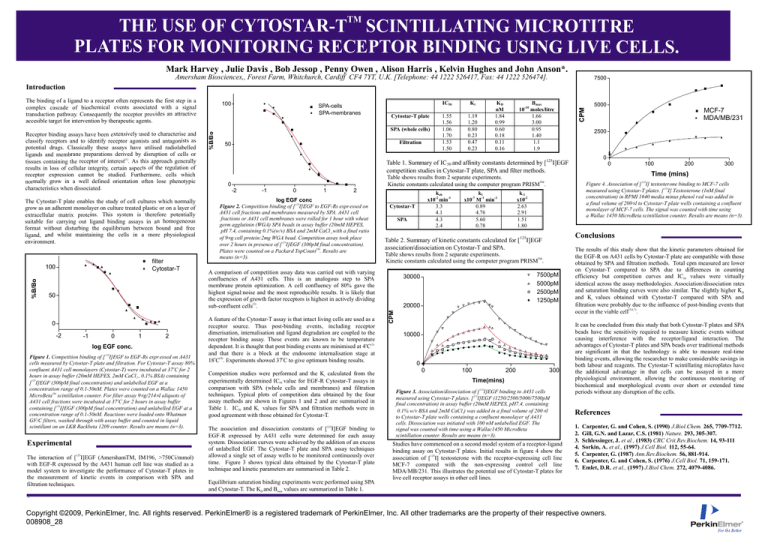
Competition studies were performed and the Ki calculated from the
experimentally determined IC50 value for EGF-R Cytostar-T assays in
comparison with SPA (whole cells and membranes) and filtration
techniques. Typical plots of competition data obtained by the four
assay methods are shown in Figures 1 and 2 and are summarised in
Table 1. IC50 and Ki values for SPA and filtration methods were in
good agreement with those obtained for Cytostar-T.
The association and dissociation constants of [125I]EGF binding to
EGF-R expressed by A431 cells were determined for each assay
system. Dissociation curves were achieved by the addition of an excess
of unlabelled EGF. The Cytostar-T plate and SPA assay techniques
allowed a single set of assay wells to be monitored continuously over
time. Figure 3 shows typical data obtained by the Cytostar-T plate
technique and kinetic parameters are summarised in Table 2.
Equilibrium saturation binding experiments were performed using SPA
and Cytostar-T. The KD and Bmax values are summarized in Table 1.
k1
x10-7 M-1 min-1
0.89
4.76
5.60
0.78
0
10000
0
0
200
100
300
Conclusions
7500pM
5000pM
2500pM
1250pM
20000
200
Figure 4. Association of [ 125I] testosterone binding to MCF-7 cells
measured using Cytostar-T plates. [ 125I] Testosterone (1nM final
concentration) in RPMI 1640 media minus phenol red was added in
a final volume of 200 µl to Cytostar-T plate wells containing a confluent
monolayer of MCF-7 cells. The signal was counted with time using
a Wallac 1450 MicroBeta scintillation counter. Results are means (n=3).
Table shows results from 2 separate experiments.
Kinetic constants calculated using the computer program PRISMTM.
30000
100
Time (mins)
k-1
x10-2
2.63
2.91
1.51
1.80
Table 2. Summary of kinetic constants calculated for [ 125I]EGF
association/dissociation on Cytostar-T and SPA.
A comparison of competition assay data was carried out with varying
confluencies of A431 cells. This is an analogous step to SPA
membrane protein optimization. A cell confluency of 80% gave the
highest signal:noise and the most reproducible results. It is likely that
the expression of growth factor receptors is highest in actively dividing
sub-confluent cells(2).
A feature of the Cytostar-T assay is that intact living cells are used as a
receptor source. Thus post-binding events, including receptor
dimerisation, internalisation and ligand degradation are coupled to the
receptor binding assay. These events are known to be temperature
dependent. It is thought that post binding events are minimised at 4oC(3)
and that there is a block at the endosome internalisation stage at
18oC(4). Experiments showed 37oC to give optimum binding results.
kob
x10-2 min-1
3.3
4.1
4.3
2.4
MCF-7
MDA/MB/231
0
Table shows results from 2 separate experiments.
TM
Kinetic constants calculated using the computer program PRISM .
0
5000
2500
Table 1. Summary of IC 50 and affinity constants determined by [125I]EGF
competition studies in Cytostar-T plate, SPA and filter methods.
CPM
The Cytostar-T plate enables the study of cell cultures which normally
grow as an adherent monolayer on culture treated plastic or on a layer of
extracellular matrix proteins. This system is therefore potentially
suitable for carrying out ligand binding assays in an homogeneous
format without disturbing the equilibrium between bound and free
ligand, and whilst maintaining the cells in a more physiological
environment.
%B/Bo
Receptor binding assays have been extensively used to characterise and
classify receptors and to identify receptor agonists and antagonists as
potential drugs. Classically these assays have utilised radiolabelled
ligands and membrane preparations derived by disruption of cells or
(1)
tissues containing the receptor of interest . As this approach generally
results in loss of cellular integrity, certain aspects of the regulation of
receptor expression cannot be studied. Furthermore, cells which
normally grow in a well defined orientation often lose phenotypic
characteristics when dissociated.
100
CPM
The binding of a ligand to a receptor often represents the first step in a
complex cascade of biochemical events associated with a signal
transduction pathway. Consequently the receptor provides an attractive
accesible target for intervention by therapeutic agents.
300
Time(mins)
Figure 3. Association/dissociation of [ 125I]EGF binding to A431 cells
measured using Cytostar-T plates. [ 125I]EGF (1250/2500/5000/7500pM
final concentration) in assay buffer (20mM HEPES, pH7.4, containing
0.1% w/v BSA and 2mM CaCl 2) was added in a final volume of 200 µl
to Cytostar-T plate wells containing a confluent monolayer of A431
cells. Dissociation was initiated with 100 nM unlabelled EGF. The
signal was counted with time using a Wallac1450 MicroBeta
scintillation counter. Results are means (n=3).
Studies have commenced on a second model system of a receptor-ligand
binding assay on Cytostar-T plates. Initial results in figure 4 show the
association of [125I] testosterone with the receptor-expressing cell line
MCF-7 compared with the non-expressing control cell line
MDA/MB/231. This illustrates the potential use of Cytostar-T plates for
live cell receptor assays in other cell lines.
The results of this study show that the kinetic parameters obtained for
the EGF-R on A431 cells by Cytostar-T plate are compatible with those
obtained by SPA and filtration methods. Total cpm measured are lower
on Cytostar-T compared to SPA due to differences in counting
efficiency but competition curves and IC50 values were virtually
identical across the assay methodologies. Association/dissociation rates
and saturation binding curves were also similar. The slightly higher KD
and Ki values obtained with Cytostar-T compared with SPA and
filtration were probably due to the influence of post-binding events that
occur in the viable cell(5,6,7).
It can be concluded from this study that both Cytostar-T plates and SPA
beads have the sensitivity required to measure kinetic events without
causing interference with the receptor/ligand interaction. The
advantages of Cytostar-T plates and SPA beads over traditional methods
are significant in that the technology is able to measure real-time
binding events, allowing the researcher to make considerable savings in
both labour and reagents. The Cytostar-T scintillating microplates have
the additional advantage in that cells can be assayed in a more
physiological environment, allowing the continuous monitoring of
biochemical and morphological events over short or extended time
periods without any disruption of the cells.
References
1.
2.
3.
4.
5.
6.
7.
Carpenter, G. and Cohen, S. (1990) J.Biol.Chem. 265, 7709-7712.
Gill, G.N. and Lazar, C.S. (1981) Nature. 293, 305-307.
Schlessinger, J. et al., (1983) CRC Crit.Rev.Biochem. 14, 93-111
Sorkin, A. et al., (1997) J.Cell Biol. 112, 55-64.
Carpenter, G. (1987) Ann.Rev.Biochem. 56, 881-914.
Carpenter, G. and Cohen, S. (1976) J.Cell Biol. 71, 159-171.
Emlet, D.R. et al., (1997) J.Biol Chem. 272, 4079-4086.
=
Copyright ©2009, PerkinElmer, Inc. All rights reserved. PerkinElmer® is a registered trademark of PerkinElmer, Inc. All other trademarks are the property of their respective owners.
008908_28

