Cerebral vasculitis - Practical Neurology
advertisement
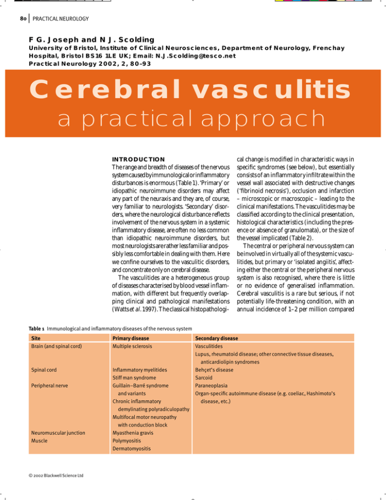
80 PRACTICAL NEUROLOGY F G. Joseph and N J. Scolding University of Bristol, Institute of Clinical Neurosciences, Department of Neurology, Frenchay Hospital, Bristol BS16 1LE UK; Email: N.J.Scolding@tesco.net Practical Neurology 2002, 2, 80–93 Cerebral vasculitis a practical approach INTRODUCTION The range and breadth of diseases of the nervous system caused by immunological or inflammatory disturbances is enormous (Table 1). ‘Primary’ or idiopathic neuroimmune disorders may affect any part of the neuraxis and they are, of course, very familiar to neurologists. ‘Secondary’ disorders, where the neurological disturbance reflects involvement of the nervous system in a systemic inflammatory disease, are often no less common than idiopathic neuroimmune disorders, but most neurologists are rather less familiar and possibly less comfortable in dealing with them. Here we confine ourselves to the vasculitic disorders, and concentrate only on cerebral disease. The vasculitides are a heterogeneous group of diseases characterised by blood vessel inflammation, with different but frequently overlapping clinical and pathological manifestations (Watts et al. 1997). The classical histopathologi- cal change is modified in characteristic ways in specific syndromes (see below), but essentially consists of an inflammatory infiltrate within the vessel wall associated with destructive changes (‘fibrinoid necrosis’), occlusion and infarction – microscopic or macroscopic – leading to the clinical manifestations. The vasculitides may be classified according to the clinical presentation, histological characteristics (including the presence or absence of granulomata), or the size of the vessel implicated (Table 2). The central or peripheral nervous system can be involved in virtually all of the systemic vasculitides, but primary or ‘isolated angiitis’, affecting either the central or the peripheral nervous system is also recognised, where there is little or no evidence of generalised inflammation. Cerebral vasculitis is a rare but serious, if not potentially life-threatening condition, with an annual incidence of 1–2 per million compared Table 1 Immunological and inflammatory diseases of the nervous system Site Brain (and spinal cord) Primary disease Multiple sclerosis Spinal cord Inflammatory myelitides Stiff man syndrome Guillain–Barré syndrome and variants Chronic inflammatory demylinating polyradiculopathy Multifocal motor neuropathy with conduction block Myasthenia gravis Polymyositis Dermatomyositis Peripheral nerve Neuromuscular junction Muscle © 2002 Blackwell Science Ltd Secondary disease Vasculitides Lupus, rheumatoid disease; other connective tissue diseases, anticardiolipin syndromes Behçet’s disease Sarcoid Paraneoplasia Organ-specific autoimmune disease (e.g. coeliac, Hashimoto’s disease, etc.) APRIL 2002 Table 2 Classification of the vasculitides according to vessel size (Scott & Watts 1994) Dominant vessel involved Large arteries Medium arteries Small vessels and medium arteries Small vessels Primary Giant cell arteritis Takayasu’s arteritis Classical polyarteritis nodosa Kawasaki disease Wegener’s granulomatosis Churg–Strauss syndrome Microscopic polyangiitis Henoch-Schönlein purpura Essential cryoglobulinaemia Cutaneous leukocytoclastic vasculitis to 39 per million for systemic vasculitis (Watts et al. 1997). MECHANISMS OF TISSUE DAMAGE In both primary and secondary CNS vasculitis, ischaemia is the cause of neurological loss of function. This results from three consequences of inflammation within the vascular wall: obstruction of the vessel lumen, increased coagulation from the effects of proinflammatory cytokines on the endothelial surface, and alterations in vasomotor tone. The development of a vasculitic process depends on interplay between cellular and humoral factors, although most authorities agree that the latter appears to be more important (Jennette et al. 1994). Antibody-dependent mechanisms Three main pathways of vascular injury are commonly invoked: direct antibody attack, immune complex mediated, and antineutrophil cytoplasmic antibody (ANCA)-related vasculitis. Direct antibody attack In some systemic vasculitides, a pathogenic role for antiendothelial cell antibodies in either injuring or activating endothelial cells has been proposed (Salojin et al. 1996), though their lack of specificity and variable rates of detection raise doubts about their importance. As they are more frequently-reported in medium and large vessel vasculitis than small vessel disease, they are in any case unlikely to be of relevance in cerebral disease. Secondary Aortitis with rheumatoid disease; Infection (e.g. syphilis) Infection (e.g. hepatitis B) Vasculitis with rheumatoid disease, systemic lupus erythematosus, Sjögren’s syndrome, drugs, infection (e.g. HIV) Drugs (e.g. sulphonamides, etc.) Infection (e.g. hepatitis C) rophages, amplification of inflammation, and the generation of lytic and injurious membrane attack complexes. Hepatitis B- and C-associated vasculitis are good examples of this process, the latter underlying many cases of cryoglobulinaemic vasculitis (Cacoub et al. 2001). Antineutrophil cytoplasmic antibodyrelated vasculitis ANCAs represent a family of antibodies directed against constituents of the neutrophil azurophil granules (Mohan & Kerr 2001; Niles 1996). Based on immunofluorescence patterns, a distinction is made between cytoplasmic ANCA (c-ANCA) that targets proteinase-3 (PR-3), associated with nearly 95% specificity for Wegener’s granulomatosis, and perinuclear ANCA (p-ANCA) directed at myeloperoxidase, less specifically found in microscopic polyangiitis and the Churg–Strauss syndrome (Mohan & Kerr 2001; Niles 1996). Cell-mediated damage Evidence for cell-mediated involvement in tissue injury in vasculitis comes in part from studying microscopic polyarteritis nodosa and Wegener’s granulomatosis (Mathieson & Oliveira 1995). In both disorders, circulating T-cells responsive to PR-3 are found, and vascular lesions contain activated T-cells and antigen-presenting MHC class II positive dendritic cells. In primary CNS and peripheral nerve vasculitic lesions, the predominant infiltrate is one of CD4-positive and CD8-positive T-lymphocytes and monocytes (Lie 1997). Immune complex mediated vasculitis THE CLINICAL FEATURES OF CEREBRAL VASCULITIS Immune complex deposition in the blood vessel wall triggers activation of the complement cascade with recruitment of polymorphs and mac- A myriad of neurological symptoms, signs or syndromes can occur in CNS vasculitis, reflecting the potential for infarction and ischaemia, © 2002 Blackwell Science Ltd 81 82 PRACTICAL NEUROLOGY Table 3 Clinical features of cerebral vasculitis Course Acute Sub-acute Chronic Relapsing-remitting CNS features Headaches Seizures (focal/generalized) Stroke-like events Encephalopathy Progressive cognitive change Movement disorders – especially chorea, myoclonus Optic nerve disease Cranial neuropathies Systemic features* Fever & night sweats Rash, especially livedo reticularis Weight loss, anorexia Oligoarthropathy *These systemic features are rather easily missed by neurologists – they must be specifically looked for. Figure 1 Suggested investigation of suspected cerebral vasculitis which may be micro- or macroscopic, focal, multifocal or diffuse, and affect any part of the brain. (Table 3.) Most accounts of the disorder describe headaches, focal or generalized seizures, stroke-like episodes with hemispheric or brainstem deficits, acute or subacute encephalopathies, progressive cognitive changes, chorea, myoclonus and other movement disorders, and optic and other cranial neuropathies – in short, there are few neurological syndromes that are not consistent with a vasculitic aetiology. Systemic features such as fever, night sweats, livedo reticularis, or oligoarthropathy may also be present but often are only revealed by direct questioning of the patient. The course is commonly acute or subacute, but chronic progressive presentations are also well described, as are spontaneous relapses and remissions. Despite the diversity of clinical presentations, three broad categories have been defined in a small study (Scolding et al. 1997), and these may help to improve recognition of the condition: • acute or subacute encephalopathy, commonly presenting as an acute confusional state, progressing to drowsiness and coma; • superficially resembling atypical multiple sclerosis (‘MS-plus’) in phenotype, with a relapsing–remitting course, and features such as optic neuropathy and brain stem episodes, but also accompanied by other features less common in multiple sclerosis, such as seizures, severe and persisting headaches, encephalopathic episodes, or hemispheric stroke-like episodes; • intracranial mass lesions with headache, drowsiness, focal signs and often raised intracranial pressure. INVESTIGATION AND DIAGNOSIS The whole host of possible neurological symptoms and signs makes cerebral vasculitis notoriously difficult to diagnose: innumerable disorders may cause a pleomorphic combination of headache, encephalopathy, strokes, seizures and focal deficits of acute or subacute onset (Table 4). A high index of suspicion along with a detailed history and physical examination, which © 2002 Blackwell Science Ltd APRIL 2002 Other vasculopathies Susac’s syndrome Homocysteinuria Ehlers–Danlos syndrome Radiation vasculopathy Köhlmeyer-Degos disease Fibromuscular dysplasia Fabry’s disease Moyamoya syndrome Amyloid angiopathy CADASIL* Marfan’s syndrome Pseudoxanthoma elasticum Viral or fungal vasculitis Infections Lyme disease AIDS Endocarditis Whipple’s disease Viral encephalitis Legionella/mycoplasma pneumonia Other immune/inflammatory diseases Sarcoidosis Lupus and anti–phospholipid syndrome Behçet’s disease Multiple sclerosis/acute dessimated encephalomyelopathy Thyroid encephalopathy Tumours and malignancy Atrial myxoma Multifocal glioma Cerebral lymphoma Paraneoplastic syndromes Cholesterol embolization syndrome Thrombotic thrombocytopaenic purpura Intracranial venous thrombosis Mitochondrial cytopathies Table 4 Some neurological and systemic disorders that may mimic cerebral vasculitis. Systemic features such as fever, night sweats, livedo reticularis, or oligoarthropathy may be present but often are only revealed by direct questioning of the patient *Cerebral autosomal dominant arteriopathy with subcortical infarcts and leukoencephalopathy. entertains a broad differential diagnosis and excludes imitators of vasculitis, is imperative. No single simple investigation is universally useful in making the correct diagnosis of cerebral vasculitis, but the relevant tests are discussed below. Blood tests and serology Routine blood tests are generally unhelpful in terms of specificity, though they can help point towards or away from the possibility of cerebral vasculitis (Calabrese & Mallek 1988; Hankey 1991; Scolding 1999a,b). Anaemia is an infrequent finding and a leukocytosis without eosinophilia is present in about 50% of pa- tients. The ESR and C-reactive protein levels are often abnormal, especially in cases secondary to systemic disease, but of course lack specificity – some include a normal ESR as a defining feature of primary angiitis of the CNS, others report moderately elevated values in two-thirds of patients (Calabrese & Mallek 1988). Serological testing is important in excluding suspected neuropsychiatric lupus or in helping to define the systemic origin of an established intracranial vasculitis, but is of little value in confirming or refuting cerebral vasculitis. ANCA assays are now routinely requested in screening for systemic vasculitides, but positivity is sometimes seen in connective tissue disorders such as lupus, © 2002 Blackwell Science Ltd 83 84 PRACTICAL NEUROLOGY and rarely in individuals without any apparent vasculitic disorder (Merkel et al. 1997). Spinal fluid examination Cerebral biopsy may provide unequivocal Cerebrospinal fluid abnormalities are non-specific, but again useful in implicating an inflammatory process within the CNS and excluding infection and malignant diseases that may present similarly. Pooled case reviews suggest a raised cell count (mainly a lymphocytosis) and protein in 50–80% (Calabrese & Mallek 1988; Hankey 1991; Scolding 1999a,b). The CSF opening pressure is raised in almost half the cases of primary angiitis of the CNS. Oligoclonal immunoglobulin bands in the CSF have been studied infrequently, but are found consistently enough (perhaps in up to 40–50%) to make their analysis worthwhile (Scolding et al. 1997). Oligoclonal band patterns, which vary substantially, perhaps even disappearing altogether during the course of disease, do help point away from multiple sclerosis when this is part of the differential diagnosis. histopathological Electroencephalography proof of vasculitis, with the potential This is neither specific nor sensitive but is abnormal in 80% of cases, often with slow-wave activity. It may provide some supportive clues in the small-vessel vasculitides affecting the brain, in excluding alternative causes, and possibly for monitoring therapy. advantage of Imaging distinguishing between primary and secondary vasculitides, and of course disclosing alternative diagnoses © 2002 Blackwell Science Ltd Magnetic resonance imaging (MRI) is a sensitive but not specific detector of vascular disease, disclosing of course the results of vascular inflammation, not inflammation itself (Harris et al. 1994). Such changes should however, prompt further studies, and MRI is certainly valuable in excluding other conditions. There may be poor correlation between MRI and angiographic findings: in one study of 50 territories affected by vasculitis on contrast angiography, at least one third were normal on MRI (Cloft et al. 1999). Other studies confirm this imperfect sensitivity, and there are (unfortunately) reported cases of proven cerebral vasculitis with normal MR imaging (Alhalabi & Moore 1994; Vanderzant et al. 1988). Single photon emission computed tomography (SPECT) appears to be a useful but nonspecific imaging tool, again mirroring but not defining a vasculitic process (Scolding et al. 1997). The value of position emission tomography (PET) scanning in this context is unclear. Magnetic resonance angiography (MRA) is finding a niche in imaging of large vessel vasculitides such as Takayasu’s arteritis, with potential to supplant conventional catheter angiography (Atalay & Bluemke 2001), but it does not have sufficient resolution to display medium or small vessel cerebral vasculitis. Establishing the diagnostic value of contrast catheter angiography is complicated by the many studies that have used this as the‘gold standard’ for confirmation. Publications with pathological evidence indicate a false negative rate for angiography of 30–40% (Calabrese & Mallek 1988; Hankey 1991), and there have been examples of patients with histologically-proven primary angiitis of the CNS and completely normal angiograms (Vanderzant et al. 1988). This APRIL 2002 may be because the affected vessels are beyond the resolution of conventional imaging. When abnormalities are present, they include segmental (often multifocal) narrowing with areas of localized dilatation or beading. Single stenotic areas in multiple vessels are more frequent than multiple stenotic areas along a single vessel segment in primary angiitis of the CNS. Retrospective series suggest a sensitivity of only 24–33% (Hankey 1991; Koo & Massey 1988; Vollmer et al. 1993; Alrawi et al. 1999), with a specificity of a similar order – an enormous number of inflammatory, metabolic, malignant or other vasculopathies mimic angiitis on angiograms. It has been suggested that clinical and angiographic features may correlate, supporting the hypothesis that segmental narrowing initially results from reversible inflammation and vasospasm, with the later irreversible changes secondary to scarring; and in consequence, that serial angiography may be useful in monitoring treatment (Alhalabi & Moore 1994). However, the procedure does carry the risk of transient (10%) or permanent neurological deficits (1%) (Hellmann et al. 1992). Although its importance has been overemphasized, it is still a valuable investigational tool with a sensitivity comparable to that of biopsy. Indium-labelled white cell nuclear scanning has a role in disclosing areas of (sometimes unsuspected) systemic inflammation. The sparse evidence supporting its use in cerebral vasculitis suggests that it is unsuccessful in identifying the majority of cases, but may usefully disclose clinically silent foci of systemic inflammation (Scolding et al. 1997). Opthalmological examination Dynamic recording of erythrocyte flow using video slit lamp microscopic recording and lowdose fluorescein angiography to examine the vasculature of the anterior ocular chamber can be a useful additional investigation. In a small study, four out of five patients had abnormal findings (Scolding et al. 1997). Typical abnormalities are marked slowing of flow, multifocal attenuation of arterioles, and erythrocyte aggregates. Fluorescein studies may confirm these changes, and demonstrate areas of small vessel infarction along with postcapillary leakage. potential advantage of distinguishing between primary and secondary vasculitides, and of course disclosing alternative diagnoses. The practicalities of achieving this are, however, difficult. Ideally an affected area in the nondominant hemisphere, or a ‘blind biopsy’ from the nondominant temporal tip (most likely to include longitudinally orientated surface vessels; Moore 1989) should be sampled. Cortical sampling alone appears to be insufficient and uninformative, with failure to obtain leptomeningeal vessels further increasing the false negative rate. It is not surprising (considering the difficulty in obtaining affected tissue) that the sensitivity is limited to (at best) approximately 70% (Calabrese & Mallek 1988; Hankey 1991; Alrawi et al. 1999). Morbidity from this invasive procedure has been estimated at 0.5–2%, though current rates may be lower. Nevertheless, the significant risk means that up to 75% of reported cases are diagnosed without histopathology (Lie 1997). A recent retrospective study of some 61 patients biopsied for suspected cerebral vasculitis has usefully illuminated this topic (Alrawi et al. 1999). No patient suffered any significant morbidity as a result of the procedure. 36% of patients were confirmed as having cerebral vasculitis, but no less usefully, and importantly, 39% biopsies showed an alternative, unsuspected diagnosis: lymphoma (6 cases), multiple sclerosis (2 cases), and infection (7 cases, including toxoplasmosis, herpes, and also two cases of cerebral abscess). Many of these nonvasculitic disorders are treatable, often indeed curable, while inappropriate treatment with steroids alone, or with more potent immunosuppressive agents would at best have no useful effect, and very often, serious adverse consequences. Biopsy failed to yield a clear diagnosis in 25% of patients in this study, though even here, biopsy arguably might not be described as ‘noncontributory’, at least decreasing the likelihood of the alternative diagnoses mentioned above. The decision not to biopsy must be balanced against anecdotal case reports where patients have been exposed to potentially harmful immunosuppressive drugs unnecessarily. CAUSES OF THE VASCULITIC PROCESS Histopathology Primary (isolated) angiitis of the CNS Cerebral biopsy may provide unequivocal histopathological proof of vasculitis, with the This curious and uncommon vasculitis was first recorded amongst ‘unknown forms of © 2002 Blackwell Science Ltd 85 86 PRACTICAL NEUROLOGY arteritis’ by Harbitz in 1922 (Harbitz 1922). It is almost exclusively confined to the brain and, less commonly, the spinal cord. The designation ‘primary’ is probably more appropriate than ‘isolated’ as autopsies have revealed extracranial involvement (e.g. pulmonary arteries and abdominal viscera; Lie 1997). Anatomically, the angiitic process is focal and segmental in distribution. Histologically, the inflammation may be granulomatous, necrotizing, or lymphocytic in character, and mixed morphologic types often occur in individual patients therefore rendering its other common title of ‘granulomatous angiitis’ difficult to sustain. In addition to being fraught by variable terminology, there are no uniform diagnostic criteria. This probably reflects the difficulty in obtaining Most authorities now ante-mortem pathological material and the need for a vigorous diagnostic approach, recognize the nonspecificity of which probably underestimates the true incidence. Calabrese defines angiographic changes, and we the disorder as ‘an acquired clinical disease characterized by CNS believe a certain diagnosis of dysfunction that remains unexplained following thorough cliniprimary CNS angiitis depends cal, laboratory, and neurological investigations; appears to be unason a positive biopsy sociated with systemic illness, and yields evidence by cerebral angiography or biopsy of CNS tissue of vasculitis confined to the CNS.’ (Calabrese & Mallek 1988.) Most authorities now recognize the nonspecificity of angiographic changes, and we believe a certain diagnosis of primary CNS angiitis depends on a positive biopsy. There is no typical and specific clinical presentation and this makes a high degree of suspicion the key to successful diagnosis. It may affect patients of any age but is most frequently reported in middle life (mean age 45), with a slight male preponderance of 4:3 (Lie 1992). Suspicion should be greater if multifocal symptoms and signs develop in a stepwise progression, and are accompanied by headache or altered mental status. Significant constitutional symptoms (especially antedating neurological dysfunction) are infrequent and should make you question the diagnosis. The importance of seriously considering and excluding alternative or coexistent disease processes cannot be overemphasized. Any brain or spinal cord vessel can be involved, including large intracranial vessels, internal carotid and vertebral arteries, but there is a predilection for the small vessels, particularly the un-named leptomeningeal vessels. © 2002 Blackwell Science Ltd Characteristic focal or segmental skip lesions cause multiple small, or sometimes large, foci of infarction or haemorrhage (Calabrese & Mallek 1988; Hankey 1991; Lie 1992). The aetiology remains speculative – it may indeed represent a nonspecific pathological reaction to a number of insults rather than a specific disease. In support of this lies the recognized (though rare) association with other disorders, such as Hodgkin’s and non-Hodgkin’s lymphomas, Sjögren’s syndrome, and infectious agents including herpes zoster and simplex, human immunodeficiency virus, HTLV-III, hepatitis C, cytomegalovirus, mycoplasma and bartonella (Lie 1996). A more favourable monophasic clinical course is suggested in the so-called ‘benign angiopathy of the CNS’. This is a syndrome with normal, or only mildly abnormal CSF, and evidence of a vasculitic picture on angiography alone (Calabrese et al. 1993). However, this concept has been questioned, in view of the recognized non-specificity of angiography, the fact that those cases not proceeding to biopsy are more likely to be the less severely affected, and children satisfying ‘benign angiopathy’ criteria often do not have a temperate, monophasic course, but have required aggressive immunotherapy (Gallagher et al. 2001). Primary systemic vasculitides with CNS disease Each of the systemic vasculitides may be complicated by cerebral involvement; often they carry their own defining characteristics. In contrast to primary angiitis of the CNS, constitutional disturbance – fever, night sweats, severe malaise, weight loss – are common and may be accompanied by a rash or arthropathy. Wegener’s granulomatosis is a necrotizing, granulomatous vasculitis primarily affecting the upper and lower respiratory tracts, often with destructive cartilaginous changes and cavitating lung lesions. Renal disease with glomerulonephritis is usual. Diagnostic criteria have been established (Table 5). Neurological involvement occurs in up to 35% of patients, but most commonly involves the peripheral nervous system (Nishino et al. 1993). Meningeal and middle ear disease may lead to significant cranial neuropathies (especially of the seventh and eighth nerves). Gadolinium-enhanced MR scanning may valuably reveal meningeal infiltration, offering a ready target for biopsy. Ocular involvement may occur with orbital pseudotumour. APRIL 2002 Cerebral small-vessel vasculitis is rare, but when of systemic inflammation, are usually sufficient it does occur it is usually responsible for enceph- for diagnosis. alopathies, seizures, and pituitary abnormalities Churg–Strauss syndrome is a disorder char– but may be indistinguishable from any other acterized by a (diagnostically valuable) hyperform of intracranial vasculitis. More likely is the eosinophilia and systemic vasculitis, occurring unique contiguous extension in individuals with recentlyof erosive granulomata from developed atopic features. the sinuses or from remote Table 5 Wegener’s Asthma and mononeuritis metastatic granulomata to the granulomatosis: American multiplex are undoubtedly the CNS. There is a high c-ANCA College of Rheumatology criteria two most frequent manifesta(proteinase-3) titre. tions of this disease. Rashes, (Leavitt et al. 1990) with purpura, urticaria, and The diagnosis should be based on at least two of the following: subcutaneous nodules, are Bloody or purulent nasal discharge common. Glomerulonephritis Nodules, cavities or infiltrates on chest X-ray may develop. It may also affect Microscopic haematuria or red cell casts coronary, splanchnic and cereGranulomatous inflammation on biopsy bral circulations. CNS involvement is evident in only about Microscopic polyangiitis has many simi- 7% (Sehgal et al. 1995; Guillevin et al. 1999). larities to Wegener’s granulomatosis, including About 50% of patients are positive for pANCA, pulmonary haemorrhage, but differs in that 25% positive for cANCA, and 25% have no anupper respiratory tract involvement is rare and tineutrophil cytoplasmic antibodies. granuloma formation is not seen. Patients usuGiant cell arteritis includes two histologially have glomerulonephritis and indeed this cally-similar but clinically-distinct diseases: vasculitis is occasionally confined to the kidney. temporal arteritis and Takayasu’s arteritis. One study found mononeuritis multiplex in Temporal arteritis is a chronic inflammatory 55% (Guillevin et al. 1999); in this study, the disorder affecting large and medium-sized arbrain was seldom affected (11%) and CNS teries, which predominantly affects postmenodisease did not contribute to mortality. There pausal women (Huston et al. 1978). A genetic are, however, infrequent reports of p-ANCA predisposition is suggested by a high incidence positive rapidly progressive glomerulonephritis in populations with Scandinavian lineage, some associated with cerebral vasculitis, requiring ag- familial accumulation, and the association with gressive therapy. the HLA-DR4 haplotype. It has an annual inciClassical polyarteritis nodosa may cause dence of 17.4 per 100 000 in the over 50-yearmedium and small-sized muscular artery in- old population, so that new onset unilateral or volvement in multiple organs, with the notable bilateral headache in this age group should alert exception of the lungs and spleen. Patients often the physician. present with renal failure and hypertension Classically it manifests as temporal head(80%). Gastrointestinal involvement occurs ache with tender, pulseless, nodular temporal in up to 50% of patients, with abdominal pain arteries, and (usually only on direct enquiry) due to visceral infarcts. Heart failure and myo- symptoms of general malaise, jaw claudicacardial infarction reflect cardiac involvement. tion and features of polymyalgia rheumatica. Neurological abnormalities are prominent Neuro-opthalmological symptoms are the (50–60%), but again mostly confined to the most widely recognized, with blindness ocperipheral nervous system. CNS involvement curring in one sixth of treated patients with the is usually only seen in established disease. It is condition (Caselli & Hunder 1994). Traditionthought that damage is initiated by immune ally, the inflammatory process is thought simply complex deposition; fibrinoid necrosis is typi- to involve the extracranial vessels and rarely to cal though not diagnostic. Although there are extend beyond the point of penetration of the no specific serological tests, about 20–30% have dura. A large study of 166 patients with biopsy hepatitis B antigen or antibody in the serum. proven temporal arteritis demonstrated neuroVisceral angiography showing aneurysms or logical involvement in 31%, describing the usual occlusions of the visceral arteries, with biopsy comprehensive range of neurological manifesproven peripheral nerve vasculitis and evidence tation: neuropsychiatric syndromes, peripheral © 2002 Blackwell Science Ltd 87 88 PRACTICAL NEUROLOGY Table 6 Diagnosing Kawasaki disease neuropathies, mononeuropathies, spinal cord lesions, neuro-otological syndromes, various pain syndromes, transient ischaemic attacks and stroke – although most authorities find almost all these pictures outside their common experience (Caselli & Hunder 1994). Infarction within the vertebrobasilar territory is relatively uncommon, but there have been isolated reports of temporal arteritis presenting as lateral medullary syndrome. The expected greater incidence of cerebrovascular disease in this group of older people may, however, be confounding. The ESR is usually significantly raised and may be used to monitor the response to steroid treatment. However, it has recently been pointed out that a ‘normal’ ESR in active disease is not excessively rare, and may perhaps be explained by an inability to mount an acute phase response, or by very localized arteritis (Salvarani & Hunder 2001). Measuring serum interleukin-6 levels is a promising alternative to the ESR. Recent work has also emphasized that a raised platelet count is a risk factor for permanent visual loss in temporal arteritis and should emphasize the need for urgent treatment (Liozon et al. 2001; Lincoff et al. 2000). Takayasu’s arteritis was originally described in young oriental women but is now globally recognized. It is alternatively named ‘pulseless disease’, because 98% of affected individuals have at least one major arterial pulse absent, as a result of the characteristic involvement of the aorta and its large branches. The disease process is initially inflammatory, and later occlusive; during this phase most of the neurological abnormalities occur. Syncope is reported in at least 50% of patients, but strokes, transient ischaemic attacks, and visual abnormalities are also seen. You should suspect this illness in a patient under the age of 40 years with symptoms of limb claudication, one or more absent pulses, systolic blood pressure difference of > 10 mmHg between each arm , and arterial bruits. Early histological features of the disease include granulomatous changes in the media At least five of the following features must be present to make the diagnosis: Fever for > 5 days Conjunctival congestion Changes to lips and oral cavity (dryness, fissuring and erythema) Changes of peripheral extremities: red palms and soles; indurative oedema; desquamation of finger tips during convalescence Macular polymorphous rash on trunk Swollen cervical lymph nodes © 2002 Blackwell Science Ltd and adventitia of the aorta and its branches, later followed by intimal hyperplasia, medial degeneration, and sclerotic adventitial fibrosis. Henoch-Schonlein purpura is an immunologically-mediated small vessel systemic vasculitis of children, affecting predominantly the skin, gastrointestinal tract, joints and kidneys. Neurological involvement, including headache and behavioural change, is well-described. More severe CNS involvement is usually explained by hypertensive or uraemic encephalopathy, steroid or cytotoxic drug therapy or electrolyte imbalance. Suspected cerebral vasculitis, manifesting mainly as headache, disturbance of consciousness, and tonic-clonic seizures, has rarely been reported after careful exclusion of alternative causes of neurological decline. Kawasaki disease, otherwise known as ‘mucocutaneous lymph node syndrome’, usually affects children under the age of 12 years. It has an annual incidence of less than 5/100 000 in the United Kingdom, but is at least 20 times as common in Japan where it was first described in 1967 (Table 6). Coronary artery aneurysms occur in onefifth of untreated cases, which may result in the most dreaded complication – myocardial infarction. Neurologically there is commonly an aseptic meningitis, but hemiplegic strokes, encephalopathy and facial palsy are also described. Pathologically, an acute systemic inflammatory vasculitis, with little or no fibrinoid necrosis, underlies the disease. Anti-endothelial cell antibodies may be involved in the pathogenesis. Skin involvement with purpura and urticaria is the most common manifestation of small vessel or cutaneous leukocytoclastic vasculitis. This is essentially a consequence of polymorphonuclear and mononuclear cell infiltrate in postcapillary venules. It is also known as hypersensitivity vasculitis because of the common presence of an allergic precipitant. Peripheral neuropathy is occasionally reported, and transient ischaemic attacks, strokes and encephalopathy only rarely. Cerebral vasculitis complicating ‘nonvasculitic’ systemic disorders Neurological or psychiatric symptoms in systemic lupus erythematosus (SLE) are common (40–50%) (Scolding 1999a, b; West 1996), but the most frequent neuropathological finding is that of a non-inflammatory vasculopathy of small arterioles and capillaries, with resulting APRIL 2002 microinfarcts and microhaemorrhages. Histopathological studies have consistently demonstrated that vasculitis of the cerebral vessels is rare, with a frequency of 7–13% (Ellis & Verity 1979; Johnson & Richardson 1968). Sarcoidosis affects the nervous system in only 5% of cases, but often has a poor prognosis (Zajicek et al. 1999). Neurosarcoidosis commonly presents with optic and other cranial neuropathies (especially involving the facial nerve) usually due to granulomatous meningeal and brainstem infiltration. Sarcoidosis may be complicated by systemic vasculitis affecting small or large calibre vessels in a similar fashion to other vasculitides, with angiographic and indeed histological evidence of cerebral vasculitis. The diagnosis can be difficult to make. Serum angiotensin converting enzyme and calcium levels are not always raised. CSF abnormalities are seen in 80%, usually with a raised protein and pleocytosis, and oligoclonal bands are positive in about 45% (McLean et al. 1995). Brain MRI shows nonspecific multiple white matter lesions or meningeal enhancement. Whole body gallium scanning can be more useful, demonstrating a characteristic pattern of uptake (particularly affecting the parotid glands and lungs). Histological diagnosis by Kveim test (where still available), or better still, biopsy of cerebral or meningeal tissue, provides the most reliable basis for treatment. Seropositive rheumatoid disease is a wellrecognized precipitant of cerebral vasculitis (Scolding 1999a, b), though skin involvement and mononeuritis multiplex are more typical manifestations. There are a few reports of cerebral vasculitis in the context of systemic sclerosis, Sjogren’s syndrome and mixed connective tissue disease, even (though rarely) without a preceding history of systemic symptoms. Cryoglobulinaemia can trigger immunecomplex deposition-triggered vasculitis (especially in association with hepatitis C infection), particularly common in mixed cryoglobulinaemia. Renal, joint and skin involvement with purpura progressing to necrotic ulceration is often seen. Peripheral neuropathy occurs in 22–32% of patients, in particular as mononeuritis multiplex, with leukocytoclastic vasculitis on biopsy. The CNS is rarely affected. Diagnosis depends on blood being collected into a plain tube and promptly transported in a vacuum flask at body temperature to the laboratory for analysis. Sarcoidosis affects the nervous system in only 5% of cases, but often has a poor prognosis Behçet’s disease is predominantly caused by vasculitis affecting small and medium-sized vessels. For diagnostic purposes, recurrent aphthous stomatitis (at least three times in one year) is essential ( International Study Group for Behçet’s Disease). This should be accompanied by any two of recurrent genital ulcers; anterior or posterior uveitis or retinal vasculitis; skin lesions (erythema nodosum, acneiform nodules, pseudofolliculitis, papular lesions); and, a positive pathergy test (nodules after pricking the skin). Neurological abnormalities occur in up to one-third of patients. Intracranial venous thrombosis, meningoencephalitis, rhomboencephalitis, and focal CNS abnormalities are typical (Akman-Demir et al. 1999). Variable clinical manifestations are noted, including those resembling multiple sclerosis. Confusion with the latter can be exacerbated by consistently abnormal but non-specific MR brain scans and evidence of intrathecal immunoglobulin synthesis. CSF analysis in neuro-Behçet’s may show increased levels of beta(2)-microglobulin, which may be a good marker of disease activity. As mentioned above, numerous infectious agents have been implicated in cerebral vasculitis (Lie 1996). At least three mechanisms may mediate microbe-related vascular damage: direct invasion, immune complex deposition, and secondary cryoglobulinaemia. An infection that deserves particular attention in this context is herpes zoster ophthalmicus. This usually affects the cerebral hemisphere ipsilateral to the eye and face involvement, probably by direct viral invasion of blood vessels (Hilt et al. 1983), producing single or multiple smoothtapered segmental narrowing on angiography. The typical clinical picture is of a monophasic illness with gradual resolution of cutaneous zoster, followed after a delay by an acute contralateral hemiparesis, hemisensory disturbance, or aphasia. The latent period may last from days to months, but is usually of the order of a few weeks. A CSF mononuclear pleocytosis and raised varicella-zoster antibody titre aid the diagnosis. Complications of shingles may affect children similarly, though there have been less frequent reports of chickenpox triggering cerebral vasculitis. Occasionally, only the spinal cord is involved in herpetic disease. On rare occasions a more generalized vasculitis may occur with ophthalmic or remote zoster infection. Chronic viral infection with parvovirus B19 has been implicated in polyarteritis nodosa, Ka© 2002 Blackwell Science Ltd 89 90 PRACTICAL NEUROLOGY wasaki disease, and Wegener’s granulomatosis, though causality is by no means proven (Lie 1996). Tuberculosis-associated vasculitis may be driven by tuberculoprotein immune complexes. Hepatitis B, Epstein-Barr virus, cytomegalo virus, Lyme disease, syphilis and malaria can all cause vasculitis by a similar mechanism, while in coccidiomycosis, vascular inflammation is either direct or via cryoglobulinaemia. Spores of the dimorphic fungus Coccidiodes immitis, endemic to the south-western United States and Northern Mexico, can be inhaled with subsequent haematogenous spread, often to the meninges. Vasculitis involving the small penetrating branches of the major cerebral vessels, and consequent deep ischaemic infarction, has been observed in up to 40% of these cases, and on rare occasion subarachnoid haemorrhage has been observed. Other implicated organisms (causing primary invasion of the vascular wall) are histoplasma and aspergillus fungi, and the protozoan toxoplasma gondii. These usually occur in immune suppressed patients, and infections such as HIV can directly trigger a cerebral vasculitis. Malignancy and cerebral vasculitis The relationship between Hodgkin’s disease and primary angiitis of the CNS has been described above. Lymphomatoid granulomatosis is a premalignant or frankly lymphomatous disorder, centred on the vessel wall. Destructive changes and secondary inflammatory infiltration of Tlymphocyte derived cells creates a histological appearance akin to true vasculitis. Cutaneous and pulmonary involvement is common, with nodular cavitating lung infiltrates. Neurological syndromes occur in 25–30% of cases and are the presenting feature in 20%, usually with multifocal punctate or linear enhancement along perivascular spaces seen on MRI (Tateishi et al. 2001). Malignant angioendotheliosis, now called intravascular lymphoma, is another rare although separate disorder; the B-cell derived neoplastic cells in this case remaining within the lumen of the affected vessel. Clinically, the neurological features may mimic vasculitis, with skin manifestations predominating – these include subcutaneous haemorrhagic or nonhaemorrhagic nodules with overlying telangiectasia or ulceration on the trunk and extremities. Lung involvement is unusual. © 2002 Blackwell Science Ltd Drug-induced cerebral vasculitis There is sufficient literature to suggest that some illicit and therapeutic drugs can result in cerebral vasculitis. The most persuasive evidence for a direct association is for amphetamines. Citron et al. described 14 patients, all of whom had abused amphetamines, with clinical and histological evidence of multisystem necrotizing vasculitis (Citron et al. 1970). Animal studies provide further support, with immediate angiographic changes after exposure to this drug. In humans, vasculitis may follow only a single dose of amphetamine but repeated exposure in young adults is the usual history (Buxton & McConachie 2000). Various other sympathomimetic agents such as ephedrine, and long-term oral methylphenidate use (chemically and pharmacologically similar to amphetamine) are also implicated. However, in many of these reports there is no tissue confirmation, and the diagnosis of ‘vasculitis’ is based on angiography – despite the fact that vasospasm can cause identical angiographic changes to those of vasculitis. This is exemplified in cocaine abuse, in which the significantly increased risk of ischaemic stroke is known to result from vasospasm affecting the large cranial arteries or those within the cortical microvasculature, and very seldom from any form of vasculitic process (Aggarwal et al. 1996). The causal mechanism is uncertain, but increased catecholamine release precipitating cerebral vasoconstriction is most likely. In cases of intravenous abuse, there is a suggestion that coinjected contaminants such as hepatitis C may be the actual perpetrator, but this would not explain the same effect after administration of therapeutic drugs by other routes. THE TREATMENT OF CEREBRAL VASCULITIS Notwithstanding the problems in recognition and diagnosis, cerebral vasculitis is a highly treatable condition for which prompt management can radically improve the outcome. Prospective randomised controlled trials are understandably difficult because of the rarity of the condition and the lack of unifying diagnostic criteria. Retrospective analyses have been our main tool, and from this has emerged significant support for the use of steroids with cyclophosphamide in confirmed cases (Scolding 2001). A reasonable induction regime is high dose steroids – probably best as intravenous methyl APRIL 2002 prednisolone, 1 g daily for 3 days – followed by require the use of alternative agents. Methoral prednisolone 60 mg/day, decreasing by otrexate, 10–25 mg on a weekly basis, may be 10 mg at weekly intervals to 10 mg/day if pos- used in conjunction with steroids, either dursible. This should be coupled with cyclophos- ing induction or maintenance. Intravenous phamide 2.5 mg/kg (lower dose in the elderly, immunoglobulin (0.4 mg/kg/day for 5 days), or in renal failure) per day. This induction com- with its good safety record, has been found usebination is suggested for 9–12 weeks, though ful in cases of systemic vasculitis, though may some doctors recommend 4–6 months. Pulsed induce only partial remission (Jayne et al. 2000; weekly intravenous cyclophosphamide appears Pritchard & Hughes 2001). Plasmapheresis may be valuable in cryto differ insignificantly in efficacy from daily oral treatment, and it may have fewer adverse oglobulinaemia. It is now generally used in effects. Careful monitoring of the blood count severe life threatening disease (e.g. pulmonary for bone marrow suppression should force a re- haemorrhage and severe glomerulonephritis) duction of the cyclophosphamide dose if there with 7–10 treatments over 14 days (Gaskin & is leucopaenia (total white count falling below Pusey 2001). Although there is little experience in patients with intracranial disease, there is evi4.0 × 109) or neutropaenia (below 2.0 × 10⁹). Cyclophosphamide is associated with haem- dence of significant improvement when used in orrhagic cystitis (a complication reduced by combination with steroids in cerebral disease adequate hydration and mesna cover), a 33-fold associated with Henoch–Schönlein purpura. increase in bladder cancer, other malignanCampath-1H is a humanized monoclonal cies, infertility, cardiotoxicity and pulmonary antibody directed against the CD52 antigen fibrosis. In a study of 145 patients treated with present on most lymphocytes. When used in this agent for Wegener’s granulomatosis (not combination with a second humanized mononecessarily neurological), and followed for clonal antibody against CD4, it has demonstraa total of 1333 patient years, nonglomerular ble long-term benefits in systemic vasculitis haematuria occurred in approximately 50%, (Mathieson et al. 1990). Interferon- can conthe majority of whom had macroscopic changes trol not only hepatitis C associated hepatitis, but consistent with cyclophosphamide-induced also cryoglobulinaemia, and vasculitis. Unforbladder injury on cystoscopy. Seven of these tunately, there is regular relapse within months (and none without preceding haematuria) of treatment withdrawal (Table 7). developed transitional cell bladder carcinoma; Although complicated immunosuppressive six had had a total cumulative dose in excess of therapy is unavoidable in many vasculitides, 100 g cyclophosphamide, and a duration of oral temporal arteritis remains a condition where treatment exceeding 2.7 years (Talar-Williams steroid-resistance is extremely rare. The role of et al. 1996). Induction Regime: 9–12 weeks (some suggest 4–6 months) The maintenance phase of high dose steroids – e.g. intravenous methyl prednisolone, 1 g/day treatment, converting to a refor 3 days together with oral cyclophosphamide 2.5 mg/kg* gime of alternate day steroids then oral prednisolone 60 mg/day after i/v methyl prednisolone; (10–20 mg prednisolone), decreasing at weekly intervals by 10 mg decrements to and substituting azathioprine 10 mg/day if possible. (2 mg/kg/day) for cyclophosMaintenance Regime: continued for a further 10 months phamide, is commenced after alternate day corticosteroids (10–20 mg prednisolone) induction, and continued azathioprine (2 mg/kg/day) substituted for cyclophosphamide. for a further 10 months; it is Methotrexate (10–25 mg once weekly) is a reasonable then gradually withdrawn. alternative for maintaining remission. Azathioprine is thought to *Cyclophosphamide starting dose usually rounded down, not up, to be less toxic, but reversible the nearest 50 mg; lower doses are required in older patients and in bone marrow suppression can renal failure; dose must be reduced if leucopaenia (total white count occur, hepatotoxicity is rare, falling below 4.0 × 10⁹) or neutropoenia (below 2.0 × 10⁹) occur. A and there is a small increased total cumulative dose in excess of 100 g of cyclophosphamide, and risk of malignancies. duration of oral treatment exceeding 2.7 years are strongly associated Deterioration, failure to with the development of bladder cancer. Consider gastric and bone respond initially, or intolerprotection, and fungal and Pneumocystis carinii prophylaxis. ance of the above regime may Table 7 Cerebral vasculitis – a common treatment regime © 2002 Blackwell Science Ltd 91 92 PRACTICAL NEUROLOGY azathioprine here is usually as a steroid-sparing agent. Fear of permanent blindness encourages most doctors to prescribe an immediate starting dose of 60–80 mg of oral prednisolone daily, although prospective studies have shown that lower doses (20 mg) may be just as effective. After 4–7 days on a high dose, gradual reduction by perhaps 5 mg weekly should be attempted to a maintenance dose of approximately 10 mg daily, using the clinical response and ESR (or plasma viscosity) as a guide. Most authorities recommend continuing steroids for 12–24 months; some patients still require steroids 2–5 years later. The importance of preventing long-term consequences of corticosteroids, in particular bone protection for osteoporosis, must be stressed. Systemic and neurological features in Behçet’s disease often respond to steroids and azathioprine, but there remains a place for thalidomide in unresponsive cases. Good results have also been reported with cyclosporin, cyclophosphamide, chlorambucil, and interferon-α, though less favourably in neuro-Behçet’s (Kaklamani & Kaklamanis 2001). For the management of intracranial venous thrombosis in this context, most advocate steroids with heparin and later warfarin. The problems posed by cerebral vasculitis remain daunting. We need a clearer understanding of the pathogenic mechanisms; we have no reliable diagnostic markers with adequate sensitivity and specificity, and we need noninvasive tools for clinical monitoring; new, safe, therapies are also required. Whether the advent of biological agents to target cytokines (such as tumour necrosis factor, interleukins, interferongamma) and costimulatory molecules (including B7-1 and B7-2) (Levine & Stone 2001) truly herald a new era in vasculitis treatment will be hard to assess unless we are able more reliably to recognize and diagnose cerebral involvement. CONCLUSIONS Cerebral vasculitis is a rare and not uncommonly fatal condition. Primary angiitis of the central nervous system is recognized where there is little or no evidence of inflammation elsewhere, but most cases are complications of systemic vasculitides and often display their individual disease characteristics. Cerebral vasculitis may also be associated with certain connective tissue disorders, various infectious diseases, or specific © 2002 Blackwell Science Ltd drugs. Interaction between antibody-dependent and cell-mediated mechanisms result in vascular injury with focal or multifocal infarction, or diffuse ischaemia affecting any part of the brain. This therefore explains the occurrence of almost any clinical picture with an acute, subacute, chronic, or relapsing and remitting course. The diagnosis is often difficult as there is no specific laboratory or imaging test. Cerebral biopsy is often regarded as definitive but is seldom performed, carries risks, and has a significant false negative rate. Angiography is more widely employed but may be non-specific and at times insensitive to small vessel vasculitis. Despite these problems, cerebral vasculitis is a highly treatable condition and prompt management can radically improve the outcome. There have been no randomised controlled trials but cyclophosphamide with steroids is usually recommended, with alternative immunosuppressant regimes in resistant cases. REFERENCES Aggarwal SK, Williams V, Levine SR, Cassin BJ & Garcia JH (1996) Cocaine-associated intracranial hemorrhage: absence of vasculitis in 14 cases. Neurology, 46, 1741–3. Akman-Demir G, Serdaroglu P & Tasci B (1999) Clinical patterns of neurological involvement in Behcet’s disease: evaluation of 200 patients. The Neuro-Behcet Study Group. Brain, 122, 2171–82 Alhalabi M & Moore PM (1994) Serial angiography in isolated angiitis of the central nervous system. Neurology, 44, 1221–6. Alrawi A, Trobe J, Blaivas M & Musch DC (1999) Brain biopsy in primary angiitis of the central nervous system. Neurology, 53, 858–60. Atalay MK & Bluemke DA (2001) Magnetic resonance imaging of large vessel vasculitis. Current Opinions in Rheumatology, 13, 41–7. Buxton N & McConachie NS (2000) Amphetamine abuse and intracranial haemorrhage. Journal of the Royal Society of Medicine, 93, 472–7. Cacoub P, Maisonobe T, Thibault V et al. (2001) Systemic vasculitis in patients with hepatitis C. Journal of Rheumatology, 28, 109–18. Calabrese LH, Gragg LA & Furlan AJ (1993) Benign angiopathy: a subset of angiographically defined primary angiitis of the central nervous system. Journal of Rheumatology, 20, 2046–50. Calabrese LH & Mallek JA (1988) Primary angiitis of the central nervous system. Report of 8 new cases, review of the literature, and proposal for diagnostic criteria. Medicine, 67, 20–39. Caselli RJ & Hunder GG (1994) Neurologic complications of giant cell (temporal) arteritis. Semin Neurology, 14, 349–53. Citron BP, Halpern M, McCarron M et al. (1970) Necrotizing angiitis associated with drug abuse. New England Journal of Medicine, 283, 1003–11. Cloft HJ, Phillips CD, Dix JE, McNulty BC, Zagardo MT APRIL 2002 & Kallmes DF (1999) Correlation of angiography and MR imaging in cerebral vasculitis. Acta Radiologica, 40, 83–7. International Study Group for Behcet’s Disease (1990) Criteria for diagnosis of Behcet’s disease. Lancet, 335, 1078–80. Ellis SG & Verity MA (1979,) Central nervous system involvement in systemic lupus erythematosus: a review of neuropathologic findings in 57 cases 1955–77. Semin Arthritis Rheum, 8, 212–21. Gallagher KT, Shaham B, Reiff A et al. (2001) Primary angiitis of the central nervous system in children: 5 cases. Journal of Rheumatology, 28, 616–23. Gaskin G & Pusey CD (2001) Plasmapheresis in antineutrophil cytoplasmic antibody-associated systemic vasculitis. Therapy Apher, 5, 176–81. Guillevin L, Cohen P, Gayraud M, Lhote F, Jarrousse B & Casassus P, (1999) Churg-Strauss syndrome. Clinical Study and long-term follow-up of 96 patients. Medicine (Baltimore), 78, 26–37. Guillevin L, Durand-Gasselin B, Cevallos R et al. (1999) Microscopic polyangiitis: clinical and laboratory findings in eighty-five patients. Arthritis Rheum, 42, 421–30. Hankey G (1991) Isolated angiitis/angiopathy of the CNS. Prospective diagnostic and therapeutic experience. Cerebrovascular Disease, 1, 2–15. Harbitz F (1922) Unknown forms of arteritis, with special reference to syphilitic arteritis and periarteritis nodosa. American Journal of Medical Science, 163, 250–72. Harris KG, Tran DD, Sickels WJ, Cornell SH & Yuh WTC (1994) Diagnosing intracranial vasculitis: The roles of MR and angiography. American Journal of Neuroradiology, 15, 317–30. Hellmann DB, Roubenoff R, Healy RA & Wang H (1992) Central nervous system angiography: Safety and predictors of a positive result in 125 consecutive patients evaluated for possible vasculitis. Journal of Rheumatology, 19, 568–72. Hilt DC, Buchholz D, Krumholz A, Weiss H & Wolinsky JS (1983) Herpes zoster ophthalmicus and delayed contralateral hemiparesis caused by cerebral angiitis. diagnosis and management approaches. Annals of Neurology, 14, 543–53. Huston KA, Hunder GG, Lie JT, Kennedy RH & Elveback LR (1978) Temporal arteritis: a 25-year epidemiologic, clinical, and pathologic Study. Annals of Internal Medicine, 88, 162–7. Jayne DR, Chapel H, Adu D et al. (2000) Intravenous immunoglobulin for ANCA-associated systemic vasculitis with persistent disease activity. QJM, 93, 433–9. Jennette JC, Falk RJ & Milling DM (1994) Pathogenesis of vasculitis. Semin Neurology, 14, 291–9. Johnson RT & Richardson EP (1968) The neurological manifestations of systemic lupus erythematosus. Medicine Baltimore, 47, 337–69. Kaklamani VG & Kaklamanis PG (2001) Treatment of Behcet’s disease – an update. Semin Arthritis Rheum, 30, 299–312. Koo EH & Massey EW (1988) Granulomatous angiitis of the central nervous system: Protean manifestations and response to treatment. Journal of Neurology, Neurosurgery and Psychiatry, 51, 1126–33. Leavitt RY, Fauci AS, Bloch DA et al. (1990,) The American College of Rheumatology 1990 criteria for the classification of Wegener’s granulomatosis. Arthritis Rheum, 33, 1101–7. Levine SM & Stone JH (2001) New approaches to treatment in systemic vasculitis: biological therapies. Best Pract Research Clinical Rheumatology, 15, 315–33. Lie JT (1992) Primary (granulomatous) angiitis of the central nervous system: a clinicopathologic analysis of 15 new cases and a review of the literature. Human Pathology, 23, 164–71. Lie JT (1996) Vasculitis associated with infectious agents. Current Opinions in Rheumatology, 8, 26–9. Lie JT (1997) Biopsy diagnosis of systemic vasculitis. Baillieres Clinical Rheumatology, 11, 219–36. Lie JT (1997) Classification and histopathologic spectrum of central nervous system vasculitis. Neurological Clin, 15, 805–19. Lincoff NS, Erlich PD & Brass LS (2000) Thrombocytosis in temporal arteritis rising platelet counts: a red flag for giant cell arteritis. Journal of Neuroophthalmology, 20, 67–72. Liozon E, Herrmann F, Ly K et al. (2001) Risk factors for visual loss in giant cell (temporal) arteritis: a prospective Study of 174 patients. American Journal of Medicine, 111, 211–7. Mathieson PW, Cobbold SP, Hale G et al. (1990) Monoclonal-antibody therapy in systemic vasculitis. New England Journal of Medicine, 323, 250–4. Mathieson PW & Oliveira DB (1995) The role of cellular immunity in systemic vasculitis. Clinical Experimental Immunology, 100, 183–5. McLean BN, Miller D & Thompson EJ (1995) Oligoclonal banding of IgG in CSF, blood–brain barrier function, and MRI findings in patients with sarcoidosis, systemic lupus erythematosus, and Behcet’s disease involving the nervous system. Journal of Neurology, Neurosurgery and Psychiatry, 58, 548–54. Merkel PA, Polisson RP, Chang Y, Skates SJ & Niles JL (1997) Prevalence of antineutrophil cytoplasmic antibodies in a large inception cohort of patients with connective tissue disease. Annals of Internal Medicine, 126, 866–73. Mohan N & Kerr GS (2001) Diagnosis of vasculitis. Best Pract Research Clinical Rheumatology, 15, 203–23. Moore PM (1989) Diagnosis and management of isolated angiitis of the central nervous system. Neurology, 39, 167–73. Niles JL (1996) Antineutrophil cytoplasmic antibodies in the classification of vasculitis. Annual Review of Medicine, 47, 303–13. Nishino H, Rubino FA, DeRemee RA, Swanson JW & Parisi JE (1993) Neurological involvement in Wegener’s granulomatosis: an analysis of 324 consecutive patients at the Mayo Clinic. Annals of Neurology, 33, 4–9. Pritchard J, Hughes RAC (2001) Intravenous immunoglobulin. Practical Neurology 1, 93–97. Salojin KV, Le TM, Nassovov EL et al. (1996) Anti-endothelial cell antibodies in patients with various forms of vasculitis. Clinical Experimental Rheumatology, 14, 163–9. Salvarani C & Hunder GG (2001) Giant cell arteritis with low erythrocyte sedimentation rate: frequency of occurence in a population-based Study. Arthritis Rheum, 45, 140–5. Scolding NJ (1999a) Cerebral vasculitis. In: Immunological and Inflammatory Diseases of the Central Nervous System. (ed. Scolding NJ), pp. 210–58. Butterworth-Heinemann, Oxford. Scolding NJ (1999b) Neurological complications of rheumatological and connective tissue disorders. In: Immunological and Inflammatory Diseases of the Central Nervous System. (ed. Scolding NJ), pp. 147–80. ButterworthHeinemann, Oxford. Scolding NJ (2001) Systemic inflammatory diseases and the nervous system. In: Contemporary Treatments in Neurology (ed. Scolding NJ), pp. 187–215. Butterworth-Heinemann, Oxford. Scolding NJ, Jayne DR, Zajicek JP, Meyer PAR, Wraight EP & Lockwood CM (1997) The syndrome of cerebral vasculitis: recognition, diagnosis and management. Quarterly Journal of Medicine, 90, 61–73. Scott DG &Watts RA (1994) Classification and epidemiology of systemic vasculitis [editorial] [see comments]. British Journal of Rheumatology, 33, 897–9. Sehgal M, Swanson JW, DeRemee RA & Colby TV (1995) Neurologic manifestations of Churg–Strauss syndrome. Mayo Clinical Proceedings, 70, 337–41. Talar-Williams C, Hijazi YM, Walther MM et al. (1996) Cyclophosphamideinduced cystitis and bladder cancer in patients with Wegener granulomatosis. Annals of Internal Medicine, 124, 477–84. Tateishi U,Terae S,Ogata A et al. (2001) MR imaging of the brain in lymphomatoid granulomatosis. AJNR American Journal of Neuroradiology, 22, 1283–90. Vanderzant C, Bromberg M, MacGuire A & McCune J (1988) Isolated smallvessel angiitis of the central nervous system. Archives of Neurology, 45, 683–7. Vollmer TL, Guarnaccia J, Harrington W, Pacia SV & Petroff OAC (1993) Idiopathic granulomatous angiitis of the central nervous system: Diagnostic challenges. Archives of Neurology, 50, 925–30. Watts RA & Scott DG (1997) Classification and epidemiology of the vasculitides. Baillieres Clinical Rheumatology, 11, 191–217. West SG (1996) Lupus and the central nervous system. Current Opinion in Rheumatology, 8, 408–14. Zajicek JP, Scolding NJ, Foster O et al. (1999) Central nervous system sarcoidosis – diagnosis and management. Quarterly Journal of Medicine, 92, 103–17. © 2002 Blackwell Science Ltd 93
