electrophoresis
advertisement
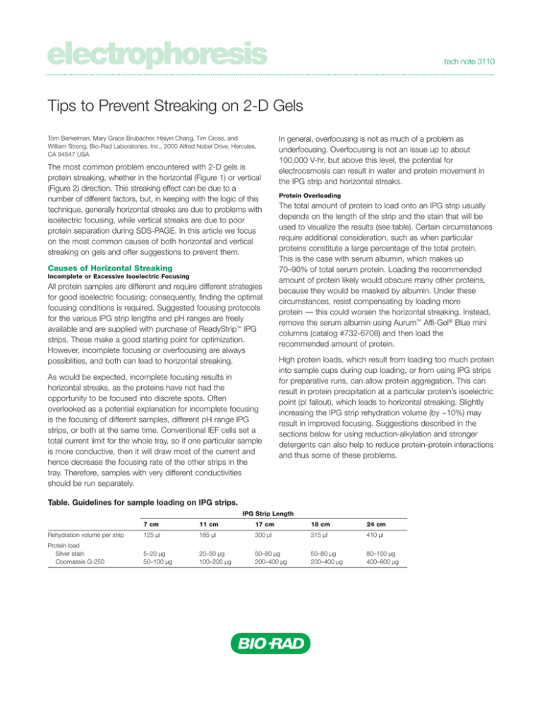
electrophoresis tech note 3110 Tips to Prevent Streaking on 2-D Gels Tom Berkelman, Mary Grace Brubacher, Haiyin Chang, Tim Cross, and William Strong, Bio-Rad Laboratories, Inc., 2000 Alfred Nobel Drive, Hercules, CA 94547 USA The most common problem encountered with 2-D gels is protein streaking, whether in the horizontal (Figure 1) or vertical (Figure 2) direction. This streaking effect can be due to a number of different factors, but, in keeping with the logic of this technique, generally horizontal streaks are due to problems with isoelectric focusing, while vertical streaks are due to poor protein separation during SDS-PAGE. In this article we focus on the most common causes of both horizontal and vertical streaking on gels and offer suggestions to prevent them. Causes of Horizontal Streaking Incomplete or Excessive Isoelectric Focusing All protein samples are different and require different strategies for good isoelectric focusing; consequently, finding the optimal focusing conditions is required. Suggested focusing protocols for the various IPG strip lengths and pH ranges are freely available and are supplied with purchase of ReadyStrip™ IPG strips. These make a good starting point for optimization. However, incomplete focusing or overfocusing are always possiblities, and both can lead to horizontal streaking. As would be expected, incomplete focusing results in horizontal streaks, as the proteins have not had the opportunity to be focused into discrete spots. Often overlooked as a potential explanation for incomplete focusing is the focusing of different samples, different pH range IPG strips, or both at the same time. Conventional IEF cells set a total current limit for the whole tray, so if one particular sample is more conductive, then it will draw most of the current and hence decrease the focusing rate of the other strips in the tray. Therefore, samples with very different conductivities should be run separately. In general, overfocusing is not as much of a problem as underfocusing. Overfocusing is not an issue up to about 100,000 V-hr, but above this level, the potential for electroosmosis can result in water and protein movement in the IPG strip and horizontal streaks. Protein Overloading The total amount of protein to load onto an IPG strip usually depends on the length of the strip and the stain that will be used to visualize the results (see table). Certain circumstances require additional consideration, such as when particular proteins constitute a large percentage of the total protein. This is the case with serum albumin, which makes up 70–90% of total serum protein. Loading the recommended amount of protein likely would obscure many other proteins, because they would be masked by albumin. Under these circumstances, resist compensating by loading more protein — this could worsen the horizontal streaking. Instead, remove the serum albumin using Aurum™ Affi-Gel® Blue mini columns (catalog #732-6708) and then load the recommended amount of protein. High protein loads, which result from loading too much protein into sample cups during cup loading, or from using IPG strips for preparative runs, can allow protein aggregation. This can result in protein precipitation at a particular protein’s isoelectric point (pI fallout), which leads to horizontal streaking. Slightly increasing the IPG strip rehydration volume (by ~10%) may result in improved focusing. Suggestions described in the sections below for using reduction-alkylation and stronger detergents can also help to reduce protein-protein interactions and thus some of these problems. Table. Guidelines for sample loading on IPG strips. IPG Strip Length 7 cm 11 cm 17 cm 18 cm 24 cm Rehydration volume per strip 125 µl 185 µl 300 µl 315 µl 410 µl Protein load Silver stain Coomassie G-250 5–20 µg 50–100 µg 20–50 µg 100–200 µg 50–80 µg 200–400 µg 50–80 µg 200–400 µg 80–150 µg 400–800 µg A treating the samples with nucleases prior to IEF (Garfin and Heerdt 2000). However, these nucleases will be present in the 2-D gel. Alternatively, nucleic acids can be removed by ultracentrifugation in the presence of spermine (Rabilloud 1996). Polysaccharides that contain sialic acid and are negatively charged can produce horizontal streaking artifacts similar to those seen with nucleic acids. Uncharged polysaccharides can also cause streaking by blocking the pores of the gel, preventing sample entry into the IPG strip, and physically impeding focusing. Ultracentrifugation is often sufficient to remove carbohydrates. B Fig. 1. Example of horizontal streaking and its prevention. Detergent was effectively removed by the ReadyPrep 2-D cleanup kit, leading to improved 2-D SDS-PAGE separation. E. coli extracts were spiked with 1% SDS. A, untreated sample; B, sample treated with the ReadyPrep 2-D cleanup kit. Sample Preparation Problems The most common cause of horizontal streaking is a problem with sample preparation. Any contaminant with a net ionic charge will affect isoelectric focusing and lead to horizontal streaking (Rabilloud 2000). Common contaminants include salt, detergents (see Figure 1), peptides, nucleic acids, lipids, and phenolic compounds. Ionic contaminants usually cause high current (around 50 µA) during isoelectric focusing. This current will limit the voltage of the isoelectric focusing cell and increase isoelectric focusing run times, as the voltage will not increase until the ionic contaminants have been depleted — a process that takes many hours (Rémy et al. 2000). This generally leads to underfocusing and streaking. Ionic contaminants can also cause excessive IPG strip heating or strip burning. To alleviate these problems, salt can be removed from the sample using dialysis, desalting columns, or the ReadyPrep 2-D cleanup kit (catalog #163-2130), which removes salts and most other ionic contaminants. Certain charged detergents, such as SDS, can coat proteins and drastically alter their net charge, ultimately affecting their isoelectric point and leading to streaking and loss of sample from the IPG strip. Nucleic acid contamination of samples also can contribute to horizontal streaking (Görg et al. 1997). Many proteins bind nucleic acids, which can generate a mixed population of protein-nucleic acid species, each with distinct isoelectric points. Nucleic acids can be removed from samples by In addition, carbamylation of polypeptides can generate charge heterogeneity in individual proteins, and although discrete trains of spots are often formed, under certain conditions — such as when broad pH gradients are used — smearing between spots can occur. Protein samples should be prepared using high-purity urea and should not be heated above 30°C. The last cause of horizontal streaking related to sample preparation is disulfide bond formation (Walsh and Herbert 1998). When disulfide bonds occur in a protein sample prior to 2-D, they occur randomly intra- and intermolecularly. Disulfide bond formation creates various protein configurations, including minor isoelectric point variants (appearing on a 2-D gel as a widening of a single protein spot) and multimers of the same protein (appearing as streaking tails on spots, phantom spots, missing spots, and smearing). Preventing disulfide bond formation eliminates these artifacts. Disulfide bond artifacts are particularly problematic in two instances: 1) analysis of basic proteins, and 2) use of longer IPG strips. Horizontal streaking due to disulfide bond formation can be reduced using the ReadyPrep reduction-alkylation kit (catalog #163-2090) in conjunction with cup loading of IPG strips (Bio-Rad bulletin 4006216). Poor Protein Solubilization Failure to completely solubilize all the proteins in a sample will result in horizontal streaking. The main components of standard sample buffers used in 2-D electrophoresis to maintain sample solubility are urea, a nonionic detergent such as Nonidet P-40 (NP-40) or a zwitterionic detergent such as CHAPS, and a reducing agent such as dithiothreitol (DTT). Other compounds can be included in this mixture, for example, thiourea (Rabilloud 1998) or novel detergents, especially those in the amido sulfobetaine family, such as ASB-14 (Rabilloud 1996), to improve the solubilization of membrane proteins. The combined effect of these reagents is to reduce horizontal streaking by better solvating the hydrophobic regions on the proteins and diminishing aggregation due either to hydrogen bonding among proteins or to interactions with the IPG gel matrix. Finally, it is always good practice to remove insoluble protein complexes by centrifugation prior to isoelectric focusing to prevent them from interfering with the entry of other proteins into the gel matrix. Electroendoosmotic Flow A Mainly a problem during isoelectric focusing of basic proteins, electroendoosmotic flow can contribute to horizontal streaking due to water transport from the cathode to the anode. The water flow slows protein migration in the opposite direction and can also cause strip dehydration at the cathode end. Solutions to this problem include replacing the paper wick at the cathode with a fresh water-soaked wick or adding organic modifiers such as glycerol, isopropyl alcohol, or methylcellulose to the rehydration/sample solution (Görg et al. 1997). Causes of Vertical Streaking Poor Protein Solubility Following the first-dimension separation, all of the proteins have moved to their isoelectric points and carry no net charge. The ionic strength of the medium is also very low, because small ions have all migrated out of the first-dimension strip. Proteins generally have minimal solubility under these conditions and are often precipitated within the gel matrix despite the presence of detergents, chaotropic agents, and other solubilizing additives. The major purpose of the equilibration step following the first dimension is to coat the proteins with SDS, giving them a negative charge and making them soluble again so that they may enter the seconddimension gel. B Protein Overloading When a large quantity of protein is loaded, or when the sample contains proteins of particularly high abundance, resolubilization during equilibration may be incomplete, resulting in vertical streaking and tailing of the most intense protein spots. This can be prevented by limiting the amount of protein loaded onto the first-dimension strip. It is often possible to compensate for a lower protein load by using a more sensitive staining technique (for example, silver stain, catalog #161-0449, or SYPRO Ruby protein gel stain, catalog #170-3125, instead of Coomassie Blue stain). In some cases, vertical streaking of abundant proteins can be prevented by prolonging the equilibration time (see Ineffective Equilibration below). Overfocusing Isoelectric precipitation increases with focusing time, so vertical streaking may be minimized by ensuring that firstdimension isoelectric focusing is not conducted for any longer than necessary. Ineffective Equilibration Equilibration should be carried out in a manner that ensures good penetration of SDS and therefore protein solubilization. The equilibration tray should be shaken or rocked to ensure continual movement of solution. Equilibration may be prolonged to as long as 45 min if necessary. This may result in the loss of some small polypeptides (<15 kD), but loss of larger proteins is generally insignificant even with prolonged equilibration. SDS should be present at a concentration of at least 2% (w/v), particularly if high (>2%) concentrations of detergent were used in the first dimension. Other components of the equilibration solution are important as well. The solution should be buffered, particularly if an alkylation step is employed, because iodoacetamide treatment generates acid. The optimal pH for equilibration is 8.8. Glycerol, which should Fig. 2. Examples of vertical streaking. A, approximately 300 µg of E. coli protein loaded onto a 17 cm pH 3–10 ReadyStrip™ IPG strip. B, a prefractionated sample of Pseudomonas putida loaded onto a 17 cm pH 3–10 ReadyStrip IPG strip. Gel image was annotated using The Discovery Series™ PDQuest™ 2-D analysis software. be present at a concentration of at least 20% (v/v), is a particularly important component of the equilibration solution, and its omission will result in vertical streaking. Protein Oxidation Oxidative crosslinking or protein refolding should be prevented during all steps of the 2-D process; protein oxidation during the second-dimension separation can result in vertical streaking. Treatment with the ReadyPrep reduction-alkylation kit (catalog #163-2090) prior to first-dimension focusing can block cysteine sulfhydryl groups and prevent their reoxidation. If the sample is not alkylated, alkylation should be performed in a second equilibration step with 2.5% iodoacetamide. If alkylation is not desired, a sufficient quantity of DTT, at least 1%, should be present in the equilibration solution. The DTT should be added to the equilibration solution immediately before use, as it may oxidize if stored. DTT is negatively charged under second-dimension conditions and will migrate ahead of proteins in the gel, ensuring a reducing environment during the separation. Poor Reagent Quality or Improperly Prepared Solutions Vertical Gaps or Vertical Blank Stripes Vertical streaking and other second-dimension problems often result from poor reagent quality or from mistakes in solution preparation. Care should be taken to use the highest-quality reagents (for example, ReadyPrep proteomics grade water, catalog #163-2091), and the pH of all buffers should be verified. Precast second-dimension gels should be used before their expiration date. Poor-quality acrylamide or old acrylamide stock solutions can cause vertical streaking due to an incompletely polymerized second-dimension gel. Reusing electrophoresis running buffer can result in poor separation and vertical streaking due to the depletion of ions and SDS in the running buffer. This practice should be avoided if vertical streaking is a persistent problem. Vertical gaps or blank stripes, for the purposes of this discussion, are different from vertical streaking. These problems can be due to trapped air bubbles in the agarose overlay on the IPG strip, excessive DTT (>50 mM) in the IPG sample buffer, or a compromised IPG strip (either insufficiently rehydrated or torn during handling). Blank stripes near the electrode, especially the cathode, can be caused by a buildup of salt. Poor Placement of the IPG Strip on the Second-Dimension Gel Vertical streaking can also be the result of gaps between the IPG strip and the second-dimension gel or damage to the IPG strip during application. The second-dimension gel should have a straight, level top edge, and care should be taken that the IPG strip is in direct contact with the second-dimension gel along its entire length. The IPG strip should not be twisted during placement. This can be prevented by ensuring that the plastic backing of the IPG strip is pressed firmly against one of the second-dimension gel plates. An agarose overlay should be used to prevent the IPG strip from coming loose or moving. Bubbles in the agarose overlay should be minimized. Uneven Pore Size in the IPG Strip Following IEF After the first-dimension separation, there are often variations in the thickness of the IPG strip. This swelling or thinning of some sections is caused by water movement in the gel (Rabilloud 2000). In thinner sections of the IPG strip, the gel pore size may have been reduced to the point that protein migration out of the IPG strips will be slowed or prevented. This can result in vertical streaks or, in severe cases, complete protein loss from trapping within the IPG strip. Water movement during first-dimension separation can be minimized by ensuring that the sample is well desalted and that focusing is not carried out any longer than necessary. Summary As alluded to at the start of this article, streaking on 2-D gels is a common and complex problem. We have identified useful remedies for some of the main causes of such streaking, although we have not presented an exhaustive list of the potential causes. In addition, the great deal of variation in the characteristics of protein samples means that each sample will behave uniquely. With such variety in the composition of protein samples, the sample preparation step becomes important to prevent frequently observed streaking patterns; this article has focused on a variety of approaches to help achieve this. For more information and for answers to other technical questions, contact us at consult.bio-rad.com References Bio-Rad Laboratories, Cup loading tray for the PROTEAN IEF cell instruction manual, Bio-Rad bulletin 4006216 Garfin D and Heerdt L (eds), 2-D Electrophoresis for Proteomics: A Methods and Product Manual, Bio-Rad bulletin 2651 (2000) Görg A et al., Very alkaline immobilized pH gradients for two-dimensional electrophoresis of ribosomal and nuclear proteins, Electrophoresis 18, 328–337 (1997) Rabilloud T, Solubilization of proteins for electrophoretic analyses, Electrophoresis 17, 813–829 (1996) Rabilloud T, Use of thiourea to increase the solubility of membrane proteins in two-dimensional electrophoresis, Electrophoresis 19, 758–760 (1998) Rabilloud T, Proteome Research: Two-Dimensional Gel Electrophoresis and Identification Methods, Springer, Berlin (2000) Rémy A et al., Focusing strategy and influence of conductivity on isoelectric focusing in immobilized pH gradients, Bio-Rad bulletin 2778 (2000) Walsh BJ and Herbert B, Setting up two-dimensional gel electrophoresis for proteome projects, ABRF News 9, 11–21 (1998) Coomassie is a trademark of BASF Aktiengesellschaft. Nonidet is a trademark of Shell International Petroleum Company. SYPRO is a trademark of Molecular Probes, Inc. Bio-Rad Laboratories, Inc. Web site www.bio-rad.com USA (800) 4BIORAD Australia 02 9914 2800 Austria (01)-877 89 01 Belgium 09-385 55 11 Brazil 55 21 2527 3454 Canada (905) 712-2771 China (86-21) 63052255 Czech Republic + 420 2 41 43 05 32 Denmark 44 52 10 00 Finland 09 804 22 00 France 01 47 95 69 65 Germany 089 318 84-0 Hong Kong 852-2789-3300 Hungary 36 1 455 8800 India (91-124)-6398112/113/114, 6450092/93 Israel 03 951 4127 Italy 39 02 216091 Japan 03-5811-6270 Korea 82-2-3473-4460 Latin America 305-894-5950 Mexico 55-52-00-05-20 The Netherlands 0318-540666 New Zealand 64 9 415 2280 Norway 23 38 41 30 Poland + 48 22 331 99 99 Portugal 351-21-472-7700 Russia 7 095 721 1404 Singapore 65-6415 3188 South Africa 00 27 11 4428508 Spain 34 91 590 5200 Sweden 08 555 12700 Switzerland 061 717-9555 Taiwan (8862) 2578-7189/2578-7241 United Kingdom 020 8328 2000 Life Science Group Bulletin 3110 US/EG Rev A 04-0240 0604 Sig 1103
