An Interaction of Electromagnetic Wave and Human Skin Membrane
advertisement
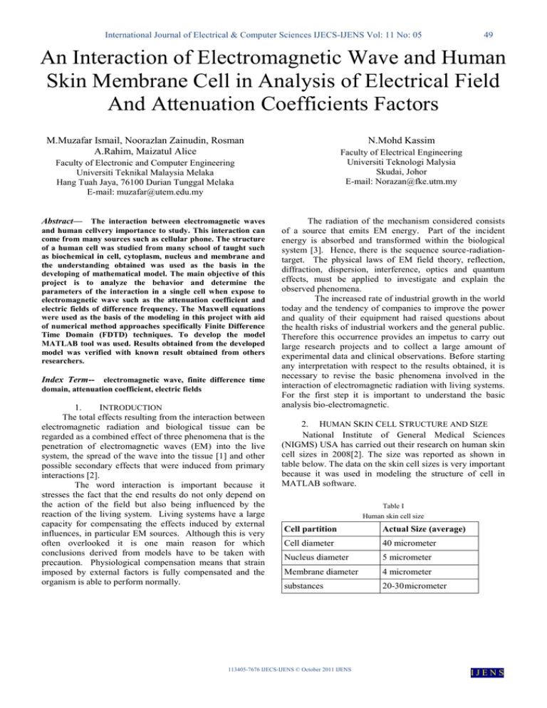
International Journal of Electrical & Computer Sciences IJECS-IJENS Vol: 11 No: 05 49 An Interaction of Electromagnetic Wave and Human Skin Membrane Cell in Analysis of Electrical Field And Attenuation Coefficients Factors M.Muzafar Ismail, Noorazlan Zainudin, Rosman A.Rahim, Maizatul Alice N.Mohd Kassim Faculty of Electrical Engineering Universiti Teknologi Malysia Skudai, Johor E-mail: Norazan@fke.utm.my Faculty of Electronic and Computer Engineering Universiti Teknikal Malaysia Melaka Hang Tuah Jaya, 76100 Durian Tunggal Melaka E-mail: muzafar@utem.edu.my Abstract— The interaction between electromagnetic waves and human cellvery importance to study. This interaction can come from many sources such as cellular phone. The structure of a human cell was studied from many school of taught such as biochemical in cell, cytoplasm, nucleus and membrane and the understanding obtained was used as the basis in the developing of mathematical model. The main objective of this project is to analyze the behavior and determine the parameters of the interaction in a single cell when expose to electromagnetic wave such as the attenuation coefficient and electric fields of difference frequency. The Maxwell equations were used as the basis of the modeling in this project with aid of numerical method approaches specifically Finite Difference Time Domain (FDTD) techniques. To develop the model MATLAB tool was used. Results obtained from the developed model was verified with known result obtained from others researchers. Index Term-- electromagnetic wave, finite difference time domain, attenuation coefficient, electric fields 1. INTRODUCTION The total effects resulting from the interaction between electromagnetic radiation and biological tissue can be regarded as a combined effect of three phenomena that is the penetration of electromagnetic waves (EM) into the live system, the spread of the wave into the tissue [1] and other possible secondary effects that were induced from primary interactions [2]. The word interaction is important because it stresses the fact that the end results do not only depend on the action of the field but also being influenced by the reaction of the living system. Living systems have a large capacity for compensating the effects induced by external influences, in particular EM sources. Although this is very often overlooked it is one main reason for which conclusions derived from models have to be taken with precaution. Physiological compensation means that strain imposed by external factors is fully compensated and the organism is able to perform normally. The radiation of the mechanism considered consists of a source that emits EM energy. Part of the incident energy is absorbed and transformed within the biological system [3]. Hence, there is the sequence source-radiationtarget. The physical laws of EM field theory, reflection, diffraction, dispersion, interference, optics and quantum effects, must be applied to investigate and explain the observed phenomena. The increased rate of industrial growth in the world today and the tendency of companies to improve the power and quality of their equipment had raised questions about the health risks of industrial workers and the general public. Therefore this occurrence provides an impetus to carry out large research projects and to collect a large amount of experimental data and clinical observations. Before starting any interpretation with respect to the results obtained, it is necessary to revise the basic phenomena involved in the interaction of electromagnetic radiation with living systems. For the first step it is important to understand the basic analysis bio-electromagnetic. 2. HUMAN SKIN CELL STRUCTURE AND SIZE National Institute of General Medical Sciences (NIGMS) USA has carried out their research on human skin cell sizes in 2008[2]. The size was reported as shown in table below. The data on the skin cell sizes is very important because it was used in modeling the structure of cell in MATLAB software. Table I Human skin cell size Cell partition Actual Size (average) Cell diameter 40 micrometer Nucleus diameter 5 micrometer Membrane diameter 4 micrometer substances 20-30 micrometer 113405-7676 IJECS-IJENS © October 2011 IJENS IJENS International Journal of Electrical & Computer Sciences IJECS-IJENS Vol: 11 No: 05 50 3. CELL MEMBRANE EFFECT The biological action of low intensity non-ionizing electromagnetic radiation on molecular and super molecular level was first established. The electromagnetic fields induce the emergence of the electric charges on cell membrane that later on would brings the additional electric potential on cellular membrane. The microwave radiation causes significant changes on different type of cell membrane characteristics, basically it induce increase of membrane permeability for sodium, potassium and chlorine ions and inhibit the active transport of these ions via lipid membranes [9]. 4. NUMERICAL SOLUTIONS FOR HUMAN CELL Numerical solutions can be categorized into the domain solution, which includes the whole domain as the operational area, and the boundary solution. The former is also called a differential solution, and the latter, an integral solution [4]. The domain solutions include the finite difference time domain (FDTD) method in which it is a simple numerical technique that is used in solving problems that are uniquely defined by three things: 1) A partial differential equation such as Laplace’s or Poisson’ equation 2) A solution region 3) Boundary and/or initial conditions A finite difference time domain method such as Poisson’s or Laplace’s equation proceeds in three steps: 1) Dividing the solution region into a grid of nodes 2) Approximating the differential equation and boundary conditions by a set of linear algebraic equations (difference equations) on grid points within solution region. 3) Solving this set of algebraic equations. Fig. 1. A typical finite difference mesh for an integrated wave propagation. The electric field profile, E(x, y) has been divided into small rectangular cells . 5. CELL STRUCTURE In the MATLAB, cell structure is designed following the standard from NIGMS cell size measurement. The cell consists of three parts which are the 3 layer membrane, chemical substances and nucleus. Fig. 2. (a): cell structure and partition The basic formulation that governs the propagation of wave in the human skin cell is a Maxwell’s equations and it derives to be equation 1 to obtaining electromagnetic field (E-field) [5]. x 2 E (i 1, j ) E (i 1, j ) 2 ( E (i, j 1) E (i, j 1)) y E (i, j ) 2 x 2 2 21 2 x 2 k0 n2 i, j 2 y (1) Lossless electromagnetic wave Fig. 2(b). three layer membrane cell, layer 1=lipid, layer 2=protein and layer 3=fat 6. ASSUMPTIONS In this project the three assumptions were made; the cell is considered as a box and has a flat surface (Figure 3(a)). In addition, the wave is propagated from only one source and transmits only on one side of a cell (Figure 3(b)). The cell will become damaged once the electromagnetic waves able to penetrate deep into the nucleus (Figure 3(c)). 113405-7676 IJECS-IJENS © October 2011 IJENS IJENS International Journal of Electrical & Computer Sciences IJECS-IJENS Vol: 11 No: 05 frequency Free space Wavelength, Propagation velocity, vp ʎ (m) constant,β (m/s) (rad/m) 2.36GHz 3x 10⁸ 0.13 49.4 3x 10 8 0.07 96.2 9GHz 3x 10 8 0.03 188 15GHz 3x 10⁸ 0.02 814 4.5GHz Fig. 3(a). 51 9. AN INTERACTION OF EM ON A HUMAN SKIN CELL From the MATLAB simulation the orange contour shows how the E-field penetrates into the human cell from layer 1 (lipid) to layer 2 (protein) and layer 3 (fat). At 2.36 GHz the E-field contour was refracted to chemicals and nucleus as shown at figure 4(a). On the contrary, at 4.5 GHz in which after penetrating the third layer (fat), the pattern of the contour propagate only chemicals in the human skin cell as shown in Figure 4(b). At 9 GHz the contour patterns are found nearly at the third layer of the cell membranes which electromagnetic waves propagating not very far away as shown in Figure 4(c). This indicates that the high frequency electromagnetic waves cannot propagate in a far distance and electric field cannot penetrate the cell other that the cell membrane. In contrast to the low frequency, the wave can penetrate more than the cell membrane. Fig. 3(b). Fig. 3(c). 7. THE PARAMETER CONSIDERED IN MATLAB DESIGN Based on the wave sources, permittivity and conductivity parameters are to be considered in the MATLAB simulation. Parameter was taken based on 4 sample frequencies which are 2.36 GHz, 4.5GHz, 9GHz and 15GHz. The permittivity and conductivity for each sample frequency is as shown in Table II. Table II permittivity and conductivity parameter Fig. 4(a). Movement of E-field at 2.36 GHz Frequencies (Hz) layer1(lipid) layer2(protein) layer3(fat) σeff (S/M) ɛr σeff (S/M) ɛr σeff (S/M) ɛr 2.36GHz 0.61 53 0.5 44.6 0.04 17.03 4.5GHz 0.72 51.3 1 0.55 42.48 0.05 15.03 9 GHz 0.87 41.4 0.59 38.9 0.11 11.3 15GHz 1.19 39 0.92 37 0.19 11 8. ELECTROMAGNETIC WAVE AT LOSSLESS MATERIAL The distance between the cellular source of electromagnetic waves and human cells was set to 10 meters. Table III shows the parameters of electromagnetic waves at a lossless material at different frequency value. Assuming that the wave moves at the velocity VP = 3x10⁸, this means that electromagnetic waves propagate very quickly at the highest frequency before entering human cells. Fig. 4(b). Movement of E-field at 4.5 GHz Table III Electromagnetic wave parameter at lossless material in different frequency. 113405-7676 IJECS-IJENS © October 2011 IJENS IJENS International Journal of Electrical & Computer Sciences IJECS-IJENS Vol: 11 No: 05 52 the frequency, it will increase the attenuation and in contrast decrease the skin depth. It will affect the value of electrical field and propagation of the electromagnetic wave. he project analysis also proof that MRI exhibit the same scenario as cellular phone and the electromagnetic wave could propagate deep into a human skin cell at 2.36GHz. Fig. 4(c). Movement of E-field at 9 GHz 10. THE VALUE OF E- FIELD FROM MATLAB SIMULATION When the electromagnetic wave propagates from a transmitter source to human skin cells, it will first interact with the cell membranes. The value of electrical field (Efield) at wave interact with cell, E0 was obtained when the E- field is at its highest in the first layer. When the wave propagates in the second layer and so on, the electrical field will decrease due to absorption of energy in human cells. E 0 vs. layer graph shows an exponential decline of the E0. When the frequency increases, E0 will decrease and the wave cannot penetrate far in cell. When electromagnetic wave penetrates the second and third layer more energy will be absorbed by the cell and it causes E0 to be reduced. Table IV and figure 4, correspondingly shows the comparison table and graph that is to compare the value of E0 at different membrane layer and frequency. 12. ATTENUATION COEFFICIENTS The attenuation constant (equation 2) , represents how fast the wave attenuates as it propagates. The units of attenuation can also be thought of as loss per meter. Increasing the effective conductivity or frequency increases the loss and hence the attenuation. Beside that when electromagnetic wave propagate to inner cell, example at layer 2 and layer 3 the value of attenuation was decrease (Figure 7). efff 2 1 ' ' 1 ' ' 2 (2) Table IV Comparison table of E0 at different membrane layer, and frequency. frequency layer1(lipid) layer2(protein) layer3(fat) 2.36GHz 0.8 0.5 0.32 4.5GHz 0.78 0.39 0.2 9GHz 0.72 0.34 0.13 15GHz 0.68 0.21 0.12 Fig. 7. Graph of attenuation, α versus layer at different frequency CONCLUSION The size of human skin cells were taken from an actual size and been modeled by using MATLAB software. From the simulation results, at 2.36 GHz the electromagnetic field was found to be larger than the other frequency values and the probability of the electromagnetic field up to the nucleus was also high and the attenuation value was decrease. At 2.36 GHz attenuation values were found lower and the skin depth was high. This shows that the electromagnetic waves can propagate over a long distance in a lower frequency. Assuming that the electromagnetic waves were able to propagate into the nucleus and the electromagnetic field was still high in the third layer of membrane, human skin cells will become damaged. The damaged cell may cause various diseases such as skin cancer and therefore very dangerous to human and likewise other inhabitant of earth. From this finding, it is strongly encourage that the telecommunications company and related industries should design their equipment at a 13. Fig. 5. Comparison figure of E0 at different membrane layer, and frequency. 11. VERIFICATION OF THE RESULT Comparing with this literature, the results obtained from this project shows that if the value of frequency increases, there will be an increase in the conductivity σ (0) and decrease the permittivity ε (x). Therefore by increasing 113405-7676 IJECS-IJENS © October 2011 IJENS IJENS International Journal of Electrical & Computer Sciences IJECS-IJENS Vol: 11 No: 05 53 higher frequency and try to avoid operating their equipment at 2.36 GHz frequency range or lower. ACKNOWLEDGEMENT I would like to thank Universiti Teknikal Malaysia Melaka for the support of my project. REFERENCES [1] [2] [3] [4] [5] [6] [7] [8] [9] [10] [11] [12] [13] [14] Cynthia furse, Douglas A. Christensen & Carl H.Durney. Basic Introduction to Bioelectromagnetic CRC Press, New York, 2009 Andre Vander Vorst, Arye Rosen, Youj Kotsuka. RF/Microwave Interaction with Biological Tissues. United State, America; 2007;pp 22-23 Frank S. Barnes, Ben Greenebaum, Biological and Medical Aspect of Electromagnetic Fields. CRC Press, New York;2007;pp45-46 C.H See, R.A Abd-Alhameed and P.S. Excell. Computational of Electromagnetic Fields In Dense Biological Cell Structure Using Modified Subgridding Of Quasi Static FDTD Method.IEEE Journal 5600-234; 2007 Keith D. Paulsen . Finite Element Modeling In Therapeutic and Diagnostic Application Proceeding-19thInternationa Conference.0-7803-4262-3/07; 2007 Gerard H. Markx .The Use Of Electric Fields In Tissue Engineering. Science and Medical Journal. Scorpus Journal;2007 Karl H.Schoenbach, Jing dong Deng, Guofen Yu, Robert H.Stark Electromagnetic Effect On Biological Cells.IEEE Journal.0-7803-6513-5;2007 Timothy E. Vaughn and Jamesc.Weaver Energy Constraints on The Creation of CellMembrane Pores By Magnetic Particle D. Poljak . Human Exposure To Electromagnetic Fields , WIT PRESS Southampton Boston;2006 Bin He .Modeling and Imaging of Bioelectrical ActivityPrinciple and Applications. Plenum Publisher, New York;2006 Schkorbatov Y.G, Kolchigi ,N.N,Grabin ,V.A, Pasiuga V.N, Kazansky O.V Cells. Effects of Electromagnetic Radiation Ultrawideband and ultrashort impulse Signals, 18-22 September, 2006 Sevastopol, Ukraine;2006 Rui Qiang, Dagang Wu, Ji chen, Shumin Wang, Donald Wilton, and Wolfgng Kainz . An Efficient Two Dimensional FDTD Method for Bioelectromagnetic Applications.IEEE Journal; 2006,pp34-35 Mohamed A.Eleiwa,Atef Z Elsherbeni. Accurate FDTD Simulation of Biological Tissue For Bioelectromagnetic IEEE Journal; 2006 Yao Xueling, Chen Jingliang, Sun Wei, Xu Chuanxiang. The Experimental Study Of Pulse Current Magnetic Fields on Human Cell.Science and medical Journal;2006 113405-7676 IJECS-IJENS © October 2011 IJENS IJENS
