MRI Pulse Sequence Acronyms: Hitachi, GE, Philips, Siemens, Toshiba
advertisement
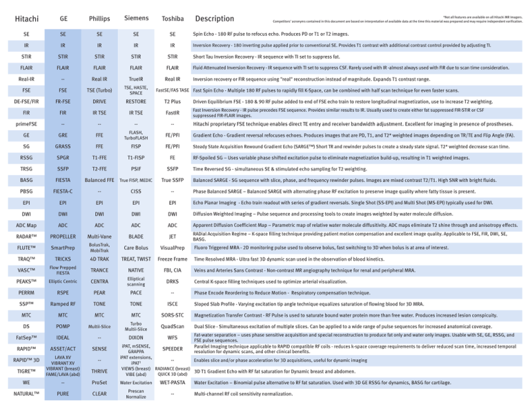
Description Hitachi GE Phillips Siemens Toshiba SE SE SE SE SE Spin Echo - 180 RF pulse to refocus echo. Produces PD or T1 or T2 images. IR IR IR IR IR Inversion Recovery - 180 inverting pulse applied prior to conventional SE. Provides T1 contrast with additional contrast control provided by adjusting TI. STIR STIR STIR STIR STIR Short Tau Inversion Recovery - IR sequence with TI set to suppress fat. FLAIR FLAIR FLAIR FLAIR FLAIR Fluid Attenuated Inversion Recovery - IR sequence with TI set to suppress CSF. Rarely used with IR -almost always used with FIR due to scan time consideration. Real-IR -- Real IR TrueIR Real IR FSE FSE TSE (Turbo) TSE, HASTE, SPACE DE-FSE/FIR FR-FSE DRIVE RESTORE FIR FIR IR TSE primeFSE -- *Not all features are available on all Hitachi MR Imagers. Competitors’ acronyms contained in this document are based on interpretation of available data at the time this material was prepared and may require independent varification. Inversion recovery or FIR sequence using "real" reconstruction instead of magnitude. Expands T1 contrast range. FastSE/FAS TASE Fast Spin Echo - Multiple 180 RF pulses to rapidly fill K-Space, can be combined with half scan technique for even faster scans. T2 Plus Driven Equilibrium FSE - 180 & 90 RF pulse added to end of FSE echo train to restore longitudinal magnetization, use to increase T2 weighting. Fast Inversion Recovery - IR pulse precedes FSE sequence. Provides similar results to IR. Usually used to create either fat suppressed FIR-STIR or CSF suppressed FIR-FLAIR images. IR TSE FastIR -- -- -- Hitachi proprietary FSE technique enables direct TE entry and receiver bandwidth adjustment. Excellent for imaging in presence of prostheses. FE/PFI Gradient Echo - Gradient reversal refocuses echoes. Produces images that are PD, T1, and T2* weighted images depending on TR/TE and Flip Angle (FA). Steady State Acquisition Rewound Gradient Echo (SARGE™) Short TR and rewinder pulses to create a steady state signal. T2* weighted decrease scan time. GE GRE FFE FLASH, TurboFLASH SG GRASS FFE FISP FE/PFI RSSG SPGR T1-FFE T1-FISP FE TRSG SSFP T2-FFE PSIF SSFP BASG FIESTA Balanced FFE True FISP, MEDIC True SSFP PBSG FIESTA-C -- CISS -- EPI EPI EPI EPI EPI Echo Planar Imaging - Echo train readout with series of gradient reversals. Single Shot (SS-EPI) and Multi Shot (MS-EPI) typically used for DWI. DWI DWI DWI DWI DWI Diffusion Weighted Imaging – Pulse sequence and processing tools to create images weighted by water molecule diffusion. ADC Map ADC ADC ADC ADC Apparent Diffusion Coefficient Map – Parametric map of relative water molecule diffusitivity. ADC maps eliminate T2 shine through and anisotropy effects. RADAR™ PROPELLER Multi-Vane BLADE JET RADial Acquisition Regime – K-space filling technique providing patient motion compensation and excellent image quality. Applicable to FSE, FIR, DWI, SE, BASG. FLUTE™ SmartPrep BolusTrak, MobiTrak Care Bolus VisualPrep TRAQ™ TRICKS 4D TRAK TREAT, TWIST Freeze Frame VASC™ Flow Prepped FIESTA TRANCE NATIVE FBI, CIA DRKS PEAKS™ Elliptic Centric CENTRA Elliptical scanning PERRM RSPE PEAR PACE -- SSP™ Ramped RF TONE TONE ISCE MTC MTC MTC MTC SORS-STC Multii-Slice Turbo Multii-Slice QuadScan DS FatSep™ RAPID™ RAPID™ 3D TIGRE™ WE NATURAL™ POMP IDEAL ASSET/ACT LAVA XV VIBRANT XV VIBRANT (breast) FAME/LAVA (abd) -PURE -SENSE -THRIVE DIXON WFS RF-Spoiled SG – Uses variable phase shifted excitation pulse to eliminate magnetization build-up, resulting in T1 weighted images. Time Reversed SG - simultaneous SE & stimulated echo sampling for T2 weighting. Balanced SARGE - SG sequence with slice, phase, and frequency rewinder pulses. Images are mixed contrast T2/T1. High SNR with bright fluids. Phase Balanced SARGE – Balanced SARGE with alternating phase RF excitation to preserve image quality where fatty tissue is present. Fluoro Triggered MRA - 2D monitoring pulse used to observe bolus, fast switching to 3D when bolus is at area of interest. Time Resolved MRA - Ultra fast 3D dynamic scan used in the observation of blood kinetics. Veins and Arteries Sans Contrast - Non-contrast MR angiography technique for renal and peripheral MRA. Central K-space filling techniques used to optimize arterial visualization. Phase Encode Reordering to Reduce Motion - Respiratory compensation technique. Sloped Slab Profile - Varying excitation tip angle technique equalizes saturation of flowing blood for 3D MRA. Magnetization Transfer Contrast - RF Pulse is used to saturate bound water protein more than free water. Produces increased lesion conspicuity. Dual Slice - Simultaneous excitation of multiple slices. Can be applied to a wide range of pulse sequences for increased anatomical coverage. Fat-water separation – uses phase sensitive acquisition and special reconstruction to produce fat only and water only images. Usable with SE, GE, RSSG, and FSE pulse sequences. Parallel Imaging technique applicable to RAPID compatible RF coils - reduces k-space coverage requirements to deliver reduced scan time, increased temporal resolution for dynamic scans, and other clinical benefits. iPAT, mSENSE, SPEEDER GRAPPA iPAT extensions, Enables slice and/or phase acceleration for 3D acquisitions, useful for dynamic imaging -iPAT2 VIEWS (breast) RADIANCE (breast) 3D T1 Gradient Echo with RF fat saturation for Dynamic breast and abdomen. QUICK 3D (abd) VIBE (abd) ProSet Water Excitation WET-PASTA CLEAR Prescan Normalize -- Water Excitation – Binomial pulse alternative to RF fat saturation. Used with 3D GE RSSG for dynamics, BASG for cartilage. Multi-channel RF coil sensitivity normalization.
