Oncogene (2004) 23, 260–266
& 2004 Nature Publishing Group All rights reserved 0950-9232/04 $25.00
www.nature.com/onc
Msh2 deficiency does not contribute to cisplatin resistance in mouse
embryonic stem cells
Nanna Claij1 and Hein te Riele*,1
1
Division of Molecular Biology, The Netherlands Cancer Institute, Plesmanlaan 121, 1066 CX Amsterdam, The Netherlands
Several reports have suggested that a defect in the DNA
mismatch repair (MMR) system not only causes resistance to methylating agents but also confers low-level
resistance to the chemotherapeutic drug cisplatin. Here we
report that in a clonogenic assay, mouse embryonic stem
(ES) cells deficient for the MMR protein MSH2 respond
similarly as wild-type cells to cisplatin. Furthermore,
restoring MSH2 expression in a cisplatin-resistant subclone selected from an Msh2/ cell population did not
sensitize cells to cisplatin. To ascertain that our observations were not the result of a mutation in the Msh2/ cells
that obscured the contribution of a defective MMR
machinery to cisplatin resistance, we made use of the
Cre-lox system to create a cell line in which the Msh2
gene can be conditionally inactivated. However, while de
novo inactivation of Msh2 rendered cells tolerant to the
methylating drug N-methyl-N0 -nitro-N-nitrosoguanidine
as expected, it did not alter the sensitivity to cisplatin. In
addition, we were not able to derive cisplatin-resistant
subclones from this freshly generated MMR-deficient cell
line. Thus, in ES cells we did not find evidence for direct
involvement of MMR deficiency in cisplatin resistance.
Oncogene (2004) 23, 260–266. doi:10.1038/sj.onc.1207015
Keywords: mismatch repair; MSH2; cisplatin; Cre-lox
Introduction
The primary function of DNA mismatch repair (MMR)
is restoring mismatched and unpaired nucleotides that
occur during DNA replication. In addition, the MMR
system mediates the cytotoxicity of several DNAdamaging drugs. Thus, cells deficient for the central
MMR repair proteins MSH2 or MLH1 are highly
tolerant to methylating agents and the base analogue 6thioguanine (Koi et al., 1994; Hawn et al., 1995;
Risinger et al., 1995; Buermeyer et al., 1999; Claij and
Te Riele, 2002). These drugs derive their cytotoxicity
from the formation of O6-methylguanine or S6-methylguanine lesion in DNA, respectively, causing mismatched base pairs (Zhukovskaya et al., 1994; Swann
et al., 1996). Recognition of such lesions by MMR
*Correspondence: H te Riele; E-mail: h.t.riele@nki.nl
Received 20 May 2003; revised 4 July 2003; accepted 10 July 2003
proteins may directly activate the apoptotic machinery.
Alternatively, it was suggested that MMR only recognizes mispairs formed during replication of these lesions,
thereby inhibiting translesion synthesis and causing cell
death. Furthermore, MMR deficiency was reported to
confer low-level resistance to the chemotherapeutic
agent cisplatin (cis-diamminedichloroplatinum). The
observation that MMR proteins can bind to oligonucleotides containing a 1,2-diguanyl intrastrand crosslink
preferably when a T is present opposite the 30 -guanine in
the crosslink, has suggested that here also MMR
counteracts translesion synthesis thereby contributing
to the cytotoxicity of cisplatin (Yamada et al., 1997).
Cisplatin is widely used for the treatment of human
malignancies; however, its clinical effectiveness is limited
by the frequent development of drug-resistant tumor cell
populations during treatment. The mechanism of
resistance is often found to be multifactorial (Kartalou
and Essigmann, 2001); for example, cisplatin-resistant
derivatives of the ovarian tumor cell line A2780 have
increased levels of glutathione, reduced drug accumulation, reduced adduct formation and increased DNA
repair (Siddik et al., 1998). Furthermore, in these
derivatives, cisplatin resistance is often accompanied
by loss of expression of the MMR protein MLH1
(Anthoney et al., 1996; Drummond et al., 1996),
suggesting that the MMR system contributes to the
cellular response to cisplatin. The hMLH1-deficient
colorectal carcinoma cell line HCT116 and the hMSH2deficient endometrial carcinoma cell line HEC59 provided additional indications for the involvement of
MMR proteins in response to cisplatin treatment.
Restoration of the repair capacity by complementation
with chromosome 3 or 2, respectively, sensitized the cells
approximately twofold to cisplatin (Aebi et al., 1996;
Fink et al., 1996). By using the transfer of whole
chromosomes, however, there remains the possibility
that other genes carried on the chromosome influence
the sensitivity; for example, in a cisplatin-resistant,
MLH1-deficient derivative of A2780 cells, correction of
MMR by chromosome 3 transfer sensitized the cells
fivefold (Durant et al., 1999). In contrast, complementation of hMLH1 by expression of the cDNA resulted in
only a 1.3-fold sensitization (Branch et al., 2000),
making the contribution of MMR deficiency to cisplatin
resistance questionable.
We have previously generated mouse embryonic stem
(ES) cells in which both Msh2 alleles were disrupted
MSH2-independent toxicity of cisplatin
N Claij and H te Riele
261
(de Wind et al., 1995). This ES cell line could provide a
more precise system to test whether loss of function of
just the MMR machinery is sufficient to cause low-level
cisplatin resistance. Fink et al. (1997) observed a 2.1fold resistance to cisplatin in these Msh2/ ES cells,
which is comparable to the level of resistance observed
in the HCT116 and HEC59 cell lines. However, here we
report that by using the same ES cells we did not observe
any differential response to the cytotoxic effects of
cisplatin between Msh2/ and wild-type cells in a
clonogenic assay. Furthermore, restoring MSH2 expression in a cisplatin-resistant derivative of Msh2/ cells
did not sensitize the cells to drug treatment. A potential
problem associated with the extensive culturing of
MMR-deficient cells may be the accumulation of
inadvertent genetic alterations that may preclude reliable assessment of the MMR defect only. To circumvent
this problem, we have made use of the Cre-lox system to
generate a cell line in which MSH2 expression can be de
novo inactivated and reactivated. This provided us with
a robust system to test the contribution of MMR to the
cytotoxicity of cisplatin.
Results
Msh2-proficient and -deficient ES cells have a similar
response to cisplatin
We have previously generated an ES cell line with a
targeted disruption of the Msh2 gene (de Wind et al.,
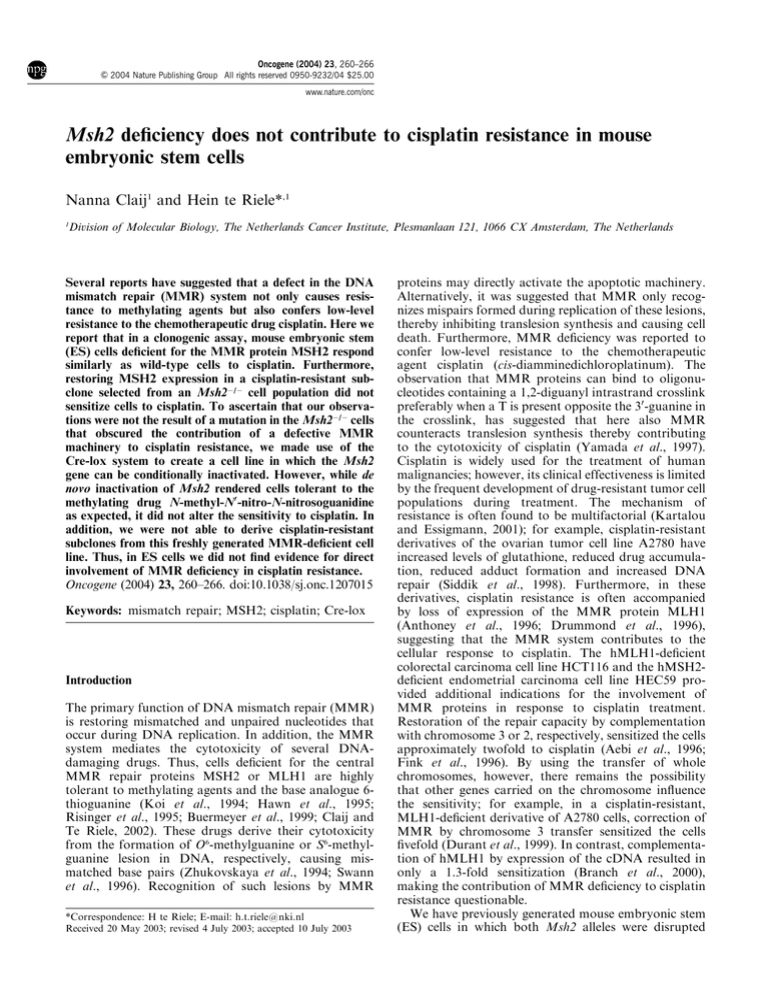
1995). In a clonogenic assay, the response of these
Msh2/ cells to cisplatin was compared to that of wildtype ES cells. Briefly, we allowed 500 cells/1.8 cm2 to
attach on a feeder layer. The next day, we treated the
cells for 1 h with different concentrations of cisplatin
and after 4 days we counted the number of surviving
colonies. By this method, we were not able to detect a
significant difference in the survival of Msh2/ and
wild-type ES cells (Figure 1).
Ectopic expression of Msh2 does not sensitize cisplatinresistant Msh2/ cells to cisplatin
As judged from the clonogenic assay in Figure 1, a
defective MMR system was not sufficient to increase cell
survival in response to cisplatin. However, after
exposure of a large number (1.5 106) of cells for 24 h
to 10 mm cisplatin, a difference in the number of
surviving Msh2/ and wild-type colonies manifested.
In one of the experiments in which sixfold more Msh2/
colonies appeared (300 versus 50 for wild-type cells),
individual colonies were picked, expanded and their
response to cisplatin was determined in a clonogenic
assay. Of the 32 Msh2/ colonies tested, 13 (which
would correspond to 122 out of 300) showed enhanced
resistance to cisplatin, while six of the 32 (i.e. nine out of
50) wild-type colonies were more resistant. All the other
clones were as sensitive as the original Msh2/ and wildtype cultures in a clonogenic assay. The level of
resistance was low, that is, not more than two- to
threefold as shown for clone Pt7 in Figure 1. Since
Figure 1 Response of Msh2-proficient and -deficient ES cells to
cisplatin. Msh2 þ / þ (squares), Msh2/ (circles) and Pt7 (triangles)
cells were exposed for 1 h to 0–40 mm cisplatin. After 4 days, the
number of surviving colonies was counted. These experiments were
performed in triplicate
Msh2/ cells have a mutator phenotype and have been
cultured for many passages since they were generated, it
is not unlikely that the Msh2/ culture contained a
small subpopulation of cells that had additional mutations affecting the response to cisplatin. A low-level
resistance may be sufficient to provide a growth
advantage in the presence of platinating agents, which
may at least partly account for the extra colonies found
in the Msh2/ population. The resistant clone Pt7 was
randomly chosen to explore whether the MMR defect
was directly involved in cisplatin resistance by collaborating with an additional mutation. By introduction of
the full-length cDNA of the Msh2 gene, MSH2 protein
expression was restored in the Pt7 cell line (Figure 2a).
This strongly sensitized the cells to 6-thioguanine as
shown for clones B and D in Figure 2b. However, reexpression of MSH2 did not affect the cisplatinresistance phenotype of the Pt7 cells (Figure 2c)
suggesting that, at least in this Pt7 cell line, MMR
deficiency is not directly involved in cisplatin resistance.
Generation of an ES cell line in which Msh2 can be de
novo inactivated
As cells with a defective MMR system have a mutator
phenotype, they may more easily accumulate mutations
that interfere with the cellular response to cisplatin.
Such mutations could obscure the contribution of
MMR deficiency to resistance, thereby causing the
discrepancy between our observations and those of
others (Drummond et al., 1996). To avoid problems of
additional genetic changes, we made use of the Cre-lox
system to generate an ES cell line in which MSH2
expression can be switched off and on again. The
previously generated Msh2-heterozygous ES cell line
Msh2-55 (de Wind et al., 1995) was used to modify
the wild-type allele with the targeting construct shown
Oncogene
MSH2-independent toxicity of cisplatin
N Claij and H te Riele
262
a
Pt7=cMsh2
B
D
C
Msh2+ +
Pt7
Ab-Msh2
Ab-Actin
in Figure 3a. Successful gene targeting produced an
Msh2lox allele in which a neomycin-resistance marker
flanked by two loxP sites (floxed neo) was inserted 2 kb
upstream of exon 12 and a single loxP site was introduced
53 bp downstream of exon 13. One of 480 G418-resistant
clones contained both the floxed neo gene and the single
loxP site targeted at the wild-type Msh2 locus, while the
knockout allele remained unchanged.
a
b
Hyg
12
13
Msh2
12
13
Msh2lox
Surviving colonies (%)
100
Neo
50
Cre recombinase
P1
12
0
0.01
0.1
1
10
P3
100
13
Msh2lox+
P1
13
[6-thioguanine], µM
c
P2
12
Msh2lox-
Msh2+ +
Msh2- Pt7
Pt7 + cMsh2 B
Pt7 + cMsh2 D
Pt7 + cMsh2 C
100
b
+6
- 6
+Cre
- 37
+37
+Cre
P1-P2
P1-P2
50
c
Ab-Msh2
Ab-Actin
0
0
10
20
30
40
[cisplatin], µM
Figure 2 The expression of MSH2 in Pt7 cells restores sensitivity
to 6-thioguanine but not to cisplatin. (a) Western blot analysis of
Pt7 cells transfected with the cDNA of Msh2 shows MSH2 protein
expression in clones B and D. Actin was used as a loading control.
(b) Msh2 þ / þ (squares), Pt7 (triangles) and Pt7 þ cMsh2 clone B
(open diamond), clone D (gray diamond) and clone C (closed
diamond) ES cell lines were exposed to increasing amounts of 6thioguanine for 1 h. After 4 days, the number of surviving colonies
was counted. Msh2/ cells (circles) were included as a 6thioguanine-resistant control. (c) The same clones were treated
with different concentrations of cisplatin. Survival curves were
simultaneously determined for all cell lines in two independent
experiments giving similar results. One is shown in (b) and (c)
Oncogene
Figure 3 De novo inactivation and reactivation of Msh2 by Cremediated recombination. (a) Previously, the Msh2 allele was
generated by insertion of a hygromycin-resistance marker (hyg)
into a unique SnaBI site within exon 12 (de Wind et al., 1995). At
the other allele indicated as Msh2lox, a single loxP site (triangle) was
introduced 53 bp downstream of exon 13. Furthermore, a floxed
Neo gene driven by the MC1 promoter was inserted 2 kb upstream
of the exon 12 with both loxP sites in the opposite orientation of
the single loxP site. Cre-mediated recombination of the loxP sites
results in deletion of the Neo gene and can cause inversion of the
exons 12 and 13 containing fragment. The exons 12–13 orientation
results in MMR-proficient Msh2lox þ / cells, the exons 13–12
orientation in MMR-deficient Msh2lox/ cells. (b) Confirmation
of the orientation of the exons 12 and 13 containing fragment by
PCR. Primer pair P1, P2 amplifies the Msh2lox þ / orientation in
clone 6 and clone 37 þ Cre; primers P1 and P3 amplify the Msh2lox/
orientation in clone 37 and clone 6 þ Cre. (c) Western blot
analysis of MSH2 protein expression in Msh2lox þ / and Msh2lox/
cells. The blot was probed with an antibody against MSH2, and an
antibody against actin was used as a loading control
MSH2-independent toxicity of cisplatin
N Claij and H te Riele
263
a
100
Surviving colonies (%)
The correctly targeted cell line was subsequently
electroporated with a Cre-encoding plasmid to express
transiently the Cre enzyme that mediates recombination
between the loxP sites. The loxP sites flanking the neo
gene were oriented in the same direction allowing
deletion of the neo gene upon Cre-mediated recombination. Indeed, 45 of 192 clones (23%) acquired G418
sensitivity after Cre expression. Since the remaining
loxP site and the single loxP site downstream of exon 13
had the opposite orientation, Cre-mediated recombination could also result in inversion of the fragment
containing exons 12 and 13. This would, in combination
with the knockout allele, result in a completely Msh2deficient cell line, tolerant to 6-thioguanine and methylating drugs. Indeed, screening of 192 Cre-exposed
clones for inversion of exons 12 and 13 by testing their
sensitivity to 6-thioguanine revealed 10 tolerant clones
(5%), of which nine were also G418 sensitive. Subsequently, the orientation of exons 12 and 13 was
determined in cell lines either sensitive or tolerant to 6thioguanine by a PCR specific for either orientation
(Figure 3b). In all cases, the PCR confirmed the
orientation we expected based on the response to 6thioguanine. MMR-proficient cells with the normal
orientation of exons 12–13 will further be designated
as Msh2lox þ /; MMR-deficient cells with exons 12–13 in
the reversed orientation will be designated as Msh2lox/.
By Western blot analysis, we were able to show that
MSH2 was expressed at high levels in Msh2lox þ / cells,
whereas the expression of MSH2 protein was completely
abolished in Msh2lox/ cells (Figure 3c).
Cre-recombinase was transiently expressed for a
second time in the Msh2lox þ / clone 6 and Msh2lox/
clone 37 to invert exons 12–13 and to reverse the MMR
status. In both cell lines, the frequency of inversion was
13% (six of 48 and 12 of 90 tested clones, respectively)
as determined by PCR. Indeed, after inversion of exons
12 and 13, clone 37 (37 þ Cre) cells regained MSH2
expression, while clone 6 (6 þ Cre) cells stained negative
for MSH2 (Figure 3c).
50
0
0.01
6-Thioguanine was used for a quick screen to identify
Msh2lox/ clones after Cre-mediated recombination. In
a more accurate clonogenic assay, we determined the
response of Msh2lox þ / (clone 6) and Msh2lox/(clone 37)
ES cells to the methylating drug N-methyl-N0 -nitro-Nnitrosoguanidine (MNNG). As shown in Figure 4a,
MSH2-deficient Msh2lox/ (37) cells were tolerant to
MNNG when compared to the MSH2-proficient
Msh2lox þ / (6) cells. Inversion of exons 12–13 in
Msh2lox þ /(6) cells by expression of Cre-recombinase,
giving Msh2lox/ (6 þ Cre) cells, greatly enhanced the
tolerance to MNNG. Likewise, by changing the
Msh2lox/ genotype of clone 37 cells to Msh2lox þ /
(37 þ Cre), the methylation-damage-tolerance phenotype was reversed, making the cells as sensitive as
Msh2lox þ / (6) cells. These data show that by using the
Cre-lox system, we can easily switch between MMR
proficiency and deficiency, thereby creating a valuable
1
10
100
[MNNG], µM
b
Msh2 lox+ - 6
100
Msh2 lox- - 6 +Cre
Msh2 lox- - 37
Msh2 lox+ - 37 +Cre
50
0
0
Msh2lox/ cells are tolerant to MNNG
0.1
10
20
30
40
[cisplatin], µM
Figure 4 Response of Msh2lox þ / and Msh2lox/ cells to DNAdamaging agents. Msh2lox þ /(6) (closed square), Msh2lox/(37)
(closed circle), Msh2lox/(6 þ Cre) (open square) and Msh2lox þ /
(37 þ Cre) (open circle) ES cell lines were exposed to increasing
amounts of the methylating drug MNNG (a) or cisplatin (b) for
1 h. After 4 days, the number of surviving colonies was counted.
Experiments were performed in triplicate
tool to evaluate the involvement of the MMR system in
the response of cells to treatment with DNA-damaging
agents.
Similar response of Msh2lox þ / and Msh2lox/ cells to
cisplatin
Next, we tested the sensitivity of de novo generated
Msh2lox/ and Msh2lox þ / cells to the cytotoxic effects of
cisplatin. The number of colonies that survived cisplatin
Oncogene
MSH2-independent toxicity of cisplatin
N Claij and H te Riele
264
treatment was equal both in a clonogenic assay
(Figure 4b) and after a 24 h exposure of 1.5 106 cells
(data not shown). Furthermore, we were not able to
identify cisplatin-resistant variants in the Msh2lox/ cell
population. These data demonstrate that in ES cells,
MMR deficiency is not directly involved in cisplatin
resistance.
Discussion
Several observations have suggested that MMR deficiency is involved in low-level cisplatin resistance. In
human ovarian tumor cell lines selected for cisplatin
resistance, the resistance phenotype was often found to
be accompanied by loss of MLH1 activity (Anthoney
et al., 1996; Drummond et al., 1996), and in ovarian
cancer, poor response to cisplatin has been correlated
with a reduction or loss of expression of MSH2 or more
frequently MLH1 (Samimi et al., 2000; Strathdee et al.,
2001; Watanabe et al., 2001). Cell lines defective for
MLH1 or MSH2 appeared to be twofold resistant to
cisplatin in comparison with corresponding MMRproficient cell lines (Aebi et al., 1996; Fink et al.,
1997). This low-level resistance was sufficient for
enrichment of an MMR-deficient cell population in
vitro (Fink et al., 1997, 1998) and a reduced response of
tumor cells in an in vivo model (Fink et al., 1997).
Furthermore, purified MSH2 protein, either alone or in
complex with its dimerization partner MSH6, can bind
to oligonucleotides containing a cisplatin adduct (Duckett et al., 1996; Mello et al., 1996; Mu et al., 1997).
Recently, the MMR proteins have been suggested to
have a function as general sensors of DNA damage with
the ability to signal directly to cell cycle checkpoints and
apoptosis. Cisplatin-induced initiation of a G1 arrest by
cyclin D1 degradation was found to be dependent on the
presence of MMR proteins (Lan et al., 2002), although
we did not find a differential reduction of cyclin D1
levels in cisplatin-treated Msh2-deficient and wild-type
mouse embryonic fibroblasts (MEFs) (unpublished
observation). Also, the cisplatin-induced activation of
c-Abl, one of the proteins of the p73-dependent
apoptosis pathway, was shown to be dependent on
functional MSH2 and MLH1 (Nehme et al., 1997; Gong
et al., 1999). However, the number of reports disputing
the involvement of the MMR system in cisplatin
resistance is increasing; for example, evidence was
obtained that the MMR-proficient ovarian carcinoma
cell line A2780, which was widely used to study cisplatin
responsiveness (Anthoney et al., 1996; Drummond et al.,
1996) contains a small pre-existing subpopulation of
MLH1-deficient cells that are significantly more resistant to cisplatin than the parental cells (Aquilina et al.,
2000). It seems that most cisplatin-resistant variants that
have been isolated over the years were derived from this
same subpopulation that contains, in addition to MLH1
deficiency, a mutation in the p53 gene (Branch et al.,
2000). Careful examination of both genetic changes
suggested that defective MMR was only a minor
contributor to cisplatin resistance, accounting for less
Oncogene
than 1.3-fold resistance and that the abrogated p53
response was the main determinant of resistance
(Branch et al., 2000). When cisplatin-resistant clones
were selected from a pure population of cisplatinsensitive, MMR-proficient A2780 cells, in all cases they
had retained normal MMR repair capacity (Massey
et al., 2003). Similarly, colon tumor cell lines selected for
cisplatin resistance did not present with defects in DNA
MMR proteins (Sergent et al., 2002). Finally, in Msh2deficient murine intestinal enterocytes, only a slightly
reduced apoptotic response to cisplatin could be
observed that did not influence the overall survival of
crypt cells (Sansom and Clarke, 2002), and indications
for the involvement of the p73 pathway were not found.
We have previously generated an Msh2-deficient ES
cell line (de Wind et al., 1995). In contrast to Fink et al.
(1997), who previously reported a twofold resistance of
these Msh2/ cells, we show here that the cells respond
similarly to the cytotoxic effects of cisplatin as wild-type
cells. However, a 24 h exposure of 1.5 106 Msh2/ and
wild-type cells to a low dose of cisplatin revealed an
increased number of surviving colonies in the Msh2/
cell population. The majority of surviving Msh2/
colonies showed a twofold resistance to cisplatin in a
clonogenic assay, suggesting that the Msh2-deficient cell
line contained a small subpopulation of cells with an
altered response to cisplatin. To explore whether in one
of the cisplatin-resistant ES cell clones, Msh2 deficiency
contributed directly to the resistance phenotype by
collaboration with an additional mutation, we restored
MSH2 expression by introducing the cDNA of the
Msh2 gene. Although the MSH2 protein level was
sufficiently high to restore the sensitivity to 6-thioguanine, the cellular response to cisplatin remained unchanged. This indicates that the cells had acquired a
genetic alteration that confers resistance to cisplatin
irrespective of the MMR status. The nature of this
genetic change remains to be identified.
By using a Cre-lox-based system in which Msh2 can
be de novo inactivated and reactivated, we generated a
set of ES cell lines in which the consequences of uniquely
Msh2 inactivation could be assessed. Again, we found
no relation between the MMR capacity of cells and their
response to cisplatin in a clonogenic assay. Furthermore, long-term exposure to cisplatin of Msh2lox þ / and
Msh2lox/ cells directly after inactivation of the Msh2
gene resulted in a similar number of surviving colonies
for the MMR-proficient and -deficient cell lines. Testing
these surviving colonies for acquired cisplatin resistance
in a clonogenic assay did not reveal resistant variants. It
remains possible that upon prolonged culturing, and in
the presence of low levels of cisplatin, resistant variants
arise more frequently in MMR-deficient cells than in
wild-type cells.
An explanation for the discrepancy between our
results and those of others who did find cisplatin
resistance in MMR-deficient cell lines, may be the
requirement for specific genetic changes that can
collaborate with MMR deficiency in affecting the
cytotoxicity of cisplatin; for example, mutated p53
(Branch et al., 2000), increased recombinational repair
MSH2-independent toxicity of cisplatin
N Claij and H te Riele
265
or increased replicative bypass may be candidates. In
yeast, data were obtained suggesting that drug resistance
mediated by loss of MMR is dependent on RAD52/
RAD1 activity, implicating the involvement of a
recombination-dependent process (Durant et al., 1999).
Enhanced replicative bypass has been observed in
human tumor cell lines with a defective MMR system
(Vaisman et al., 1998). Furthermore, enhanced expression and activity of DNA polymerase b was found to
correlate with cisplatin resistance (Mamenta et al., 1994;
Canitrot et al., 1998).
In conclusion, we did not find any evidence for a
direct contribution of MMR to the cytotoxicity of
cisplatin in ES cells. This observation is particularly
important, as previous work has suggested that the
MMR status of tumor cells may be an important
determinant of the outcome of platinum-based chemotherapy. In view of our data, such a conclusion
should be considered with caution.
Materials and methods
Clonogenic assays
ES cell lines were seeded onto feeder layers of MEFs, mitotically
inactivated by 25 Gy of irradiation, at a density of 500 cells/
1.8 cm2. The next day, the cells were treated for 1 h with 0–40 mm
MNNG (Sigma), 0–100 mm 6-thioguanine (Sigma) or 0–60 mm
cisplatin (Platosin, Pharmachemie, The Netherlands). After 4
days, densely packed surviving colonies were counted under a
microscope. For MNNG, during the whole procedure from 1 h
prior to exposure, 20 mm O6-benzylguanine (kindly provided by
R Moschel) was present in the medium to inhibit the removal of
methyl groups from the O6-position of guanine by endogenous
methyltransferase activity.
Selection for cisplatin-resistant clones
ES cell lines were seeded onto gelatin-coated tissue culture
plates at a density of 1.5 106 cells/60 cm2. The following day,
the cells were exposed for 24 h to 10 mm cisplatin. After 2
weeks, the number of surviving colonies was counted. A
number of these colonies were picked, expanded and their
cisplatin sensitivity was determined in a clonogenic assay.
Msh2 cDNA expression
The full-length cDNA of Msh2 was cloned behind the EF1a
promoter. A puromycin-resistance marker driven by the PGK
promoter was inserted into the same vector upstream of EF1acMsh2. The vector was linearized before electroporation into
the cisplatin-resistant Msh2-deficient cell line Pt7. Puromycin
(2.2 mg/ml)-resistant colonies were picked and expanded. As
cells expressing a high level of MSH2 are sensitive to 6thioguanine, individual clones were plated on a 96-well plate at
a density of 500 cells per well and treated with 60 mm 6thioguanine for 1 h the next day. After 7 days, in 35% of the
wells, all cells had died, identifying clones possibly expressing a
high level of MSH2.
Western blot analysis
The MSH2 protein level was determined by Western blot
analysis, using an antibody against MSH2 (de Wind et al.,
1998). An antibody against actin (I-19, Santa Cruz) was used
as a loading control.
Generation of an ES cell line in which Msh2 can be de novo
inactivated
In a 12.5 kb BamHI genomic fragment of the mouse Msh2
locus containing exons 12 and 13 (de Wind et al., 1995), a
single loxP site was inserted into the NsiI site located 53 bp
downstream of exon 13. Subsequently, the G418-resistance
marker neomycin (MC1neo) flanked by loxP sites was
introduced into the HindIII site 2 kb upstream of exon 12,
with the loxP sites in the opposite orientation of the single
loxP site. This targeting vector was introduced into the Msh2heterozygous ES cell line Msh2-55 as described (de Wind et al.,
1995). Southern blot analysis of EcoRI-digested DNA from
G418 (250 mg/ml)-resistant colonies, using probes flanking the
targeting construct, identified a modified wild-type allele
containing both the floxed MC1neo gene and the single loxP
site in one of 480 clones.
Cre-recombinase-mediated deletion of MC1neo and inversion of
exons 12 and 13
Into the correctly targeted cells, a plasmid containing Crerecombinase driven by the CMV promoter was electroporated,
together with a plasmid containing a puromycin-resistance
marker (PGKpur) in the ratio of 10 : 1. Cells were selected for
uptake of DNA with 1.8 mg/ml puromycin from 20 to 72 h after
electroporation (Taniguchi et al., 1998). Puromycin-resistant
clones were screened for deletion of the neo gene by testing for
G418 sensitivity (250 mg/ml) and for inversion of the fragment
containing exons 12 and 13 by selecting 6-thioguanine (10 mg/
ml) resistance. The orientation of the exons 12 and 13
containing fragment was confirmed by PCR. The Msh2lox þ /
orientation was detectable in a reaction with primer P1 in exon
13 (50 -GCATCCTTGCTCGAGTCG-30 ) and primer P2 specific for the polylinker sequence downstream of the 30 loxP site
(50 -CTGCAAAATCAGATCCCCG-30 ).
The
inverted
Msh2lox/ orientation was determined by PCR with the same
P1 primer and a primer P3 specific for the upstream polylinker
sequence of the remaining 50 loxP site (50 -GCAGGAATTCGATATCAAGC-30 ).
An Msh2lox þ /clone (6) and an Msh2lox/ clone (37) were
electroporated for a second time with Cre-recombinase and
PGKpur to invert exons 12 and 13, thereby changing the
MMR status.
Acknowledgements
We thank Judith Martens for cloning the loxP site downstream of exon 13, Valerie Doodeman and Yvonne van Klink
for technical assistance, Jos Jonkers for providing us with
CMV-Cre, loxP and floxed Neo plasmids, R Moschel for the
gift of O6-benzylguanine, and Marjolein Sonneveld for helpful
comments on the manuscript. We acknowledge support from
the Dutch Cancer Society (Grant NKI 98-1838) and the
European Committee (Grant ENV4-CT97-0469).
References
Aebi S, Kurdi-Haidar B, Gordon R, Cenni B, Zheng H, Fink
D, Christen RD, Boland CR, Koi M, Fishel R and Howell
SB. (1996). Cancer Res., 56, 3087–3090.
Anthoney DA, McIlwrath AJ, Gallagher WM, Edlin
AR and Brown R. (1996). Cancer Res., 56, 1374–
1381.
Oncogene
MSH2-independent toxicity of cisplatin
N Claij and H te Riele
266
Aquilina G, Ceccotti S, Martinelli S, Soddu S, Crescenzi M,
Branch P, Karran P and Bignami M. (2000). Clin. Cancer
Res., 6, 671–680.
Branch P, Masson M, Aquilina G, Bignami M and Karran P.
(2000). Oncogene, 19, 3138–3145.
Buermeyer AB, Wilson-Van Patten C, Baker SM and Liskay
RM. (1999). Cancer Res., 59, 538–541.
Canitrot Y, Cazaux C, Frechet M, Bouayadi K, Lesca C,
Salles B and Hoffmann JS. (1998). Proc. Natl. Acad. Sci.
USA, 95, 12586–12590.
Claij N and Te Riele H. (2002). Oncogene, 21, 2873–2879.
de Wind N, Dekker M, Berns A, Radman M and Te Riele H.
(1995). Cell, 82, 321–330.
de Wind N, Dekker M, van Rossum A, van der Valk M and Te
Riele H. (1998). Cancer Res., 58, 248–255.
Drummond JT, Anthoney A, Brown R and Modrich P. (1996).
J. Biol. Chem., 271, 19645–19648.
Duckett DR, Drummond JT, Murchie AI, Reardon JT,
Sancar A, Lilley DM and Modrich P. (1996). Proc. Natl.
Acad. Sci. USA, 93, 6443–6447.
Durant ST, Morris MM, Illand M, McKay HJ, McCormick
C, Hirst GL, Borts RH and Brown R. (1999). Curr. Biol., 9,
51–54.
Fink D, Nebel S, Aebi S, Zheng H, Cenni B, Nehme A,
Christen RD and Howell SB. (1996). Cancer Res., 56,
4881–4886.
Fink D, Nebel S, Norris PS, Aebi S, Kim HK, Haas M and
Howell SB. (1998). Br. J. Cancer, 77, 703–708.
Fink D, Zheng H, Nebel S, Norris PS, Aebi S, Lin TP, Nehme
A, Christen RD, Haas M, MacLeod CL and Howell SB.
(1997). Cancer Res., 57, 1841–1845.
Gong JG, Costanzo A, Yang HQ, Melino G, Kaelin Jr WG,
Levrero M and Wang JY. (1999). Nature, 399, 806–809.
Hawn MT, Umar A, Carethers JM, Marra G, Kunkel TA,
Boland CR and Koi M. (1995). Cancer Res., 55,
3721–3725.
Kartalou M and Essigmann JM. (2001). Mutat. Res., 478,
23–43.
Koi M, Umar A, Chauhan DP, Cherian SP, Carethers JM,
Kunkel TA and Boland CR. (1994). Cancer Res., 54,
4308–4312.
Oncogene
Lan Z, Sever-Chroneos Z, Strobeck MW, Park CH, Baskaran
R, Edelmann W, Leone G and Knudsen ES. (2002). J. Biol.
Chem., 277, 8372–8381.
Mamenta EL, Poma EE, Kaufmann WK, Delmastro DA,
Grady HL and Chaney SG. (1994). Cancer Res., 54, 3500–
3505.
Massey A, Offman J, Macpherson P and Karran P. (2003).
DNA Repair, 2, 73–89.
Mello JA, Acharya S, Fishel R and Essigmann JM. (1996).
Chem. Biol., 3, 579–589.
Mu D, Tursun M, Duckett DR, Drummond JT, Modrich P
and Sancar A. (1997). Mol. Cell. Biol., 17, 760–769.
Nehme A, Baskaran R, Aebi S, Fink D, Nebel S, Cenni B,
Wang JY, Howell SB and Christen RD. (1997). Cancer Res.,
57, 3253–3257.
Risinger JI, Umar A, Barrett JC and Kunkel TA. (1995). J.
Biol. Chem., 270, 18183–18186.
Samimi G, Fink D, Varki NM, Husain A, Hoskins WJ,
Alberts DS and Howell SB. (2000). Clin. Cancer Res., 6,
1415–1421.
Sansom OJ and Clarke AR. (2002). Oncogene, 21, 5934–5939.
Sergent C, Franco N, Chapusot C, Lizard-Nacol S, Isambert
N, Correia M and Chauffert B. (2002). Cancer Chemother.
Pharmacol., 49, 445–452.
Siddik ZH, Mims B, Lozano G and Thai G. (1998). Cancer
Res., 58, 698–703.
Strathdee G, Sansom OJ, Sim A, Clarke AR and Brown R.
(2001). Oncogene, 20, 1923–1927.
Swann PF, Waters TR, Moulton DC, Xu Y-Z, Zheng Q,
Edwards M and Mace R. (1996). Science, 273, 1109–1111.
Taniguchi M, Sanbo M, Watanabe S, Naruse I, Mishina M
and Yagi T. (1998). Nucleic Acids Res., 26, 679–680.
Vaisman A, Varchenko M, Umar A, Kunkel TA, Risinger JI,
Barrett JC, Hamilton TC and Chaney SG. (1998). Cancer
Res., 58, 3579–3585.
Watanabe Y, Koi M, Hemmi H, Hoshai H and Noda K.
(2001). Br. J. Cancer, 85, 1064–1069.
Yamada M, O’Regan E, Brown R and Karran P. (1997).
Nucleic Acids Res., 25, 491–495.
Zhukovskaya N, Branch P, Aquilina G and Karran P. (1994).
Carcinogenesis, 15, 2189–2194.


