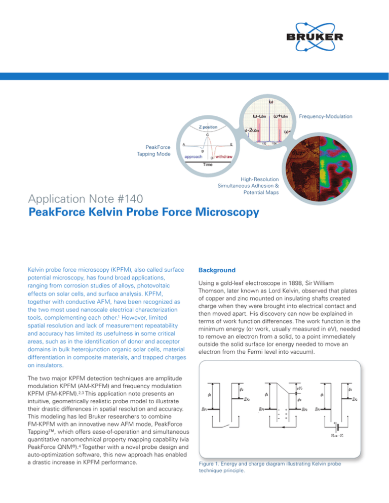
Frequency-Modulation
PeakForce
Tapping Mode
High-Resolution
Simultaneous Adhesion &
Potential Maps
Application Note #140
PeakForce Kelvin Probe Force Microscopy
Kelvin probe force microscopy (KPFM), also called surface
potential microscopy, has found broad applications,
ranging from corrosion studies of alloys, photovoltaic
effects on solar cells, and surface analysis. KPFM,
together with conductive AFM, have been recognized as
the two most used nanoscale electrical characterization
tools, complementing each other.1 However, limited
spatial resolution and lack of measurement repeatability
and accuracy has limited its usefulness in some critical
areas, such as in the identification of donor and acceptor
domains in bulk heterojunction organic solar cells, material
differentiation in composite materials, and trapped charges
on insulators.
The two major KPFM detection techniques are amplitude
modulation KPFM (AM-KPFM) and frequency modulation
KPFM (FM-KPFM).2,3 This application note presents an
intuitive, geometrically realistic probe model to illustrate
their drastic differences in spatial resolution and accuracy.
This modeling has led Bruker researchers to combine
FM-KPFM with an innovative new AFM mode, PeakForce
Tapping™, which offers ease-of-operation and simultaneous
quantitative nanomechnical property mapping capability (via
PeakForce QNM®).4 Together with a novel probe design and
auto-optimization software, this new approach has enabled
a drastic increase in KPFM performance.
Background
Using a gold-leaf electroscope in 1898, Sir William
Thomson, later known as Lord Kelvin, observed that plates
of copper and zinc mounted on insulating shafts created
charge when they were brought into electrical contact and
then moved apart. His discovery can now be explained in
terms of work function differences. The work function is the
minimum energy (or work, usually measured in eV), needed
to remove an electron from a solid, to a point immediately
outside the solid surface (or energy needed to move an
electron from the Fermi level into vacuum).
Figure 1. Energy and charge diagram illustrating Kelvin probe
technique principle.
When two different conductors are brought into electrical
contact, for example via an external wire contact, electrons
will flow from the one with lower work function to the one
with higher work function, equalizing the Fermi energies.
If they are made into a parallel plate capacitor, equal and
opposite charges will be induced on the surfaces. The
potential established between these two surfaces is called
the contact potential difference (CPD), contact potential,
or surface potential, which equals the work function
difference of the two materials. Measuring the CPD is thus
quite simple. An external potential (also called the backing
potential) is applied to the capacitor until the surface
charges disappear. At this point, the external potential
equals the CPD. The various Kelvin probe techniques
developed thus far differ mainly only on how this chargefree state is detected.
In 1932, William Zisman of Harvard University introduced
the vibrating electrode technique and the “nulling”
concept.5 Vertically vibrating the tip over a sample causes
the capacitance to vary as the distance changes. This
induces charge to flow, giving rise to an AC current. The
backing potential, at which AC current is at a minimum or
ideally 0, “nulled,” is found to equal the CPD. This technique
leads to development of systems that automatically track
shifts in contact potential due to changes in the work
function of the sample.
J.M.R. Weaver and coworkers were the first to combine
Kelvin method with AFM.6 They embraced the nulling
concept in finding the charge-free point, and capitalized
on AFM’s unique capability to detect small forces and
force gradients. The central idea is that the electric force
and electric force gradient between the two plates of a
capacitor will become “0,” when charge disappears. It is
fitting, for this reason, to call the technique Kelvin probe
force microscopy (KPFM), as Nonnenmacher did in 1991.7
KPFM opened the door to measuring CPD, therefore
work function, in the nanometer regime. AM-KPFM and
FM-KPFM are based on electric force and electric force
gradient detection respectively.
Electric Force and Electric Force Gradient
A conductive probe and a conductive sample form a
capacitor. The electrostatic force between is:
1 ∂C
Fel = −
(∆V ) 2
2 ∂z
where Fel is the electric force, and ∆V is the potential
difference between the probe and the sample. ∆V is the
sum of the intrinsic CPD, an externally applied DC voltage
VDC and ac voltage VAC:
∆V = V DC − VCPD + V AC sin(ωt )
Combining the above two equations, we arrive at:
Fel =
1
1 ∂C
∂C
∂C
2
2
((VDC − VCPD ) 2 + VAC ) +
(VDC − VCPD )VAC sin(ωt ) +
VAC cos(2ωt )
2
4
∂z
∂z
∂z
DC Term
2
ω Term
2ω Term
The above equation states that the applied ac bias at
frequency ω is causing the electric force to modulate at
both ω and 2ω, which can be measured directly using
cantilever deflection. Figure 2 shows the forms prescribed
by the above equation. Most relevant is the fact that
oscillation amplitude at ω (shown as amplitude 1) drops to 0
when VCD=VCPD, the very idea of “nulling” electric force to
find surface potential in amplitude modulation KPFM.
Figure 2. DC deflection (top), amplitudes at frequency ω (center) and
2ω (bottom) when the DC tip bias is swept while an ac bias with
frequency ω is superimposed, corresponding to the DC term, ω term
and the 2ω term described in equation 1.
The electric force gradient is associated with electric force,
Therefore,
F 'el =
∂ 2C
∂ 2C
1
1 ∂ 2C
2
2
VAC cos(2ωt )
((VDC − VCPD ) 2 + VAC ) +
(VDC − VCPD )VAC sin(ωt ) +
2
2
∂z
∂z
z 2
2
4 ∂
DC Term
ω Term
2ω Term
Similarly, VCD=VCPD when the modulation amplitude of
the electric force gradient at ω drops to 0, the basis for
“nulling” the electric force gradient to find the surface
potential in frequency modulation KPFM.
AM-KPFM through Electric Force Detection
The modulated electric force, by the application of an ac
bias between the tip and sample, can be conveniently
measured using the oscillation of the cantilever. The ac bias
frequency is typically selected to be the resonant frequency
of the AFM cantilever for enhanced sensitivity afforded by
cantilever’s quality factor Q. The KPFM feedback, using the
oscillation amplitude as input, adjusts a DC bias until the
oscillation amplitude drops to 0, when VDC equals CPD.
Figure 4. Amplitude vs. frequency plot of the vertical deflection
signal recorded with high-speed data capture when a MESP-RC
probe is shaken, near a sample surface, by the tapping piezo
at its resonant frequency while an ac bias of 2kHz is applied,
illustrating the emergence of sidebands due to ac bias-induced
frequency modulation.
Figure 3. AM-KPFM diagram. An ac bias with a frequency ω,
typically the resonant frequency of the cantilever, is applied between
the probe and the sample, giving rise to an alternating electric force
between the probe and sample that causes the probe to oscillate.
FM-KPFM through Electric Force Gradient Detection
The detection of electric force gradient is less straight
forward. The force gradient changes the effective spring
constant of the cantilever. When placing a conductive
cantilever in an electric field, its effective spring constant
is the sum of its natural spring constant k and the electric
force gradient, the same effect of connecting two springs
in parallel.
In practical implementations, side band amplitude is
rarely directly measured at the sideband frequency using
a single lock-in amplifier. A more common method uses
two cascaded lock-in amplifiers, with the first one locking
at the resonant frequency, the phase output of which
is fed into the second lock-in, which locks at the ac bias
frequency. The amplitude output of the second measures
the amplitude sum of ω ± ωm, which is then used for KPFM
feedback (see Figure 5).
We know the resonant frequency of a cantilever depends
on the spring constant:
Therefore the electric force gradient will cause the resonant
frequency to change:
When the electric force gradient is modulated, as caused
by an ac bias, the resonant frequency of the cantilever
will be modulated. As implied in the equation, resonant
frequency will be modulated at both the ac bias frequency
ω and its second harmonic 2ω. If one is mechanically
shaking the cantilever at its resonant frequency ω, and
simultaneously applying an ac bias at frequency ωm, usually
only a few kHz, the modulation of the resonant frequency
gives rise to two pairs of sidebands at ω ± ωm, and ω ± 2ωm
(see Figure 4). The amplitude of the sideband measures
the resonant frequency modulation amplitude; using the
amplitude of the side band at ω ± ωm for KPFM feedback,
and adjusting the DC bias until they disappear leads to the
point of VCD=VCPD.
Figure 5. FM-KPFM diagram. An ac voltage is applied to the tapping
piezo to oscillate the AFM cantilever at its resonant frequency ω.
An ac bias at frequency ωm, usually a few kHz, is applied between
the probe and the sample, modulating the resonant frequency. Two
cascaded lock-in amplifiers are used to detect the sum amplitude of
sideband pair ω ± ωm.
Probe Modeling—Spatial Resolution and Accuracy
of AM-KPFM and FM-KPFM
Probe modeling illuminated our understanding of the spatial
resolution and accuracy provided by the two KPFM modes.
We utilized a realistic yet straightforward approach to m
odeling the probe in an attempt to understand how much
3
of the tip needs to be included to account for (1) half of
the total electric force in AM-KPFM, or (2) half of the total
electric force gradient in FM-KPFM.
We set out with the capacitor model of probe and sample,
and approximated the probe as consisting of a microcantilever and a tip cone with a point end (see Figure 6).
As a good conductor, the potential is the same all over
the probe, and charges are only present on the surfaces.
The integrated capacitance of the cantilever and tip
cone can be analytically expressed. Electric force and
electric force gradient are deduced from the first and
second derivatives of capacitance. (See Appendix I for the
mathematic deductions.)
decays to around 200nm, which demonstrates the sharp
dependence of spatial resolution on probe-sample distance.
While the above modeling is only a first order
approximation, it is useful for conceptual understanding and
it does reach the same conclusion as Colchero, who used
a different set of assumptions.2 It has been recognized
that the electric force from the tip cone and cantilever
predominate over that from the small tip-apex, therefore,
AM-KPFM is not at all a quantitative nanoscale measuring
tool. Only FM-KPFM, where dominating electric force
gradient signal comes from the tip apex, offers high spatial
resolution and accurate local CPD information. The effect of
tip geometry on spatial resolution is only secondary.
100%
90%
FM z=5 nm
80%
Cone Contribution%
70%
FM z=50 nm
60%
50%
40%
30%
FM z=5
z=50nm
nm
AM
20%
Figure 6. Model of a KPFM Probe with a rectangular cantilever. The
capacitance of the cantilever is an integration of capacitance of each
tiny rectangle along the cantilever. As the same force would cause
different deflection depending on its distance to the base of the
lever, the capacitance is normalized to recognize this.
Figure 7 shows the simulation result of the widely used
SCM-PIT probe, which has the following nominal geometry:
cantilever 225µm long, 30µm wide, tip 10µm tall, and
cone half angle of 22.5°. The relative contribution of the
tip cone, integrated to height h, versus total interaction, is
plotted against tip height. To quantify the spatial resolution
of KPFM, we adopted the similar definition used by
Cohchero,2 the radius of the ring at the height up to which
the integral contribution of the tip cone accounts half the
total electric force in case AM-KPFM; or half the total
electric force gradient for FM-KPFM. For AM-KPFM, when
lift height is 5nm, the contribution of the whole tip cone
remains below 50%, indicating its spatial resolution is
dominated by the width of the cantilever-on the micrometer
scale, also implying the CPD obtained is not local, but a
convolution over the large area covered by the cantilever.
This calls its accuracy into question, except on large
uniform samples. On the other hand, for FM-KPFM, when
the tip is lifted 5nm above the surface, half of the signal is
gathered from up to 15nm above the tip end, corresponding
to a diameter of 12nm. This suggests a possible resolution
of 10nm may be achieved with FM-KPFM. The CPD
information collected is local to the area right underneath
the tip affording credible accuracy. Lifting the tip higher, for
instance, 50nm above the surface, the spatial resolution
4
10%
AM z=50 nm
0%
1
10
100
1,000
10,000
Height /nm
Figure 7. The contribution of the tip cone integrated up to height h
over total electric force (blue) and electric force gradient (purple) are
plotted versus height from tip end for a SCM-PIT probe. Geometries
are: cantilever 225µm long, 30µm wide, tip 10µm tall, and cone half
angle of 22.5°. The “BLUE” plots are electric force at tip-sample
separation 5nm and 50nm. The “purple” plots are electric force
gradient at tip-sample separation 5nm and 50nm.
Modes Overview and Comparison
KPFM, as a combination of the Kelvin probe technique and
AFM, makes surface potential measurements accessible
on the nanometer scale. KPFM traditionally operates in
conjunction with TappingMode™, however, there are many
reasons to combine it with the tried and proven PeakForce
Tapping mode (see Bruker application note AN135). The four
possible combinations are charted in Table 1 (the naming
of each combination will be used through this application
note). Some of these combinations may be done either
in a single-pass fashion, where AFM imaging and KPFM
measurement run simultaneously on one scan line; or in a
dual-pass (or lift-mode) fashion, where AFM imaging mode
is run on the first pass and KPFM on the second pass using
lift-mode.
PeakForce KPFM™, the combination of PeakForce
Tapping mode and FM-KPFM, integrates the benefits
manifestation that the dominant contribution is from the tip
apex. Thus, the cantilever contribution becomes negligible.
The data confirms that FM-KPFM offers much higher
lateral resolution, due to the minimal contribution from the
cantilever, and much better accuracy than AM-KPFM.
Table 1. Chart summarizing the four major combinations between
two major KPFM modes, AM-KPFM and FM-KPFM, and two major
AFM modes, TappingMode and PeakForce Tapping. All modes are
implemented in a dual-pass fashion (lift-mode), except KPFM-FM,
which is done in single-pass.
and capabilities of PeakForce Tapping (ease of use and
simultaneous PeakForce QNM capability) and the superior
spatial resolution and accuracy of FM-KPFM. It is best
done in a dual-pass fashion. It is possible to implement
PeakForce KPFM in a single-pass fashion but not without
sacrificing performance, as interference from PeakForce
Tapping can sometimes be severe. KPFM-FM is done
in a single-pass fashion. Though it can also be done in a
dual-pass fashion, there is no benefit that is not covered
by PeakForce KPFM. It is helpful to clear some confusion
in the literature, where single-pass KPFM is sometimes
improperly used to refer exclusively to KPFM-FM, albeit
KPFM-AM can also be done in a single-pass fashion.
Figure 8. Height (top) and potential profiles using PeakForce
KPFM-AM (middle) and PeakForce KPFM (bottom) on the Bruker
KPFM standard sample, on the same location and with the same tip
(PFQNE-AL).
PeakForce KPFM and PeakForce KPFM-AM
Bruker offers a standard KPFM sample, which is patterned
with Au, Si, and Al strips. Both the Al and Au films are
50nm in thickness deposited on an n-doped silicon
substrate. In theory, when the tip is on any one of the
three different regions, a constant potential value (CPD)
should be read. A staircase potential profile is therefore
expected across the three different materials. There is a
noticeable slope, however, present on each of the stairs of
the potential profile acquired with PeakForce KPFM-AM.
This can be readily explained by the probe modeling
described above. The potential data is a convolution from
the tip, the tip cone, and the cantilever. Although the tip
is on one single material, the relatively gigantic cantilever
covers other materials and thus contributes a large portion
of the total value. For instance, when the tip is on the the
Al/Si interface, only half of the cantilever is over Al, and
with the majority of the other half over Si (and smaller
portion extending over the Au). The final measured value
ends up a weighted average of the work function of the
three materials. As the tip moves away from the edge,
more contribution comes from the Al, and less from
Si and Au, giving rise to the slope. This is true of all of
the interfaces, and not only speaks of the poor lateral
resolution of AM-KPFM, but also its inaccuracy as a result
of convolution. In contrast, the stairs on the potential
profile acquired using PeakForce KPFM are largely level, a
Figure 9. PeakForce KPFM and PeakForce KPFM-AM potential maps
and cross section profile of a Sn-Pb alloy (60:40 by weight, at right is
the topography (4µm scan).
Data on a Sn-Pb alloy further illustrates this point. The
topography reveals different domains in the alloy (see
Figure 9, right). They are clearly resolved on both potential
maps acquired with PeakForce KPFM-AM (see Figure
9, left) and PeakForce KPFM (see Figure 9, middle) with
little difference perceivable by qualitative evaluation of
the potential maps. Their cross section, however, reveals
a stark contrast. PeakForce KPFM gives a difference of
240mV between the two selected domains, consistent
with the work function difference between Sn (4.42eV) and
Pb (4.25eV), whereas KPFM-AM gives only a difference of
97mV, which is less than half of the expected theoretical
difference, and half of what is seen from PeakForce KPFM.
Again, this highlights the better accuracy of FM-detection
5
KPFM over AM-detection KPFM. The lateral resolution is
thus defined as the smallest dimension on which a certain
accuracy (90%) can be achieved, the distinction here being
between whether you can resolve the difference and
whether you can resolve the difference accurately.
values, however, differ by about 100mV, which is not yet
fully accounted for.
Mechanical Property Mapping: The adhesion channel from
PeakForce KPFM shows a distinct contrast between the
Sn and Pb domains and also has good correlation with
potential data, rendering additional assurance in domain
identification. On the other hand, little contrast is shown
on the phase map by TappingMode. The simultaneous
quantitative nanomechanical property mapping (PeakForce
QNM) is an outstanding capability that only PeakForce
Tapping can offer. This is further illustrated on a polymer
film comprised of polystyrene (PS) and low-densitypolyethylene (LDPE) cast on a silicon substrate. In addition
to the potential map, modulus, deformation, and adhesion
data were obtained.
Figure 10. Height (left) and PeakForce KPFM potential (right) map
of single-walled carbon nanotubes (SWCNTs) laid on a Si substrate.
A single strand CNT (pointed to by the upper arrow) sticks out of a
CNT bundle (pointed to by the lower arrow), which can be resolved
in potential map, but no longer with accuracy as the dimension is too
small to give dominant contribution in FM-KPFM.
Figure 10 is a height and PeakForce KPFM potential map
of single-walled carbon nanotubes (SWCNTs) laid on a Si
substrate. The 30nm part suggests a CNT bundle, with
a single strand CNT (~2nm in diameter) sticking out at
the top, which can be seen on the potential map. Note
a 30mV difference from the substrate is obtained on the
single strand CNT, which is only a third of that read on the
CNT bundle (105mV). PeakForce KPFM can indeed resolve
nanometer features, but is no longer accurate. The smaller
dimension contributes only a non-dominant portion to the
total electric force gradient, with the rest coming from
the substrate, ending up with a much smaller contrast
due to signal convolution. Note an even smaller value
would be read when using PeakForce KPFM-AM, as the
contribution from the tip-apex diminishes. This exemplifies
that PeakForce KPFM has nanometer resolving power, but
is only accurate on features 10nm or larger.
PeakForce KPFM and KPFM-FM
PeakForce KPFM is a combination of PeakForce Tapping
mode AFM with FM-KPFM, whereas KPFM-FM is a
combination of TappingMode AFM with FM-KPFM. In
principle, their major difference lies solely on the AFM
side. Where PeakForce Tapping affords ease of use,
simultaneous quantitative and distinctive mechanical
properties, TappingMode provides phase contrast and
resulting in data ambiguity. Little difference is expected
in KPFM performance, and this is largely true, with some
exceptions. Figure 11 shows the potential maps on Sn-Pb
acquired with PeakForce KPFM and KPFM-FM on the same
spot with the same probe (PFQNE-AU, prototype). The
contrasts between Sn (lighter) and Pb (darker) domains
measured using the two modes are indeed almost
identical, 240mV and 235mV respectively. Their absolute
6
Figure 11. PeakForce KPFM and KPFM-FM Potential Maps and cross
section profiles (top) of the Sn-Pb (60:40 by weight), and at bottom
are the respective adhesion and phase imaging (4µm scan).
Artifacts in TappingMode KPFM-FM: Figure 11 shows that
the tapping phase image from KPFM-FM does not display
much contrast on the Sn-Pb alloy sample. While this can be
interpreted as a shortcoming of TappingMode in revealing
mechanical properties, a small phase contrast proves
mandatory for faithful KPFM measurement. Frequency
modulation KPFM uses phase as a means of frequency
shift detection and any sharp phase change from tip-sample
direct contact can be a source of error leading to artifacts in
the potential map. In light of this, if there is a sharp phase
contrast, care must be taken to ensure that the potential
map is least affected. One measure is to use light tapping
(a default setting in Bruker’s implementation), that is, to
Figure 12. PeakForce KPFM data including height, potential and quantitative mechanical property maps such as Young’s modulus,
deformation and adhesion on a polymer blend consisting of polystyrene (PS) and low-density-polyethylene (LDPE) (10µm scan).
use the maximum possible tapping amplitude setpoint.
Our experience suggests light tapping is beneficial in
most cases. It may not, however, be sufficient to eliminate
possible artifacts for certain samples when running singlepass-based KPFM-FM. Phase contrast sometimes can be
so strong that single-pass KPFM-FM simply cannot remove
phase cross-talk (e.g., on polymers with strong dipoles). In
this case, one has to turn to dual-pass based techniques,
where sufficient lift-height can be used for the tip to clear
the sample surface to eliminate phase shift due to direct
tip-sample contact.
PeakForce KPFM proves advantageous on this regard,
and can also be used to identify possible artifacts. By
incrementally changing the lift height, if potential contrast
shows an abrupt change in response to a small change
in lift height, one can determine beyond doubt that the
potential contrast obtained below a certain lift-height
contains artifacts. This is exemplified in the imaging of a
brush polymer sample on mica where, when the lift height
is changed from 25nm to 27nm (a relatively small change
compared to the polymer chain width of 5nm), instead of a
possible blurring of the potential contrast on the chains as
the tip goes up, the chain structure disappeared altogether
(see Figure 13). The chain pattern seen at smaller lift
heights can therefore be attributed to artifacts. PeakForce
KPFM provides a means to identify and eliminate possible
artifacts from tip-sample direct contact.
Figure 13. Sequential PeakForce KPFM maps of a brush polymer sample on mica substrate versus lift height. An abrupt change in potential
contrast is observed when lift height increases from 25nm to 27nm, suggesting that the potential contrast of the polymer chains seen at a lift
height of 25 nm or below are artifacts, caused by phase shift from direct tip-sample contact. Scan size is 250nm. The inset at the upper-left
corner is the height image.
7
Potential / V
0.95
0.90
0.85
0.80
0.75
0.70
0.65
0.60
0.55
0.50
0.45
0.40
0.35
0.30
0.25
0.20
0.15
0.10
0.05
0.00
-0.05
-0.10
-0.15
-0.20
-0.25
-0.30
Average
(V)
Std Dev
(V)
Maximum
(V)
Minimum
(V)
Au-Al
0.825
0.019
0.847
0.796
Au
0.639
0.018
0.617
0.670
Al
-0.185
0.020
-0.159
-0.222
9 KPFM Probes
Figure 14. PeakForce KPFM data obtained using nine different PFQNE-AL (Bruker) probes on the Bruker KPFM standard sample. The value
on Al (grey triangle), Au (gold triangle) and their differences (blue diamond) are plotted. Standard deviation of each is listed at right, which is
on the order of 5x improvement over traditional KPFM measurements.
It is worth noting that KPFM-FM does have some perceived
advantage when imaging features smaller than a few
nanometers, as the average tip-sample separation can
be maintained smaller than can be practically achieved
with lift-mode-based PeakForce KPFM. This, however,
does not offset the substantial advantages that PeakForce
KPFM offers.
Interpreting KPFM Phase: In addition to KPFM potential
data, KPFM phase data is simultaneously obtained and can
prove useful. It reflects the dielectric constant contrast of
the sample. Referring to equation 2 above, one finds the
second derivative of capacitance, which has dependence
on dielectric constant of the material between the tip and
the conductive portion of the sample, can be obtained from
the 2ω term. It can also be obtained from the DC term,
especially when VCD=VCPD, which is the case when KPFM
is actively running. This is equivalent to electrostatic force
microscopy (EFM) phase imaging, with the advantage that
work function and dielectric information are separated,
while the standard EFM phase imaging is a mix of work
function and dielectric information
Repeatability
While repeatability of relative contrast within one KPFM
image has been reasonably good, even with the same
type of probe, absolute values can vary hundreds of mV
from experiment to experiment. Improving measurement
consistency is a crucial step toward quantitative work
function measurement. With a new probe design and
optimized instrumentation, we have found ways to reduce
probe-to-probe measurement scatter to below a standard
deviation of 50mV. This represents a major advance for
8
quantifying KPFM, which in turn expands KPFM’s utility in
ever more demanding applications.
Table 2 lists some of the major sources responsible
for measurement inconsistency. Even with a sound
Source of Uncertainty
Operating Frequency
Tip-Sample Seperation
Tip Work Function Change
Due to Tip Wear
Electrochemical Reaction
Under Bias
Bruker Solution
Tight parameter control (ScanAsyst-KPFM):
Thermal tune for resonance frequency
Fixed oscillation amplitude
Optimal phase setting
Probe Design:
Single tip material
Proprietary way to limit DC currrent flow
Table 2. Possible sources for KPFM measurement inconsistency.
electronic design that has little electronic cross-talk,
operating parameters and probe variation can cause data
fluctuation. These include (1) whether ac drive is right on,
and always on, the resonant frequency of the cantilever;
(2) whether tip-sample separation is kept the same from
run to run; (3) whether the work function of the tip, which
is essentially the reference point, remains unchanged;
and (4) whether the sample is undergoing oxidation,
adsorption of molecules, or alteration due to possible
electrochemical reactions (as current may flow between
the tip and sample).
To overcome these uncertainties, a ScanAsyst® KPFM
operation mode was implemented, which resembles
ScanAsyst in that it provides automated imaging
optimization, but differs in its technical implementation.
In ScanAsyst KPFM, the algorithm seeks the resonant
frequency of the cantilever using thermal tune to ensure
the accuracy and consistency of the operating frequency.
The cantilever is always driven to a free-air oscillation
amplitude of 20nm when performing either PeakForce
KPFM or KPFM-FM, and the drive phase is always
accurately and automatically set. A proprietary probe design
eliminates possible DC current flow between the tip and
sample, and the uncoated silicon tip ensures no significant
work function change will occur from tip wear (as often
happens with metal coated probes). These enhanced
capabilities have greatly improved KPFM repeatability;
Figure 15 showcases the repeatability obtained with nine
different PFQNE-AL probes on the Bruker KPFM standard
sample. A standard deviation of less than 20mV has been
achieved in the potentials measured on an Al strip, an Au
strip, and in their potential difference. When the tip work
function is properly calibrated, an accurate work function
of the sample can be deduced. In addition, the ScanAsyst
KPFM mode brings ease-of-use to the operation.
PeakForce KPFM opens up room for improvement,
as opposed to KPFM-FM based on TappingMode.
TappingMode requires the cantilever to have a sufficiently
large spring constant to overcome capillary forces from
water layers that are often present on sample surfaces.
It also requires the Q to be not so large as to limit
TappingMode bandwidth. PeakForce Tapping removes
these limitations, allowing the use of cantilevers with a
much smaller spring constant and larger Q, enabling room
for probe optimization. Currently, PFQNE-AU probes are
twice as sensitive as the commonly used SCM-PIT probes.
Probes with higher Q/k ratio can be designed to further
improve PeakForce KPFM sensitivity.
Platforms and Accessories
All KPFM modes are available on Bruker Dimension Icon®
and MultiMode® 8 AFMs, and can be used in conjunction
with a variety of accessories.
Sensitivity
Frequency modulation KPFM was first realized under
ultrahigh vacuum (UHV) in 1998,8 and was not introduced
into ambient AFM until about ten years later. In addition
to the environment change that can affect KPFM
measurement fidelity, there is also a dramatic difference in
detection sensitivity. This is immediately noticeable when
you compare AM-KPFM and FM-KPFM data in an ambient
setting. The latter is usually much noisier, an evidence of
lack of sensitivity. It is therefore helpful to understand what
governs FM-KPFM detection sensitivity. Mathematical
deduction is not included in this application note, but as
previously mentioned, the electric force gradient can be
viewed as an addition to the natural spring constant of the
cantilever. For a smaller spring constant, the same electric
force gradient would mean a larger relative change, causing
a larger frequency shift and thus a stronger signal. The Q
simply makes the phase versus frequency plot steeper,
further enhancing the signal:
KPFM Sensitivity
∝ k
It is noteworthy that under UHV (i.e., without air damping)
the Q of standard EFM probe can be hundreds of
times larger than in air, thereby affording ample signal
amplification of the FM-KPFM signal. Measurement
sensitivity has rarely been an issue and high quality data
is regularly attained. Sensitivity is neither an issue for
AM-KPFM in ambient conditions, as the signal itself,
convoluted from the tip apex, tip cone and the cantilever,
is often large enough with conventional EFM probes. The
low Q in air, however, imposes a practical challenge for
FM-KPFM detection, where the signal is often weak as
the majority of the signal is only from the tip apex and the
lower part of the tip cone (the same reason FM-KPFM has
better spatial resolution). A large Q/k ratio, meaning smaller
spring constant and larger Q, is demanded of the probe.
Figure 15. Turnkey glove-box enabling all AFM functions to be
performed in a 1ppm level oxygen and water environment. Shown
inside is a Dimension Icon AFM, a version of glove-box for the
MultiMode 8 is also available.
Environmental Control: Most notably, for samples that
require stringent environmental control (e.g., lithium battery
anode or cathode materials, organic photovoltaic or light
emitting devices), a turn-key 1ppm-level of oxygen and
water capable glove box (customized for best performance)
can be used. The glove box solution proves beneficial
to measurement repeatability of samples subject to
change when exposed to ambient air, including most
semiconductor materials and metals.
Backside Illumination: The Photoconductive AFM module,
offered on the Dimension Icon system, is compatible
with industry-standard Newport solar simulators to
enable even, backside sample illumination across the
scan area. This allows KPFM potential mapping while
illuminating with achievable intensities equivalent to 300
suns, enabling study of static and dynamic photovoltaic
responses to illumination of organic solar cells/materials.
Figure 16 shows PeakForce KPFM data on a MDMO
(poly[2-methoxy-5-(3’,7’-dimethyloctyloxyl)]-1,4-phenylene
9
Figure 16. PeakForce KPFM Potential map (third from left) of MDMO-PCBM bulk heterojunction solar cell on an ITO substrate in dark (bottom
half) and under illumination with an intensity equivalent to 300 suns (top half). A 535mV downshift of work function is seen under illumination,
a 3D rendering and histogram of potential data is at the right. Sample courtesy of Dr. Philippe Leclere, University of Mons.
vinylene)-PCBM ([6,6]-phenyl C61-butyric acid methyl ester)
bulk heterojunction solar cell. Work function down-shifts
535mV under illumination simulating 300 suns intensity.
Therefore, the amplitude at ac bias driving frequency ω and
the second harmonic 2ω are:
KPFM-HV Mode
KPFM-HV stands for High-Voltage KPFM mode, which
measures electrostatic potential beyond ±10V. KPFM-HV
is a dual-pass technique, with PeakForce Tapping as the
base AFM mode. Question arises as why one would need
to measure high voltage. KPFM, as we have learned,
measures the work function difference between the
sample and tip. When we look through the work function
table of the elements,9 the highest work function is 5.93eV
(Osmium) and lowest 2.14eV (Cesium), so the maximum
possible work function difference will never exceed 3.79V.
All the KPFM techniques described above cover the range
up to ±10V, and therefore suffice for any work function
measurement task. Demand arises when it comes to
measuring trapped (immobilized) charges in insulators.
For instance, when one walks on a carpet on a dry day,
static charge can accumulate up to tens, hundreds, or
even thousands of volts. The KPFM-HV mode meets the
need for measuring high voltage electrostatic potentials.
This technique is no longer using a KPFM feedback loop,
nor a high voltage source as one may logically think. It
deviates from the force or force-gradient-nulling concept
central to the aforementioned KPFM techniques. It is
done with existing hardware common to an AFM, and
through calculation.
We adopt the concept that the sample and tip form a
capacitor. We further assume the electric force follows the
same expression:
Fel =
1
1 ∂C
∂C
∂C
2
2
(∆φ 2 + VAC ) +
VAC cos(2ωt )
∆φVAC sin(ωt ) +
∂z
2
4
∂z
∂z
DC Term
ω Term
2ω Term
where ∆ϕ is the electrostatic potential difference at the tip
in reference to the sample.
10
Aω =
∂C
∆φV AC
∂z
A2ω =
1 ∂C
V AC 2
4 ∂z
We obtain the electrostatic potential difference:
∆φ =
Aω
1
V
4 ac A2ω
and
∂C
=
∂z
4 A2ω
V AC 2
dC/dz reflects the dielectric constant variations across
the sample.
Implementation: KPFM-HV is implemented in a dualpass fashion. On the first pass, PeakForce Tapping is
used to obtain topography and mechanical properties. On
the second pass (lift-mode), PeakForce Tapping drive is
stopped, and an ac bias with a frequency lower than half
of the cantilever resonant frequency is applied between
the tip and sample. The vertical deflection signal is
simultaneously fed to two synchronized but separate lock-in
amplifiers; one to detect the amplitude and phase at the
drive frequency, and the other the amplitude at the second
harmonic. Calculation is carried out in the background to
obtain electrostatic potential difference Δφ, and dC/dz. This
technique allows one to measure voltage in the range of
±200V with an error less than 15%. Higher voltage (up to
±1000V) can be measured with accuracy only limited by
nonlinearity of photodetectors in the AFM. This technique
is available on MultiMode 8 AFMs (no additional hardware
required) and Dimension Icon AFMs (a PeakForce HV
module is required to minimize measurement distortion
from electronic interference).
Examples: Some polymers can be charged through contact
electrification. Simply contacting and then separating two
similar or dissimilar polymer pieces, or contacting it to
and then separating it from a conducting surface, induces
charge on surfaces being contacted. Figure 19 shows the
potential map (left), phase (middle), and a 3D rendering of
the potential map of a PDMS (polydimethylsiloxane) film.
The sample is prepared by casting a PDMS film about
0.5mm in thickness on a silicon wafer. A piece is peeled off
the silicon substrate and then its underside is imaged. The
potential map shows the electrostatic potential, reflecting
electrostatic charge density (absolute value), and sign,
which is learned from the phase data. The 3D rendering
shows a minimum of -99V at the center of a negatively
charged area, with positive charges having an electrostatic
potential as high as 156V.
Figure 17. KPFM-HV diagram, in the first pass, surface topography
and mechanical properties are obtained in PeakForce Tapping; in
the second pass, an ac bias at a frequency lower than half of the
cantilever resonant frequency is applied between the probe and
the sample, which causes the probe to oscillate at frequency ω and
its second harmonic 2ω. The electric potential between the tip and
sample is calculated based on the oscillation amplitude at frequency
ω and 2ω. Note that no KPFM feedback is involved.
PDMS samples can be easily obtained, in fact, a PDMS
gel-pad is often found in AFM probe packaging. Simply
cut off a piece of gel-pad from the bottom of an AFM
probe box, and use the contact electrification technique
to have it charged. Charge can also be introduced through
triboelectric charging (one type of contact electrification,
through rubbing), for instance, by repeatedly scanning the
same area for some extended period of time. Figure 19
Figure 18. KPFM-HV images of PDMS (polydimethylsiloxane) films after being charged by peeling off from the silicon substrate upon which
it was cast. The HV Potential data (left) shows the electrostatic potential and the sign of charge; HV Phase data (middle) denotes negative
charge with negative phase, and positive charge with positive phase. At the right is a 3D rendering of the potential map, the negative
charge gives rise to an electrostatic potential of -99V at its center, and some of the adjacent positive charges go as high as 158V.
Figure 19. KPFM-HV potential map on PDMS, encasing five previously scanned sites in a square-center arrangement. Sequential images from
left to right reveal that the negative charges are accumulating, spreading out and merging with each other as scanning goes on.
11
KPFM Modes
Probe Choices
PeakForce KPFM
PeakForce KPFM-AM
KPFM-FM
KPFM-AM
KPFM-HV
1st
PFQNE-AL
PFQNE-AL
PFQNE-AL
PFQNE-AL
TAP150A
2nd
SCM-PIT
ScanAsyst-Air HR
SCM-PIT
SCM-PIT
SCM-PIT
3rd
None
SCM-PIT
None
None
MESP-RC
Table 3. Probe Selection Guide for Each KPFM Mode.
(left) shows five previously scanned sites with negative
charges. Sequential imaging indicates the negatively
charged areas spreading out and merging with each other
as scanning continues.
KPFM-HV mode provides a simple means to measure
electrostatic potential of immobilized charges as high as
±200V.
Probe Selection Guide
Table 3 is a probe selection guide for each mode, as of
the date this note is published. For up-to-date information,
please follow the guidance provided in the NanoScope®
software. Other probes not in the list may also work,
but their performance in relation to KPFM measurement
sensitivity, accuracy, and/or repeatability may not
be guaranteed.
Conclusion
Bruker offers a suite of new KPFM modes, three of
which are based on the breakthrough PeakForce Tapping
technology with a dual-pass implementation:
PeakForce KPFM, using Frequency Modulation detection;
PeakForce KPFM-AM, using Amplitude
Modulation detection;
PeakForce KPFM-HV, using Amplitude Modulation and
extending the accessible potential range by more than a
factor of ten.
PeakForce KPFM, achieved by combining PeakForce
Tapping with frequency-modulation KPFM exhibits the most
outstanding performance:
It retains the high spatial resolution and accuracy of
FM-KPFM (probe modeling reveals that FM-KPFM
offers a resolution down to 10nm without compromising
measurement accuracy, in stark contrast to the
micrometer-scale resolution offered by AM-KPFM).
It achieves high repeatability through tight parameter
control using the ScanAsyst KPFM operation algorithm
and novel probe design.
It leverages PeakForce QNM to produce simultaneous
mechanical and electrical information, enhancing its
material identification power.
It promises further enhancement to FM-KPFM sensitivity
(PeakForce Tapping lifts the restrictions on probe
selection imposed by TappingMode, and cantilevers with
lower spring constant and higher Q can be employed for
improved sensitivity).
Ultimately, PeakForce KPFM has brought us a major step
forward toward high-resolution, quantitative work function
measurements. It is poised to meet the challenges
in application areas that demand ever higher spatial
resolution and better accuracy and repeatability. With
proven applications on metal, semiconductor, organic
materials, the superior capabilities of PeakForce KPFM also
promise benefits for bio-materials, and other advanced
application areas.
12
For Fm/ωm < 0.5
Appendix I: Mathematics about
Frequency Modulation
The modulation of the resonant frequency, corresponding
to the electric force gradient modulation at the driving
frequency of the ac bias is therefore:
ω~ ≈ −
ω ∂ 2C
2k ∂z 2
definetion
(V DC − VCPD )V AC sin(ω m t ) →
Fm sin(ω m t )
~
Where ω is the modulated frequency component, Fm
denotes the frequency modulation amplitude. For the
cantilever mechanically driven to oscillate at its resonant
frequency, applied with an ac bias with frequency ωm, the
cantilever motion can be written:
So, feeding the phase output of the first lock-in amplifier
to a second lock-in amplifier locking at frequency ωm, the
amplitude output of the second lock-in will be Fm/ωm,
exactly the frequency modulation index (some scaling
factor may exist).
Appendix II: Mathematics on Probe Modeling
Probe modeling starts with the capacitor model for
the probe and sample, and approximates the probe as
consisting of a micro-cantilever and a tip cone with a point
end (see Figure 7). As a good conductor, the potential is the
same all over the probe, and charges are only present on
the surfaces. The integrated capacitances of the cantilever
and tip cone can be analytically expressed. Electric force
and electric force gradient can be deduced from the first
and second derivatives of the capacitance.
Lever Contribution to Electric Force and
Force Gradient
This equation states that, in addition to the oscillation at
the resonant frequency, a pair of sidebands will appear at
ω± ωm. This is very useful for understanding the oscillation
spectrum. Figure 4 is the spectrum when an MESP-RC
probe is mechanically driven at 133kHz, while an ac bias
of 2kHz is applied. The amplitude of the sideband reflects
the amplitude of the frequency modulation. The ratio of the
sum of first side-peak pair versus the center peak height
gives the frequency modulation index Fm/ωm. For example,
0.1 would mean the frequency modulation amplitude is
200Hz (0.1 x 2kHz). The FM-KPFM feedback is to adjust DC
bias so the first sideband pair disappears, at which point
VCPD is found.
In practical implementation, sideband amplitude is rarely
directly measured at the sideband frequency using a
single lock-in amplifier. A more common method uses two
cascade lock-in amplifiers, with the first one locking at the
resonance frequency, the phase output of which is fed to
the second lock-in, which locks at the ac bias frequency.
The following equation provides insight in this approach:
For simplicity, we assume the cantilever is parallel to the
sample, ignoring the tilt of the cantilever. Recognizing
that the electric force at the end of the lever causes more
deflection than the same force at the base of the cantilever,
the capacitance is normalized in proportion to its length
from the base:
where, W is the width and L is the length of the cantilever,
H is the height of the tip cone, z is the distance from the tip
end to the sample, and ε is the dielectric constant.
The electric force from the lever is therefore:
and the electric force gradient is:
F ' Lever =
V2
WL
ε
2 (H + z) 3
The phase is:
13
Therefore, the electric force from the cone up to height h is:
and the electric force gradient from the cone up to height
h is:
1. C. Li, S. Minne, B. Pittenger, A. Mednick, M. Guide, and
T-Q. Nguyen, “Simultaneous Electrical and Mechanical
Property Mapping at the Nanoscale with PeakForce
TUNA,” Bruker application note AN132 Rev. A0 (2011).
2. J. Colchero, A. Gil, and A.M. Baro, “Resolution
Enhancement and Improved Data Interpretation in
Electrostatic Force Microscopy,” Phys. Rev. B 64(24),
245403 (2001).
3. U. Zerweck, C. Loppacher, T. Otto, S. Grafström, and
L.M. Eng, “Accuracy and Resolution Limits of Kelvin
Probe Force Microscopy,” Phys. Rev. B 71,
125424 (2005).
4. B. Pittenger, N. Erina, and C Su, “Quantitative
Mechanical Property Mapping at the Nanoscale with
PeakForce QNM,” Bruker application note AN128, Rev.
A0 (2010).
5. W.A. Zisman, “A New Method of Measuring Contact
Potential Differences in Metals”, Rev. Sci. Instrum. 3,
367 (1932).
6. J.M.R. Weaver and D.W. Abraham, “High Resolution
Atomic Force Microscopy Potentiometry”, J. Vac. Sci.
Technol. B 9(3), 1559-61 (1991).
7. M. Nonnenmacher, M.P. O’Boyle, and H.K.
Wickramasinghe, “Kelvin Probe Force Microscopy,” Appl.
Phys. Lett. 58, 2921-23 (1991).
8. S.Kitamura and M. Iwatsuki, “High-resolution imaging
of contact potential difference with ultrahigh vacuum
noncontact atomic force microscope,” Appl. Phys. Lett.
72(24), 3154-56 (1998).
9. D.R. Lide, “Table of Work Functions of Elements,” CRC
Handbook of Chemistry and Physics, 89th edition. Boca
Raton, Fla. London: 12-114 (2008).
Authors
Chunzeng Li, Stephen Minne, Yan Hu, Ji Ma, Jianli He, Henry Mittel,
Vinson Kelly, Natalia Erina, Senli Guo, Thomas Mueller
(Bruker Nano Surfaces)
Bruker Nano Surfaces Divison
Santa Barbara, CA · USA
+1.805.967.1400/800.873.9750
productinfo@bruker-nano.com
www.bruker.com
14
ScanAsyst, and TappingMode are trademarks of Bruker Corporation. All other trademarks are the property of their respective companies. AN140, Rev. A1
The surface of the tip cone is treated as a stack of rings.
The total capacitance is the sum of the capacitance of each
ring, which is approximated by the projected area of the ring
and its vertical distance from the sample surface:
References
©2013 Bruker Corporation. All rights reserved. Dimension Icon, MultiMode, NanoScope, PeakForce KPFM, PeakForce QNM, PeakForce Tapping,
Tip-Cone Contribution to Electric Force and
Force Gradient

