15
A closer look at proteolysis and MHC-class-II-restricted
antigen presentation
Ana-Maria Lennon-Duménil, Arnold H Bakker, Paula Wolf-Bryant,
Hidde L Ploegh* and Cécile Lagaudrière-Gesbert
Antigen presentation by MHC class II molecules relies on the
action of endocytic proteases, which are differentially expressed
in antigen-presenting cells and are regulated by different
components of the immune system. Endocytic enzymes process
and convert exogenous antigens into peptidic determinants
capable of interaction with MHC class II molecules. Chemical
and genetic tools have recently been developed to study the role
of lysosomal proteases in antigen presentation.
Addresses
Department of Pathology, Harvard Medical School, 200 Longwood
Avenue, Boston, MA 02115, USA
*e-mail: ploegh@hms.harvard.edu
Current Opinion in Immunology 2002, 14:15–21
0952-7915/02/$ — see front matter
© 2002 Elsevier Science Ltd. All rights reserved.
Abbreviations
AEP
asparaginyl endopeptidase
APC
antigen-presenting cell
Cat
cathepsin
DC
dendritic cell
GILT
γ-IFN-inducible lysosomal thiol reductase
Ii
invariant chain
Introduction
MHC class II molecules present antigenic peptides to
CD4+ T cells. These peptides result from the internalization and processing of antigens in the endocytic pathway of
antigen-presenting cells (APCs), where they meet class II
molecules en route to the cell surface [1,2]. The
generation of the full set of antigenic peptides and their
loading onto class II molecules both require the action of
endocytic proteases [3–9]. The invariant chain (Ii), which
directs class II molecules into the endocytic pathway and
protects them from binding peptides before they reach the
appropriate endosomal compartment, must be proteolysed
in a multi-step process to be ultimately replaced by the
peptide antigen. Here, we focus on how professional APCs
regulate their proteolytic machinery to optimize antigen
presentation by class II molecules, and on tools recently
developed to identify the endocytic hydrolases involved in
this process.
Endocytic proteases in APCs
A variety of proteolytic enzymes — including cysteine,
aspartic and serine proteases, and metalloproteases —
reside in the endocytic pathway of most cell types. Those
prevalent in APCs are the aspartic protease cathepsin (Cat)
D and the cysteine proteases CatB, F, H, L, S and Z, and
AEP (asparaginyl endopeptidase) [3,6]. These hydrolases
are synthesized in the endoplasmic reticulum as proenzymes,
equipped with a propiece that occupies their active-site
cleft and maintains them in an inactive state. During transport along the endocytic pathway, the propiece is removed,
either autocatalytically or by the action of another
protease, to generate the mature, active form of the
enzyme [10,11]. The architecture of the active site
determines the type of substrate they process, some being
endopeptidases (CatE, D, F, L and S, and AEP) whereas
others preferentially display exopeptidase activity (CatB,
H and Z) [10,11]. With the exception of AEP, which
cleaves only after asparagine residues [12], most endocytic
proteases have broad cleavage specificity. The biochemical
characteristics of endocytic cysteine proteases have been
reviewed recently [11].
Genetic and chemical tools designed to analyze the
activity of endocytic proteases in APCs
Mice in which individual cathepsin genes have been
knocked out have overcome some of the limitations of
in vitro studies that utilize purified enzymes to assign a
particular role to individual endocytic proteases in antigen
presentation by class II molecules. Indeed, in vitro, both
CatB and CatD remove Ii from its complex with class II
either in crude cell-lysates or in more purified preparations. However, the proteolytic digestion of Ii in APCs
isolated from mice that lacked either CatB or CatD
remained unaffected [13]. In contrast, analysis of mice
deficient in CatS and CatL identified these as two key
enzymes required for Ii degradation [14–17]. The final
step of Ii proteolysis was found to be mediated by CatS in
B cells and dendritic cells (DCs), whereas CatL performs
this cleavage in cortical thymic epithelial cells (cTECs)
[14–17]. Several reviews [5–8] have covered this topic
recently and therefore sequential proteolysis of Ii will not
be further discussed here.
Chemical inhibitors are important tools that complement
the protease-knockout mice in the study of class-II-associated proteolysis. In the case of cysteine proteases,
inhibitors have been produced to visualize individual
enzymatic activities in distinct types of APCs. This was
achieved by developing electrophilic substrate-analogs
that can undergo a nucleophilic attack by the cysteine
residue in the protease active site [18]. This reaction
modifies the enzyme so that it now is covalently and
irreversibly attached to its inhibitor. By labeling these
inhibitors with radioactive (125I) or affinity (biotin) tags,
they can be used as probes to visualize and purify individual cysteine protease activities from cell lysates of APCs
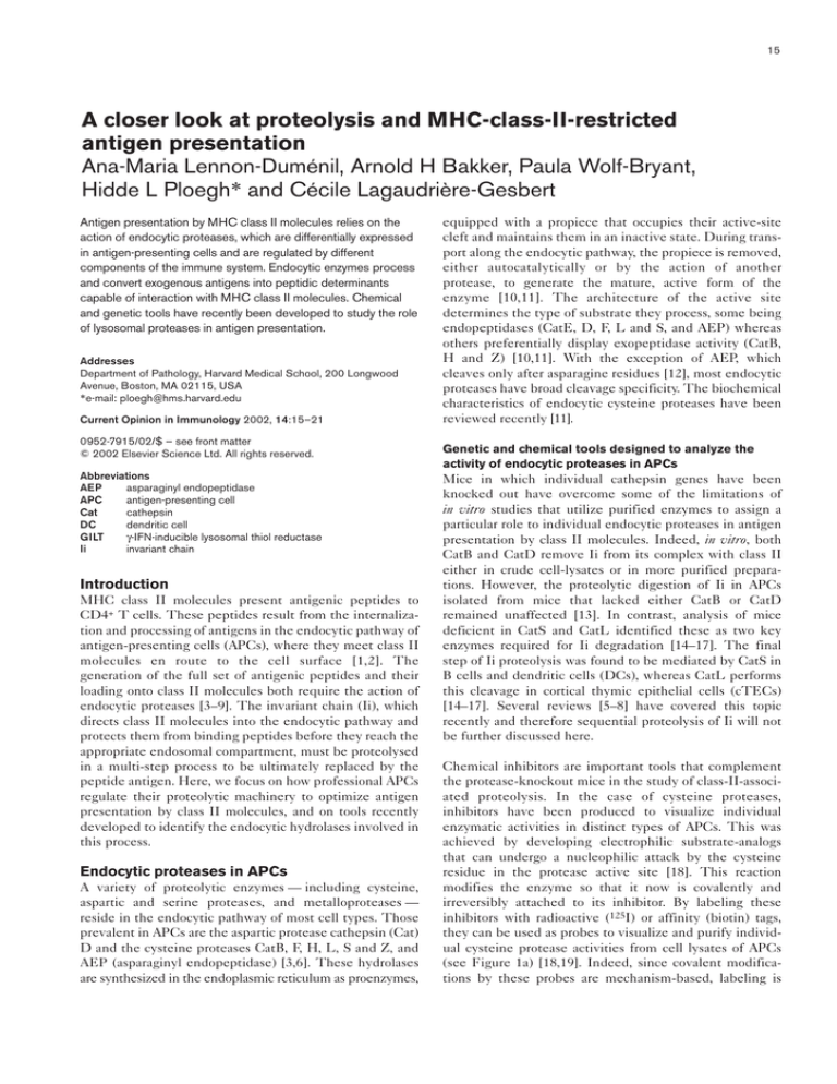
(see Figure 1a) [18,19]. Indeed, since covalent modifications by these probes are mechanism-based, labeling is
16
Antigen processing and recognition
Figure 1
(a)
Autoradiogram (or
streptavidin) blot
Probe–125I
Streptavidin
beads
Coomassie- or silver-stain
+ protein identification
Probe–biotin
Pull-down
Probe–fluorochrome
Multiplexing
Cell lysis
Coupling
Electrophoresis
(b)
Probe–fluorochrome
Microscopy
(c)
(d)
C S F H K V B L
APC
surface
Bead, antigen
or pathogen
Streptavidin
Probe–biotin
Protease
Phagosome
Early endosome
Late endosome
a
Tr
Lysosome
n
sp
or
Tools to analyze activity of cysteine proteases
in APCs. (a) Active-site-directed probes can
be used in different ways to screen APC
lysates for active proteases. Cell lysates can,
for example, be incubated with iodinated or
biotinylated probes. Here, and in (b) but not
(c), black circles represent proteases of
interest and colored circles represent probes
bound to these proteases. Electrophoresis of
these lysates followed by autoradiography or
by streptavidin-blotting (top right) reveals the
level of activity of the different proteases that
have bound to the probe. Alternatively,
proteases that react with biotinylated probes
can be pulled down with streptavidin-coated
beads and stained using Coomassie- or silverstaining. This purification can be used for
identification of unknown proteases. In
another strategy, probes coupled to different
fluorochromes can be used for ‘multiplexing’
(see also [d], below). (b) Fluorescent activesite-directed probes can also be used for the
analysis of protease activity and localization in
intact cells. (c) Coupling biotinylated probes
(red) to streptavidin-coated beads, antigens or
pathogens (black circles) allows analysis of
the proteolytic environment of antigens after
internalization into an APC. After entering the
endocytic pathway, the complex will bind the
active cysteine proteases (shown in various
colors) that it encounters. The components of
a resulting complex that contains several
molecules of one protease (blue) are shown in
detail. Lysis of the APC followed by
electrophoresis and streptavidin blotting
reveals the presence and levels of activity of
the different proteases that have bound to the
probe inside the cell. (d) An example of
‘multiplexing’. Probes optimized for specific
cathepsins were coupled to different
fluorochromes. Purified cathepsins (as
indicated by their letters on the top of the
panel) were bound to these probes and
separated by electrophoresis. The image in
(d) was kindly provided by Matthew Bogyo
and is reproduced, with permission, from [24••].
t
proportional to the enzymatic activity of the protease
targeted. For a given protease, different labeling intensities
thus correspond directly to differences in activity levels.
As described below, these probes have been used to
assess protease profiles in various types of professional
APCs and under different conditions of stimulation (e.g.
in response to cytokines or in mice deficient for Ii or
endocytic proteases) [17,18,20,21••,22••,23]. In addition,
active-site-directed probes can be coupled to fluorescent
moieties with distinct emission spectra to be used
for ‘multiplexing’ active-site labeling experiments or
for localization of active enzymes in living cells
(Figure 1a,b,d) [24••]. Together, these chemical tools
should allow the construction of a more complete record
of the hydrolases relevant for antigen presentation.
The regulation of endocytic protease activity in APCs
Tissue-specific expression
Certain cathepsin genes are expressed in a tissue-specific
manner, allowing differential protease activity in the
various types of APCs [3–9]. CatS, for example, is present
predominantly in bone-marrow-derived APCs (B cells,
DCs and macrophages) [6,17], whereas CatL is poorly
represented in B cells and DCs, but is present rather in
macrophages and cTECs [17]. CatF and CatZ are also
preferentially active in macrophages and bone-marrowderived DCs [20]. It has been shown that CatF can cleave
Proteolysis and MHC-class-II-restricted antigen presentation Lennon-Duménil et al.
17
Figure 2
(a)
ss
ss
ss
ss
Macropinocytosis Phagocytosis Receptor-mediated uptake
Antigen
ss
ss
ss
Cell membrane
Unfolding
Unlocking
GILT
AEP
CatB?
Degradation
Lo
(b)
ad
in g
(d)
Proteases
Toxin
p41–CatL
Export
Cystatins
(f)
Trimming
A schematic overview of antigen processing
in the endocytic pathway of APCs.
(a) Antigens can be internalized through
different modes of uptake (macropinocytosis,
phagocytosis and receptor-mediated uptake).
(b) Once in the endocytic pathway, GILT
unfolds the antigens by breaking disulfide
bonds, after which unlocking of the antigen is
the first step towards degradation and loading
onto MHC class II molecules. (c) Pathogens
can introduce toxins (green squares) and
peptidic inhibitors (black helices) that directly
or indirectly prevent proteolytic activity,
whereas (d) cystatins (colored helices) are
endogenous peptidic inhibitors. (e) Cytokines
can have both stimulatory and inhibitory
effects on endocytic protease activity.
(f) Mature CatL can be chaperoned by
binding of p41 to its active site. This
p41–CatL complex can be secreted.
(g) Secreted active CatL could play a role in
extracellular antigen processing as well as in
extracellular-matrix degradation.
MHC class II
Cell membrane
(c)
Pathogen
(e) Cytokines
(g)
Extracellularmatrix degradation
Current Opinion in Immunology
Ii in vitro, similar to CatS [20], whereas no specific role in
class-II-restricted antigen presentation has been attributed
yet to CatZ. Finally, the capacity of DCs to convert internalized antigens in T-cell epitopes is under strict control
of their developmental stage [25••]. This has led to the
suggestion that DCs may adjust their proteolytic levels to
regulate the production of antigenic determinants during
maturation [26].
impairing presentation by class II molecules [21••]. In
contrast, the proinflammatory cytokines IL-6, TNFα and
IL-1β decrease the endosomal pH [21••,27]. This results
in an increase of the activity of some endocytic proteases
and of class-II/peptide loading [21••,27], demonstrating
that the balance of pro- and anti-inflammatory cytokines
can directly affect the antigen presentation capacity of
APCs by regulating the activity of endocytic proteases.
The strength of proteolysis in the endocytic pathway
of APCs can also be regulated by a variety of external
and internal stimuli, including cytokines, pathogen
products and endogenous competitive inhibitors, including components of the class II pathway itself (Figure 2).
Endogenous competitive inhibitors
Regulation by cytokines
Competitive endogenous cysteine protease inhibitors that
bind tightly and reversibly to the enzyme’s active site can
also be found in APCs [11,21••,28]. They can be classified
into three types: the propiece of the enzyme itself, the
cystatin family of inhibitors and a fragment of the p41 (exon 6b)
isoform of Ii that resembles a thyroglobulin domain [3,4,11].
Factors such as cytokines regulate endocytic proteolysis at
different levels, by modifying the biosynthesis, stability or
activity of the enzyme. For example, in macrophages,
γ-IFN can both upregulate transcription of cathepsin
genes [4,5] and promote the maturation of proforms to
active enzymes [22••]. In addition, the activity of endocytic
proteases can be modulated by changes in the endosomal
pH. The cytokine IL-10 has been shown to raise the pH of
APC endosomes, thus attenuating the levels of hydrolase
activities present in those acidic compartments and
Both the propieces and the cystatins bind the enzyme’s
active site in reverse orientation to prevent their own
proteolysis [29]. There is no evidence that the propiece
liberated by cleavage from one protease can have an
inhibitory effect ‘in trans’ on a distinct hydrolase. The
inhibitors from the cystatin family have been found to be
involved in many physiological processes and diseases by
providing protection from inappropriate proteolysis
[10,11]. In terms of specificity, cystatins can target CatB, F,
18
Antigen processing and recognition
K, L and S, AEP and CatH with variable affinity constants
(nanomolar for exopeptidases and picomolar for endopeptidases) [10,11]. Cystatin C has been suggested as an
inhibitor of CatS activity during maturation of DCs [28].
Indeed, changes in CatS activity were shown to correlate
with a redistribution of class II molecules at the surface of
DCs during activation, suggesting a role for cystatins in the
control of antigen presentation in professional APCs [28].
However, this observation remains controversial since no
defect in class II surface expression was observed in
mature DCs from CatS-knockout mice [30••,31].
The significance of the chaperone function of p41
Competitive inhibitors of cysteine proteases can also
exhibit a chaperone function in vivo: the p41 isoform of Ii
stabilizes mature CatL in bone-marrow-derived APCs
[22••]. Previous studies showed that the p41-specific
64-amino-acid fragment binds non-covalently to the active
site of mature CatL [32−35]. Moreover, in vitro studies
demonstrated that this segment of p41 inhibits the enzymatic activity of CatL [32−35]. Mutant mice deficient for
Ii or expressing either p31 or p41 were used to study the
functional significance of the CatL/p41 interaction in vivo.
Contrary to expectations, CatL expression and activity are
strongly reduced in macrophages isolated from Ii-deficient
mice [22••]. In the absence of p41, mature CatL is
degraded, suggesting that p41 protects CatL from
pre-mature destruction [22••]. Therefore, p41 is not merely
an inhibitor of CatL enzymatic activity, but serves as a
chaperone to help maintain a pool of mature enzyme in
late-endocytic compartments of APCs. Whether this can
be generalized to other protease–inhibitor complexes
remains to be established.
In addition, active-site labeling experiments suggested
that CatS and CatB are responsible for degradation of
mature CatL when p41 is absent, indicating — perhaps
not surprisingly — that cathepsins can regulate the activity
of one another. Indeed, Honey et al. [36•] demonstrated
that, in the absence of CatS, the levels of mature CatL
are considerably increased. Therefore it is likely that
cathepsins modulate each other’s activity by contributing
to their turnover and/or maturation process.
What is the specific function of the pool of CatL
complexed to p41? One possibility is that CatL–p41
complexes are packaged into lysosomal secretory vesicles
to be released into extracellular space, since — unlike free
mature CatL — the enzyme complexed to p41 can survive
in a neutral pH environment [35]. In agreement with this
hypothesis, release of mature CatL takes place only from
macrophages that express p41 (A-M Lennon-Duménil,
unpublished data). Secreted active CatL could play a role
in the degradation of the extracellular matrix to promote
cellular migration during inflammation [37,38], or in the
generation of antigenic peptides, to be loaded on the
empty class II molecules that are reported to be present at
the surface of some APCs [39]. Regulation of proteolysis in
macrophages and DCs is relevant not only for antigen
presentation, but also for the modification of the microenvironment of these APCs. By regulating the ability of
APCs to secrete active CatL, Ii — a component of the
class II machinery — would directly participate in the
initiation of the inflammation process. Concerted regulation
of antigen presentation, migration of the APCs and
recruitment of effector cells at the site of inflammation is
essential to ensure an efficient immune response.
Regulation by pathogens
Endocytic proteases are likely to be targeted by pathogens
capable of evading the immune system. Indeed,
Bm-CPI-2 — a cystatin-like gene product recently identified from the filarial nematode parasite Brugia malayi —
was demonstrated to inhibit AEP activity and interfere
with the presentation of tetanus toxin epitopes (see below)
[40••]. This could equally be true for several molecules
encoded in other parasites and shown to resemble protease
inhibitors. Intracellular pathogens like Helicobacter pylori
can also inhibit endocytic hydrolases since they encode
toxins capable of neutralizing the endosomal pH [41].
Processing of exogenous antigens in
endocytic compartments of APCs
Upon internalization into the APC, exogenous antigens
travel along the endocytic pathway and meet a variety of
enzymes charged with processing them. The APC must
ensure that the antigen is not completely destroyed in this
process but is instead broken down into polypeptides
of variable length capable of interacting with class II
molecules. Indeed, class II molecules must access a
diverse repertoire of antigenic determinants in order to
optimize T-cell activation. The diversity of this peptide
repertoire depends both on the enzymes encountered by
antigens and on the receptivity of class II molecules. Both
these parameters in turn depend on the type of compartment(s) to which the antigen is targeted for processing and
loading onto class II molecules.
Unfolding of antigen and accessibility to proteases
Exposure to acidic pH is the first step in antigen processing,
initiating unfolding of the protein. Reduction of inter- and
intra-molecular disulfide bonds is necessary for complete
denaturation [42], in order to facilitate access of substrate to
proteolytic enzymes. GILT (γ-IFN-inducible lysosomal thiol
reductase) is an enzyme capable of catalyzing disulfide-bond
reduction at low pH, suggesting that it is involved in
class-II-restricted antigen-presentation (Figure 2) [43••,44].
Additional features of the antigen can affect accessibility of
proteolytic enzymes. These include its glycosylation state,
interaction with its receptor upon internalization, and early
binding to class II molecules [2,9,45–47]. Indeed, the open
ends of the class II binding groove are permissive to interaction with long polypeptides, with the immunogenic epitope
anchored into the groove. This ensures that the T-cell epitope
is protected from destruction whereas the extremities of the
peptide can undergo further trimming (Figure 2) [9,46–50].
Proteolysis and MHC-class-II-restricted antigen presentation Lennon-Duménil et al.
Antigen unlocking and degradation
Polypeptides bound to class II molecules typically consist
of a core sequence with ragged amino- and carboxyl-termini,
suggesting that more than one protease is responsible for
cleavage of the antigen: (an) initial cleavage(s) by
endopeptidases would be necessary to ‘unlock’ the antigen
and allow further trimming of the ends by exopeptidases
(Figure 2) [3,51•]. This ‘unlocking function’ was attributed
to the cysteinyl protease AEP, essential for proteolysis of
the carboxy-terminal domain of tetanus toxin antigen
(termed TTCF) [12]. Indeed, cleavage of TTCF at a
single site by AEP is key for the production of all TTCF
antigenic peptides for presentation to T cells [12,51•].
The pathway of degradation of a radiolabeled immune
complex (125I-F[ab′]2) — internalized via Fcγ-receptors in
bone-marrow-derived APCs from the different cathepsinmutant mice — is compatible with the picture emerging
from the processing of TTCF by AEP: degradation is
initiated by discrete proteolytic steps that are performed
by a limited number of endocytic proteases [23]. In the
case of 125I-F(ab′)2, the initial cleavages involve CatB and
generate a discrete high-molecular-weight processing
intermediate. In contrast, complete degradation of
125I-F(ab′) also requires CatS activity [23]. Impeding the
2
protease responsible for antigen unlocking should hinder
further presentation of all T-cell epitopes. The identification of additional proteases responsible for unlocking of
antigens will be essential if this knowledge is to be applied
to manipulation of immunological processes such as
induction and maintenance of tolerance.
Many endocytic proteases have been implicated by in vitro
studies (using purified enzymes or protease inhibitors)
in antigen degradation; however — as for Ii processing —
such analyses do not necessarily reflect what takes place
ex/in vivo. For example, in vitro, CatB and D can generate
T-cell epitopes from intact antigens [52,53], although only
a modest shift in the efficiency of presentation of these
antigenic determinants is observed ex vivo in APCs from
CatB- or CatD-deficient animals [13]. Even APCs from
CatS- or CatL-knockout mice, which clearly display a
defect in proteolysis of Ii, are still able to stimulate T cells
[15,16]. However, since these studies are carried out ex vivo
(with purified APCs and T-cell hybridomas) they do not
exclude a role for these proteases in vivo. Future experiments using infectious agents in protease-deficient mice
should help resolve this issue.
Direct analysis of the proteolytic environment of
antigens upon internalization
The mechanisms by which antigens are internalized are
diverse and tightly regulated. The mode of antigen uptake
depends on both the type of antigen and the type of APC,
and dictates the endocytic compartment to which the antigen will be targeted [2,54–56]. Given the fact that each
endocytic vesicle is likely to vary in its proteolytic content,
targeting of the antigen after uptake will determine the
19
type of proteases to which it will be exposed, and therefore
the repertoire of antigenic peptide that will result from its
degradation. Development of tools aimed to analyze the
proteolytic environment to which antigens are exposed
upon internalization will therefore help us to understand
the rules that govern the process of antigen degradation.
Internalization of antigens can be nonspecific, involving
phagocytosis or fluid-phase endocytosis, or specific,
through receptors expressed on the surface of APCs
(Figure 2) [54–57]. The main function of such receptors is
to target and concentrate the antigen in intracellular
compartments competent for processing and interaction
with class II molecules, resulting in a productive immune
response. Moreover, the specificity of some of these
receptors ensures recognition of rare antigens.
Even though it is now clear that the mode of antigen
uptake determines the endocytic compartment in which
the antigen ends up, little is known about the proteolytic
environment to which antigens are actually exposed during
trafficking into the APC. Do antigens meet proteases
immediately after internalization? Is the proteolytic
environment of antigens different for distinct APCs and/or
internalization modes? Do features of the antigen itself or
extracellular stimuli affect the proteases to which it is
exposed in the APC? The work of Desjardins and collaborators [58••], who used latex beads to isolate phagosomes
from macrophages and analyzed their protein content,
may be an inspiration to address these questions. The
combination of this technique with the use of the activesite-directed probes immobilized on latex beads should
allow the direct identification of the active proteases
contained in the phagosome during its maturation (see
Figure 1c). Furthermore, by engineering probes that could
be coupled to pathogens or soluble antigens, it should be
possible to directly sense the proteolytic environment to
which they are exposed after internalization into the APC.
Conclusions
Recently developped genetic and chemical tools have
allowed important progress in elucidating the proteolytic
events associated with MHC-class-II-restricted antigen
presentation. We now have a reasonably good understanding of the rules that govern Ii proteolysis, but the pathways
of antigen degradation remain less well defined. Indeed,
essential questions such as the exact identification of the
intracellular compartments and enzymes responsible for
degradation of antigens that have been differentially
internalized in APCs remain to be resolved.
Sensing the proteolytic environment to which intracellular
antigens are exposed could help us to address these
questions. This approach could also allow to understand
additional points relevant for immunology such as, for
example, escape of intracellular pathogens that are
internalized by macrophages from destruction. Indeed,
pathogens can not only inhibit phagolysosomal fusion, but
also frequently encode protease-inhibitor homologs that
20
Antigen processing and recognition
could directly affect the activity of surrounding hydrolases.
Identification of the proteases to which autoantigens are
exposed may also help clarify the contribution of antigenprocessing enzymes to the development of autoimmunity.
This, together with analyzing the immune response of
mice deficient for endocytic proteases, will help understand how the activity of these enzymes influences the
outcome of the immune response.
Acknowledgements
The authors thank Jose A Villadangos and Edda Fiebiger for crictical
reading of the manuscript.
References and recommended reading
Papers of particular interest, published within the annual period of review,
have been highlighted as:
• of special interest
•• of outstanding interest
1.
Wolf PR, Ploegh HL: How MHC class II molecules acquire peptide
cargo: biosynthesis and trafficking through the endocytic
pathway. Annu Rev Cell Dev Biol 1995, 11:267-306.
2.
Watts C: Capture and processing of exogenous antigens for
presentation on MHC molecules. Annu Rev Immunol 1997,
15:821-850.
3.
Watts C: Antigen processing in the endocytic compartment. Curr
Opin Immunol 2001, 13:26-31.
4.
Chapman HA, Riese RJ, Shi GP: Emerging roles for cysteine
proteases in human biology. Annu Rev Physiol 1997, 59:63-88.
5.
Chapman HA: Endosomal proteolysis and MHC class II function.
Curr Opin Immunol 1998, 10:93-102.
6.
Riese RJ, Chapman HA: Cathepsins and compartmentalization in
antigen presentation. Curr Opin Immunol 2000, 12:107-113.
7.
Nakagawa TY, Rudensky AY: The role of lysosomal proteinases in
MHC class II-mediated antigen processing and presentation.
Immunol Rev 1999, 172:121-129.
8.
Villadangos JA, Bryant RY, Deussing J, Driessen C, LennonDumenil A-M, Riese RJ, Roth W, Saftig P, Shi GP, Chapman HA et al.:
Proteases involved in MHC class II antigen presentation. Immunol
Rev 1999, 172:109-120.
9.
Villadangos JA, Ploegh HL: Proteolysis in MHC class II antigen
presentation: who’s in charge? Immunity 2000, 12:233-239.
10. McGrath ME: The lysosomal cysteine proteases. Annu Rev
Biophys Biomol Struct 1999, 28:181-204.
11. Turk V, Turk B, Turk D: Lysosomal cysteine proteases: facts and
opportunities. EMBO J 2001, 20:4629-4633.
12. Manoury B, Hewitt EW, Morrice N, Dando PM, Barrett AJ, Watts C:
An asparaginyl endopeptidase processes a microbial antigen for
class II MHC presentation. Nature 1998, 396:695-699.
13. Deussing J, Roth W, Saftig P, Peters C, Ploegh HL, Villadangos JA:
Cathepsins B and D are dispensable for major histocompatibility
complex class II-mediated antigen presentation. Proc Natl Acad
Sci USA 1998, 95:4516-4521.
14. Riese RJ, Wolf PR, Bromme D, Natkin LR, Villadangos JA, Ploegh HA,
Chapman HA: Essential role for cathepsin S in MHC
class II-associated invariant chain processing and peptide
loading. Immunity 1996, 4:357-366.
15. Nakagawa TY, Brissette WH, Lira PD, Griffiths RJ, Petrushova N,
Stock J, McNeish JD, Eastman SE, Howard ED, Clarke SR et al.:
Impaired invariant chain degradation and antigen presentation
and diminished collagen-induced arthritis in cathepsin S null
mice. Immunity 1999, 10:207-217.
16. Shi GP, Villadangos JA, Dranoff G, Small C, Gu L, Haley KJ, Riese R,
Ploegh HL, Chapman HA: Cathepsin S required for normal MHC
class II peptide loading and germinal center development.
Immunity 1999, 10:197-206.
17.
Nakagawa T, Roth W, Wong P, Nelson A, Farr A, Deussing J,
Villadangos JA, Ploegh J, Peters C, Rudensky AY: Cathepsin L:
critical role in Ii degradation and CD4 T cell selection in the
thymus. Science 1998, 280:450-453.
18. Bogyo M, Verhelst S, Bellingard-Dubouchaud V, Toba S,
Greenbaum D: Selective targeting of lysosomal cysteine
proteases with radiolabeled electrophilic substrate analogs.
Chem Biol 2000, 7:27-38.
19. Greenbaum D, Medzihradszky KF, Burlingame A, Bogyo M: Epoxide
electrophiles as activity-dependent cysteine protease profiling
and discovery tools. Chem Biol 2000, 7:569-581.
20. Shi GP, Bryant RA, Riese R, Verhelst S, Driessen C, Li Z, Bromme D,
Ploegh HL, Chapman HA: Role for cathepsin F in invariant chain
processing and major histocompatibility complex class II peptide
loading by macrophages. J Exp Med 2000, 191:1177-1186.
21. Fiebiger E, Meraner P, Weber E, Fang IF, Stingl G, Ploegh H,
•• Maurer D: Cytokines regulate proteolysis in major
histocompatibility complex class II-dependent antigen
presentation by dendritic cells. J Exp Med 2001, 193:881-892.
This article describes how pro- and anti-inflammatory cytokines modulate
MHC-class-II-restricted antigen presentation in human DCs by modifying
the activity of endocytic proteases. The data demonstrate that IL-1β and
TNFα enhance CatS and CatB activity, resulting in an increase of the
formation of MHC-class-II–peptide complexes and presentation to T cells. This
appears to be due to the ability of pro-inflammatory cytokines to acidify the pH
of endocytic compartments. In contrast, IL-10 downregulates the activity of
CatS and CatB and prevents acidification of DC endosomal compartments.
22. Lennon-Dumenil AM, Roberts RA, Valentijn K, Driessen C,
•• Overkleeft HS, Erickson P, Peters J, Bikoff E, Ploegh HL,
Wolf-Bryant P: The p41 isoform of invariant chain is a chaperone
for cathepsin L. EMBO J 2001, 20:4055-4064.
This work documents the in vivo significance of the CatL/p41 interaction
described in [27]. The results demonstrate that CatL expression and activity are
strongly reduced in professional APCs that lack Ii. In the absence of p41, a
considerable fraction of lysosomal, active CatL is proteolyzed, suggesting that
p41 protects the enzyme from premature destruction. Therefore, p41 is not
merely an inhibitor of CatL enzymatic activity, but serves as a chaperone to help
maintain a pool of mature enzyme in late-endocytic compartments of APCs.
23. Driessen C, Lennon-Dumenil AM, Ploegh HL: Individual cathepsins
degrade immune complexes internalized by antigen-presenting
cells via Fcγγ receptors. Eur J Immunol 2001, 31:1592-1601.
24. Greenbaum D, Baruch A, Hayrapetian L, Zsuzsanna D, Burlingame A,
•• Medzihradszky K, Bogyo M: Chemical approaches for functionally
probing the proteome. Mol Cell Prot 2001, in press.
This work describes the development of fluorescent active-site-directed
probes that can be used in vitro to characterize individual cysteine protease
activities in cell lysates and in vivo to investigate their intracellular localization by microscopy. In addition, the authors used these active-site-directed
probes to screen for selective inhibitors of the different cysteine proteases,
showing that their approach can be applied to identify potential pharmacological targets and the corresponding inhibitors.
25. Inaba K, Turley S, Iyoda T, Yamaide F, Shimoyama S, Reis e Sousa C,
•• Germain RN, Mellman I, Steinman RM: The formation of
immunogenic major histocompatibility complex class II-peptide
ligands in lysosomal compartments of dendritic cells is regulated
by inflammatory stimuli. J Exp Med 2000, 191:927-936.
This work uses an antibody that specifically recognizes MHC class II
molecules in association with an antigenic peptide, to demonstrate that the
formation of productive MHC-class-II−peptide complexes is developmentally
controlled during differentiation of DCs. Even though immature DCs can
efficiently internalize antigens, MHC-class-II−peptide complexes are found
exclusively in cells that have received an inflammatory stimulus.
26. Mellman I, Steinman RM: Dendritic cells: specialized and regulated
antigen processing machines. Cell 2001, 106:255-258.
27.
Drakesmith H, O’Neil D, Schneider SC, Binks M, Medd P, Sercarz E,
Beverley P, Chain B: In vivo priming of T cells against cryptic
determinants by dendritic cells exposed to interleukin 6 and
native antigen. Proc Natl Acad Sci USA 1998, 95:14903-14908.
28. Pierre P, Mellman I: Developmental regulation of invariant chain
proteolysis controls MHC class II trafficking in mouse dendritic
cells. Cell 1998, 93:1135-1145.
29. Stubbs MT, Laber B, Bode W, Huber R, Jerala R, Lenarcic B, Turk V:
The refined 2.4 Å X-ray crystal structure of recombinant human
stefin B in complex with the cysteine proteinase papain: a novel
type of proteinase inhibitor interaction. EMBO J 1990,
9:1939-1947.
Proteolysis and MHC-class-II-restricted antigen presentation Lennon-Duménil et al.
30. Villadangos JA, Cardoso M, Steptoe RJ, van Berkel D, Pooley J,
•• Carbone FR, Shortman K: MHC class II expression is regulated in
dendritic cells independently of invariant chain degradation.
Immunity 2001, 14:739-749.
As variance with the paper by Pierre and Mellman [28], this article
demonstrates that DCs from CatS-knockout mice display wild-type levels of
surface-peptide-loaded MHC class II molecules and undergo normal
maturation. The authors propose a model in which the differences in surface
expression of MHC class II molecules between immature and mature DCs
result from different endocytosis rates of membrane MHC-class-II−peptide
complexes and are independent of Ii proteolysis.
31. Driessen C, Bryant RA, Lennon-Dumenil AM, Villadangos JA,
Bryant PW, Shi GP, Chapman HA, Ploegh HL: Cathepsin S controls
the trafficking and maturation of MHC class II molecules in
dendritic cells. J Cell Biol 1999, 147:775-790.
32. Bevec T, Stoka V, Pungercic G, Dolenc I, Turk V: Major
histocompatibility complex class II-associated p41 invariant chain
fragment is a strong inhibitor of lysosomal cathepsin L. J Exp Med
1996, 183:1331-1338.
33. Guncar G, Pungercic G, Klemencic I, Turk V, Turk D: Crystal structure
of MHC class II-associated p41 Ii fragment bound to cathepsin L
reveals the structural basis for differentiation between cathepsins
L and S. EMBO J 1999, 18:793-803.
34. Fineschi B, Sakaguchi K, Appella E, Miller J: The proteolytic
environment involved in MHC class II-restricted antigen
presentation can be modulated by the p41 form of invariant chain.
J Immunol 1996, 157:3211-3215.
35. Ogrinc T, Dolenc I, Ritonja A, Turk V: Purification of the complex of
cathepsin L and the MHC class II-associated invariant chain
fragment from human kidney. FEBS Lett 1993, 336:555-559.
36. Honey K, Duff M, Beers C, Brissette WH, Elliott EA, Peters C,
•
Maric M, Cresswell P, Rudensky AY: Cathepsin S regulates the
expression of cathepsin L and the turnover of GILT in B
lymphocytes. J Biol Chem 2001, 276:22573-22578.
This paper shows that CatS controls the turnover and activity of other endocytic
enzymes, including CatL and GILT. Changes in CatS activity alter the levels
of both CatL and GILT, and will therefore directly and indirectly affect the
processing and loading of antigens for presentation by MHC class II molecules.
37.
Felbor U, Dreier L, Bryant RA, Ploegh HL, Olsen BR, Mothes W:
Secreted cathepsin L generates endostatin from collagen XVIII.
EMBO J 2000, 19:1187-1194.
38. Punturieri A, Filippov S, Allen E, Caras I, Murray R, Reddy V, Weiss SJ:
Regulation of elastinolytic cysteine proteinase activity in normal
and cathepsin K-deficient human macrophages. J Exp Med 2000,
192:789-799.
39. Santambrogio L, Sato AK, Fischer FR, Dorf ME, Stern LJ: Abundant
empty class II MHC molecules on the surface of immature
dendritic cells. Proc Natl Acad Sci USA 1999, 96:15050-15055.
40. Manoury B, Gregory WF, Maizels RM, Watts C: Bm-CPI-2, a cystatin
•• homolog secreted by the filarial parasite Brugia malayi, inhibits
class II MHC-restricted antigen processing. Curr Biol 2001,
11:447-451.
This work shows that the parasite product Bm-CPI-2 — which is homologous to
the cysteine protease inhibitors from the cystatin family — can inhibit AEP activity in vitro and in vivo, preventing TTCF unlocking and the presentation of TTCF
T-cell epitopes. This suggests that intracellular pathogens capable of producing
such molecules may be able to modify the activity of proteolytic enzymes in the
host APC and therefore to prevent recognition by the immune system.
41. Molinari M, Salio M, Galli C, Norais N, Rappuoli R, Lanzavecchia A,
Montecucco C: Selective inhibition of Ii-dependent antigen
presentation by Helicobacter pylori toxin VacA. J Exp Med 1998,
187:135-140.
42. Collins DS, Unanue ER, Harding CV: Reduction of disulfide bonds
within lysosomes is a key step in antigen processing. J Immunol
1991, 147:4054-4059.
43. Arunachalam B, Phan UT, Geuze HJ, Cresswell P: Enzymatic
•• reduction of disulfide bonds in lysosomes: characterization of a
gamma-interferon-inducible lysosomal thiol reductase (GILT).
Proc Natl Acad Sci USA 2000, 97:745-750.
This paper describes the first thiol reductase to be identified, GILT, as being
capable of catalyzing disulfide-bond reduction at acidic pH. GILT resides in
21
late-endocytic compartments and its expression profile parallels that of MHC
class II molecules (constitutive in professional APCs and γ-IFN-inducible in
other tissues), suggesting its involvement in antigen processing.
44. Phan UT, Arunachalam B, Cresswell P: Gamma-interferoninducible lysosomal thiol reductase (GILT). Maturation,
activity, and mechanism of action. J Biol Chem 2000,
275:25907-25914.
45. Surman S, Lockey TD, Slobod KS, Jones B, Riberdy JM, White SW,
Doherty PC, Hurwitz JL: Localization of CD4+ T cell epitope
hotspots to exposed strands of HIV envelope glycoprotein
suggests structural influences on antigen processing. Proc Natl
Acad Sci USA 2001, 98:4587-4592.
46. Moudgil KD, Sekiguchi D, Kim SY, Sercarz EE: Immunodominance
is independent of structural constraints: each region within hen
eggwhite lysozyme is potentially available upon processing of
native antigen. J Immunol 1997, 159:2574-2579.
47.
Watts C, Antoniou A, Manoury B, Hewitt EW, McKay LM, Grayson L,
Fairweather NF, Emsley P, Isaacs N, Simitsek PD: Modulation by
epitope-specific antibodies of class II MHC-restricted
presentation of the tetanus toxin antigen. Immunol Rev 1998,
164:11-26.
48. Germain RN: MHC-dependent antigen processing and peptide
presentation: providing ligands for T lymphocyte activation. Cell
1994, 76:287-299.
49. Castellino F, Zappacosta F, Coligan JE, Germain RN: Large protein
fragments as substrates for endocytic antigen capture by MHC
class II molecules. J Immunol 1998, 161:4048-4057.
50. Deng H, Apple R, Clare-Salzer M, Trembleau S, Mathis D, Adorini L,
Sercarz E: Determinant capture as a possible mechanism of
protection afforded by major histocompatibility complex class II
molecules in autoimmune disease. J Exp Med 1993,
178:1675-1680.
51. Antoniou AN, Blackwood SL, Mazzeo D, Watts C: Control of antigen
•
presentation by a single protease cleavage site. Immunity 2000,
12:391-398.
This reference and [12] identify the endocytic cysteine protease, AEP, as a
key enzyme for the generation of T-cell epitopes from TTCF. By showing that
AEP cleaves TTCF at a single site and that by blocking this unique cleavage
event one can prevent the presentation of all TTCF T-cell epitopes, the
authors introduce the concept of ‘antigen unlocking’, which is explained in
the text of our review.
52. Van Noort JM, Jacobs MJ: Cathepsin D, but not cathepsin B,
releases T cell stimulatory fragments from lysozyme that are
functional in the context of multiple murine class II MHC
molecules. Eur J Immunol 1994, 24:2175-2180.
53. Vidard L, Rock KL, Benacerraf B: Diversity in MHC class II
ovalbumin T cell epitopes generated by distinct proteases.
J Immunol 1992, 149:498-504.
54. Van Bergen J, Ossendorp F, Jordens R, Mommaas AM, Drijfhout JW,
Koning F: Get into the groove! Targeting antigens to MHC class II.
Immunol Rev 1999, 172:87-96.
55. Amigorena S, Bonnerot C: Role of B-cell and Fc receptors in
the selection of T-cell epitopes. Curr Opin Immunol 1998,
10:88-92.
56. Lanzavecchia A: Mechanisms of antigen uptake for presentation.
Curr Opin Immunol 1996, 8:348-354.
57.
Bakke O, Nordeng TW: Intracellular traffic to compartments for
MHC class II peptide loading: signals for endosomal and
polarized sorting. Immunol Rev 1999, 172:171-187.
58. Garin J, Diez R, Kieffer S, Dermine JF, Duclos S, Gagnon E,
•• Sadoul R, Rondeau C, Desjardins M: The phagosome proteome:
insight into phagosome functions. J Cell Biol 2001,
152:165-180.
This article describes a proteomic analysis of the phagosome formed by
macrophages after internalization of latex beads. This very systematic and
complete study identified >140 proteins that are present in the phagosome
at different stages of biogenesis. More specifically, this work shows that the
distinct endocytic proteases are gradually incorporated into the phagosome
during maturation, rather than being delivered in bulk.


