Laser Scanning Confocal Microscope
FV1200 ®
FLUOVIEW®
FV1200:
the freedom to choose your own path.
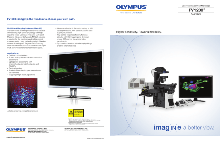
Multi-Point Mapping Software (MMASW)
Standard raster scanning modes are not capable
of measuring high speed physiology with high
signal-to-noise. Olympus’ innovative Multi-point
Mapping Advanced Software (MMASW) provides
a solution for the most demanding high speed
measurements. Using methods similar to Random
Access Scanning and Targeted Path Scanning,
users have the freedom to choose their own rapid
multi-point measurement or stimulation paths.
• Measure cell network fluctuations at up to 101
cycles per second, with up to 50,000 Hz data
output per position
• Map cellular responses to simultaneous
stimulus with ROI mapping and Olympus’
unique SIM scanner for optogenetics
experiments
• Synchronize detection with electrophysiology
or other external devices
Applications:
• Calcium ion fluctuations
• Pulsed multi-point or multi-area stimulation
experiments
• Optogenetic experiments with
channelrhodopsin, halorhodopsin, and
GCaMPs
• Electrophysiology
• Studies of functional output over cells and
cell networks
• Mapping of light reactive positions
Artistic rendering using Bitplane Imaris
©2012 Olympus America Inc. All rights reserved.
• Olympus, FV and IX are registered trademarks of Olympus Corporation,
Olympus America Inc., and/or their affiliates, in the U.S. and/or other countries. All
other third party marks are property of their respective owners.
• Images on monitors are simulated.
• Specifications and appearances are subject to change without any notice or
obligation on the part of the manufacturer.
OLYMPUS AMERICA INC.
3500 Corporate Parkway, Center Valley, PA 18034-0610, U.S.A.
OLYMPUS CANADA INC.
OLYMPUS LATIN AMERICA INC.
5301 Blue Lagoon Drive, Suite 290 Miami, FL 33126, U.S.A.
25 Leek Crescent, Richmond Hill, Ontario L4B 4B3
www.olympusamerica.com
Printed in USA FV1200BROCHURE13.01
Higher sensitivity. Powerful flexibility.
Laser Scanning Confocal Microscope FV1200
The FV1200 has been designed specifically for live cell imaging with high sensitivity
detection, near-IR laser capability and high light throughput.
Imaging of living tissues demands the highest levels of sensitivity which allows for reduced laser power, phototoxicity and
photobleaching, and innovative approaches to measurement of fluorescent indicators. The new FV1200 brings sensitivity
and power to your research through a range of innovative technology and sensitivity improvements, allowing you to
capture the most critical elements of your biological samples with speed, precision and reliability.
New High Sensitivity Gallium Arsenide Phosphide
(GaAsP) Detector Unit
Live cell imaging requires some of the highest sensitivity
detection possible, and the new 2 channel GaAsP
confocal detector is ideal for this purpose.
• 2 channels of hand selected GaAsP detectors with
quantum efficiency up to 45%
• Peltier cooling for minimal dark noise
• High signal-to-noise detection
• Along with standard 3 channel PMTs, allows up to
5 simultaneous fluorescent imaging channels when
combined with 748nm laser
• Provides high sensitivity while allowing researchers
flexibility for high dynamic range imaging with standard
PMTs
High Reflection Silver Coated Scanning Mirrors
Anti-oxidization silver coating ensures durability and
improves reflection efficiency in both excitation and
emission, increasing the reflectance in the visible range
by 5-15%, and IR reflectance up to 22%. Silver coating
improves reflection efficiency, especially in the near–IR
range. For laser scanning confocal systems, emitted
fluorescence is “descanned,” meaning that laser excitation
intensity will increase as well as collected fluorescence
intensity, providing important improvements in sensitivity.
The improvement in the near-IR range is particularly
important given the new 748nm laser available with the
FV1200 for researchers using long wavelength excitation
dyes. Available laser lines include 405nm, 440nm, 458nm,
488nm, 515nm, 543nm, 559nm, 635nm, and 748nm.
ZDC3
• IX3-ZDC focus detection and tracking can be performed
via the innovative touch panel independent of software
focus
• Search function supported by a cell-safe, near-infrared
laser enabling instant focusing on samples
• Capable of both single shot and continuous focus
modes
Multi-Alkali PMT
New IX83 Microscope
• Touch panel control for easy switching of observation
conditions
• Greater frame stability and rigidityinfrared laser enabling
instant focusing on samples
• Enhanced remote focus and one touch autofocus with
ZDC
IX3-ZDC Optical Path Diagram
Comparison of galvano mirror Silver vs. Aluminum
*Reflectance of two Galvo mirrors
GaAsP - PMT
Signal-to-noise is improved through a
combination of both higher sensitivity and
lower dark noise. Greatest improvements
will be seen with dim samples/regions.
Red arrows indicate synapsin signal made
clear by GaAsP.
Take advantage of Olympus exclusive and unique objectives and robust autofocus with
the IX83ZDC on the highly flexible yet stable new IX83 microscope. True simultaneous
stimulation with the FV1200 SIM scanner and ultra-high speed measurement technology
which is faster and more sensitive than resonance scanners makes the FV1200 an ideal
instrument for interactive live cell imaging and fluorescence measurements.
Optimized Olympus Exclusive 30x, 40x and 60x
Silicone Objectives
• Increased numerical apertures and refractive index
matching ideal for live cell and tissue imaging
• Long term time lapse with silicone oil that does not dry
out like water immersion
Olympus Exclusive SIM Scanner for Simultaneous
Stimulation
• Interact with your cells and tissue in real time with true,
precise simultaneous multicolor stimulation
• Unique tornado scanning for highest efficiency of FRAP
and photoactivation
• Use with Multi-point mapping for ultimate speed in
optogenetics


