Journal of Minimally Invasive Gynecology (2006) 13, 398 – 402
Fertility after treatment of Asherman’s syndrome stage
3 and 4
Hervé Fernandez, MD, Fadheela Al-Najjar, MD, Aurélia Chauveaud-Lambling, MD,
René Frydman, MD, and Amélie Gervaise, MD
From the Department of Obstetrics and Gynecology, Hôpital Antoine Béclère, Assistance Publique-Hôpitaux de Paris,
Clamart cedex, France (all authors).
KEYWORDS:
Asherman’s
syndrome;
Operative
hysteroscopy;
Placenta accreta;
Reproductive
outcome.
Abstract
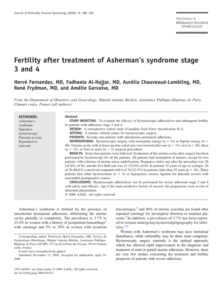
STUDY OBJECTIVE: To evaluate the efficacy of hysteroscopic adhesiolysis and subsequent fertility
in patients with adhesions stage 3 and 4.
DESIGN: A retrospective cohort study (Canadian Task Force classification II-2).
SETTING: A tertiary referral center for hysteroscopic surgery.
PATIENTS: Seventy-one patients with intrauterine permanent adhesions.
INTERVENTIONS: Hysteroscopic surgery with monopolar energy (n ⫽ 31) or bipolar energy (n ⫽
40). Uterine cavity with at least one free ostial area was restored after one (n ⫽ 31), two (n ⫽ 20), three
(n ⫽ 15), or four or more (n ⫽ 5) surgical procedures
RESULTS: Sixty-four patients were followed. Evaluation of the uterine cavity after surgery has been
performed by hysteroscopy for all the patients. All patients had resumption of menses, except for two
patients with a history of uterine artery embolization. Pregnancy index rate after the procedure was 28
(43.8%) of 64, and the live birth rate was 21 (32.8%) of 64. In patients 35 years of age or younger, 20
of 30 (66.6%) conceived compared with 8 of 34 (23.5%) in patients older than 35 years (p ⫽ .01). Three
patients had either hysterectomy (n ⫽ 2) or hypogastric arteries ligation for placenta accreta with
uneventful postoperative course.
CONCLUSIONS: Hysteroscopic adhesiolysis can be performed for severe adhesions stage 3 and 4
with safety and efficacy. Age is the main predictive factors of success: the pregnancies were at risk of
abnormal placentation.
© 2006 AAGL. All rights reserved.
Asherman’s syndrome is defined by the presence of
intrauterine permanent adhesions, obliterating the uterine
cavity partially or completely. The prevalence is 3.7% to
23.4% in women with a history of postpartum hemorrhage1
with curettage and 5% to 39% in women with recurrent
Corresponding author: Professeur Hervé Fernandez, MD, Service de
Gynécologie-Obstétrique, Hôpital Antoine Béclère, Assistance PubliqueHôpitaux de Paris (AP-HP), 157 rue de la Porte-de-Trivaux. 92141 Clamart
cedex, France.
E-mail: herve.fernandez@abc.aphp.fr
Submitted November 17, 2005. Accepted for publication April 24,
2006.
1553-4650/$ -see front matter © 2006 AAGL. All rights reserved.
doi:10.1016/j.jmig.2006.04.013
miscarriages,2 and 40% of uterine synechia are found after
repeated curettage for incomplete abortion or retained placenta.3 In addition, a prevalence of 2.7% has been reported in women undergoing hysterosalpingography for infertility.4,5
Women with Asherman’s syndrome may have menstrual
disturbance, while subfertility may be there main complaint.
Hysteroscopic surgery currently is the optimal approach,
which has allowed rapid improvement in the diagnosis and
treatment of total or partial uterine adhesions. However, there
are very few studies concerning the treatment and fertility
prognosis of patients with severe adhesions.
Fernandez et al
Table 1
Fertility and Asherman’s syndrome
399
Etiology of intrauterine adhesions and symptoms at initial consultation
Etiology
No. (%)
Symptom
D&C for elective abortion
D&C for missed or incomplete abortion
D&C for postpartum hemorrhage
D&C for molar pregnancy
Myomectomy
Uterine embolization
30
18
5
1
15
2
Subfertility
Subfertility
Subfertility
Subfertility
Subfertility
Subfertility
(42)
(25.4)
(7)
(1.4)
(21.1)
(2.8)
and
and
and
and
and
hypomenorrhea
amenorrhea
amenorrhea
recurrent pregnancy loss
amenorrhea
D&C ⫽ dilatation and curettage.
We previously published a study of 31 patients with
severe adhesions treated by hysteroscopy.6 We found two
factors increased subsequent fertility: the role of age (⬍ 35
years) and the interest to repeat the procedures to restore a
normal cavity. Since this first short-term study, we introduced a new surgical technique with Versapoint electrode
(Versapoint Electro-Surgical System, Gynecare, Inc.,
Menlo Park, CA ) without cervical dilatation.
The aim of this study was to evaluate the safety and
efficacy of this new hysteroscopic adhesiolysis surgical
technique in the treatment of 40 new patients and to evaluate the results of 71 consecutive patients with severe Asherman’s syndrome by observing the complete elimination of
all synechia in the uterine cavity, postoperative resumption
of menses, and pregnancy rate in a retrospective case report
series.
Materials and methods
From January 1990 through December 2003, 302 patients underwent hysteroscopy treatment for intrauterine adhesions (IUA). We reviewed all operative reports and selected all patients with severe adhesions, with partial
agglutination of uterine wall (stage 3) or total agglutination
(stage 4) and both ostial area occluded (stage 3 and 4
according to the European Society of Hysteroscopy).7 Seventy-one patients (23%) were selected according to these
criteria.
Median age was 36.1 years ⫾ 6.1 (range 26 – 47). Mean
parity was 0.6 ⫾ 0.9 (range 0 –3). Risk factors for cavity
obliteration were the following: history of at least one dilatation and curettage (D&C) (range 1–5) for elective abortion in the first trimester (30 patients [42.3%]); at least one
D&C (range 1– 4) for missed abortion or incomplete abortion in the first trimester (18 patients [25.4%]); and D&C for
postpartum hemorrhage (5 patients [7%]). One patient
(1.4%) had curettage for molar pregnancy. Fifteen patients
(21.1%) had myomectomy involving hysterotomy, with six
of them performed in another hospital, and two patients
(2.8%) underwent uterine embolization for fibroid uterus in
absence of rational indication (Table 1). The interval between the causal procedure and hysteroscopy treatment of
IUA unfortunately was not available.
Eight patients (11.3%) were referred to our tertiary care
reproductive center after first inefficient procedure. There
was no case of tuberculosis endometritis in this series.
Of the 71 patients, 31 patients (43.6%) reported amenorrhea, 19 (26.7%) profound hypomenorrhea, and 21
(29.7%) normal cycles (exclusively in cases of stage 3).
Fifty patients experienced infertility (70.4%), and 21 patients (29.6%) suffered recurrent pregnancy loss. Diagnosis
was made by hysterosalpingography and confirmed by hysteroscopy in all cases.
Surgical technique
Hysteroscopy was performed under general or epidural
anesthesia in the early proliferative phase of the menstrual
cycle in the patients who were menstruating.
For 31 patients (43%) operated on from 1990 through
1997, a 9-mm resectoscope equipped with hysteroscopic
monopolar knife (Karl Storz GmbH, Tuttlingen, Germany)
was introduced into the blind reduced cavity, obtained after
prudent dilatation of the cervix by Hegar’s dilators. Glycine
was used as distending medium. Starting in 1998, we used
Versapoint bipolar electrode exclusively with normal saline
solution to distend the uterine cavity (n ⫽ 40 [57%]). No
cervical dilatation was needed with Versapoint 5F electrode. In both methods, fluid balance was recorded in all
women.
Treatment was performed by making several myometrial
incisions 4-mm deep: two or three lateral incisions from the
fundus to the isthmus on both sides and two or three transversal incisions of the fundus. Procedure was stopped at that
point, even if ostial areas were not visible. A simultaneous
laparoscopy was performed only in three patients with a
history of pelvic inflammatory disease or ectopic pregnancy, to observe the distal tubal status. In our large experience, ultrasound and laparoscopic control are not used to
avoid uterine perforation with hysteroscopy, due to absence
of usefulness in monitoring this surgery. Prophylactic antibiotics amoxicillin and clavulanic acid at the dose of 2 g
(SmithKline Beecham, Nanterre, France) were given routinely at the induction of anesthesia. No intrauterine contraceptive device was inserted, because no significant advantages has been noted when compared with hormonal
therapy alone.8 Postoperative estrogen therapy (estradiol 4
400
Journal of Minimally Invasive Gynecology, Vol 13, No 5, September/October 2006
Table 2
Results according to surgical technique
Perforation
Normal cavity after
initial procedure
After 2 procedures
After 3 procedures
After 4 or more
Lost to follow-up
Resectoscope
(n ⫽ 31)
Versapoint
(n ⫽ 40)
4
16
3
15
7
7
1
3
13
8
4
4
mg daily; Laboratories Cassenne, Puteaux, France) was
given to all patients for 2 months to plan postoperative
diagnostic hysteroscopy.
The anatomic result defined by complete elimination of
synechia with restoration of normal size and shape of the
uterus was checked in all patients by an outpatient hysteroscopy without anesthesia. Hysteroscopy, even if it misses
small areas of adhesions, conversely to hysterosalpingography visualizes the restoration of endometrium—the cavity
that appears to be the main prognostic factor—and can
easily rupture filmy adhesion during this procedure. Subsequent fertility was studied by calling all patients by telephone. We considered only the first pregnancy after index
surgery.
We evaluated the results between the two surgical techniques. The 2, test modified by Yates correction when
appropriate, and Fisher’s exact test were used for statistical
evaluation, and p ⬍.05 was considered to be statistically
significant. Where appropriate, we used means, standard
deviation, and CIs for normally distributed data; and for
skewed data, we used medians and ranges.
Results
Seventy-one patients underwent a total of 136 hysteroscopic procedures. The mean operating time for the procedure was 25.4 ⫾ 6.2 minutes (95% CI 24.35–26.45). All
patients were discharged from the hospital on the day of
surgery. Complications were noted in seven patients (5.1%)
of 136 hysteroscopies involving perforation occurring dur-
Table 3
ing cervical dilatation (n ⫽ 4) or during the procedure (n ⫽
3). No perforation occurred during the first surgery using
Versapoint electrode. If perforation occurred, the procedure
was terminated, and patients were operated on 2 months
later without any difficulty, and no difference in subsequent
fertility was observed.
A completed elimination of all synechia was obtained
after the initial procedure by outpatient control hysteroscopy in 31 patients (43.6%), and these patients were allowed to attempt pregnancy; nevertheless in 16 of these 31
patients (51.6%), filmy adhesions easily ruptured during
postoperative hysteroscopy. In the remaining 40 patients,
postoperative hysteroscopy diagnosed the persistence of
moderate or severe IUA and justified a second operative
hysteroscopy with direct synechialysis. No difference was
observed between the two surgical techniques.
Finally reconstruction of normal uterine cavity was realized after one (n ⫽ 31), two (n ⫽ 20), three (n ⫽ 15), or four
or more (n ⫽ 5) surgical procedures despite the heterogeneous quality of endometrium mucosa.
Menstruation was restored in all patients with history of
amenorrhea and oligomenorrhea, with the exception of two
patients who underwent uterine artery embolization for uterine myomas.
Seven patients were lost to follow-up after control hysteroscopy. The median follow-up time was 41 months
(range 6 – 86) for the remaining 64 patients. The results in
accordance with surgical techniques are presented in Table
2, and no difference was observed.
Twenty-eight index pregnancies were obtained in 64
patients (43.8%) with no difference between surgical techniques, and the outcomes were as follows: 3 had first trimester missed abortions, 4 had second trimester fetal losses,
and 21 (32.8%) had live births. Three out of four second
trimester losses occurred after resectoscope technique. All
patients conceived spontaneously with the exception of
three who underwent a first cycle of in vitro fertilization and
embryo transfer (Table 3).
Nine (42.9%) of 21 patients with a history of pregnancy
loss and 12 (24%) of 50 patients with infertility had live
births. In patients aged 35 years or younger, 20 out of 30
conceived (66.6%) compared with 8 out of 34 (23.5%) in
patients aged more than 35 years (p ⬍.05) (Table 4). In 21
patients with live births, there were 12 vaginal deliveries at
Obstetrical history and pregnancy outcome after synechialysis with subsequent pregnancy and live birth rate
Obstetrical history and recent obstetric outcome after synechialysis
Pregnancy rate No. (%)
Live birth rate No. (%)
Infertility
Recurrent pregnancy loss
Spontaneous pregnancy
Assisted pregnancy by IVF
Current pregnancy missed abortion
Current pregnancy fetal loss in second trimester
18/50
10/21
25/28
3/28
3/28
4/28
12/50
9/21
20/25
1/3
_
_
IVF ⫽ in vitro fertilization.
(36)
(47.6)
(89.3)
(10.7)
(10.7)
(14.3)
(24)
(42.9)
(80)
(33.3)
Fernandez et al
Table 4
Fertility and Asherman’s syndrome
Pregnancy rate related to age
No. of pregnancies
Live birth
ⱕ 35 years No. (%) (n ⫽ 30)
⬎ 35 years No. (%) (n ⫽ 34)
p
20 (66.6)
16 (53.5)
8 (23.5)
5 (14.7)
⬍.001
.371
term and 9 cesarean sections (CS). Three CS were performed for breech presentation with fetal distress, and another three CS were performed for fetal distress with cephalic presentation. Caesarean sections were performed in
two patients with previous one and two CS, respectively, for
placenta accreta. The last CS was performed for chorioamnionitis at 30 weeks’ gestation.
Severe complications occurred in 3 (14.3%) of 21 patients. Two patients had hysterectomy for placenta accreta
(patients with previous one and two CS, respectively) without postoperative complications. The third patient had CS
for Candida albicans chorioamnionitis at 30 weeks’ gestation, after preterm rupture of membrane. Hemostasis was
obtained by hypogastric arteries ligation with an uneventful
postoperative course. The newborn required respiratory
support initially. The pregnancy rate was 14 (48.4%) of 31,
10 (50%) of 20, and 4 (20%) of 20, respectively, after one,
two, and three or more surgical procedures.
Discussion
Management of uterine synechia improved progressively
during the last 10 years by the widespread use of hysteroscopic surgery, which helps in diagnosis of uterine synechia
and restoration of normal size and shape to the uterus, which
is essential to carry a pregnancy to term. With this largest
series published of patients with severe adhesions, we confirmed the role of age and the interest to repeat the procedures until restoration a normal cavity are predictive factors
of subsequent fertility. Moreover, in 90% of pregnancies in
our series occurred by natural means.
Various hysteroscopic adhesiolysis techniques were described and published in the last decades, either division of
adhesion by hysteroscopic scissors9 or by using the resectoscope.10 In our study, pregnancy to term was achieved in
32.8% patients, and normal uterine cavity and menstruation
were achieved in all patients with the exception of two
patients with history of uterine artery embolization for uter-
Table 5
401
ine myomas who continue to have amenorrhea. Coccia et al11
achieved a 33.3% pregnancy at term rate by using pressure
lavage under ultrasound guidance in adhesiolysis. The new
Versapoint technique has been used in the last few years and
demonstrated its efficacy in treating intrauterine pathologies.12–15 We observed only 1 out of 40 second trimester
fetal losses with Versapoint, whereas the rate was 3 out of
31 after resectoscope techniques. Versapoint could prevent
iatrogenic cervical incompetence due to cervical dilatation
realized for resectoscope techniques. This difference can be
due to absence of cervical dilatation and/or less mucosa
destruction by bipolar energy.
From the above studies, the rate of pregnancy at term
after synechialysis was almost the same among the various
techniques of hysteroscopic surgery (Table 5). All these
series can be compared because, whatever the classification
used in each of these series, we always considered severe
adhesions.
However, the Versapoint technique has improved safety
by avoiding the complication of fluid overload and cervical
laceration or uterine perforation due to cervical dilatation in
patients with stenosed cervix or nullipara patients,14 and we
observed no perforation with this technique. Moreover, the
risk of excessive fluid absorbed is theoretically possible but
the short time of the procedure, always less than 30 minutes,
decreases the risk of this complication.
There are probably few indications left for laparotomy,
even in the treatment of severe IUA. Repeated hysteroscopic procedures as described16,17 and in our series allowed the re-establishment of a normal cavity in all patients,
and 20% fertility after three or more procedures in spite of
a higher risk of uterine perforation, whichever surgical technique was used during the repeat procedures.
Many studies fail to present their results according to the
severity of the adhesion. Therefore different techniques are
difficult to compare. So in our study we treated severe
Asherman synechia stage 3 and 4, and we obtained a 32.8%
live birth rate after adhesiolysis. The live birth rate observed
demonstrates the difficulty of having a normal endometrium
Delivery rate after adhesiolysis using various hysteroscopic methods
Study
9
Valle and Sciarra
Chen et al10
Capella-Allouc et al6
Coccia et al11
Our series (2004)
No. of patients
Hysteroscopic method
Pregnancy at term (%)
47
23
28
3
71
Resectoscope with scissors
Resectoscope with Laminaria (MedGyne, Lombard, IL)
Monopolar knife
Pressure lavage under ultrasound guidance
Versapoint and resectoscope
15
8
9
1
21
(31.9)
(34.9)
(32.1)
(33.3)
(32.8)
402
Journal of Minimally Invasive Gynecology, Vol 13, No 5, September/October 2006
suitable for nidation even after restoration of a normal
uterine cavity in patients with stage 4 adhesions, which
corresponds to what was reported by Valle and Scierra9
where the severity of adhesions affected the chance of
pregnancy after adhesiolysis.
When the patient’s age was considered, we found that 20
(66.6%) of 30 patients aged 35 years or younger conceived,
compared with 8 (23.5%) of 34 patients older than 35 years.
These results confirm that it is worthwhile repeating hysteroscopic treatments until a normal uterine cavity is restored, especially for young women age 35 years or less. For
women older than 35 years, the principal aim of the treatment should be resumption of normal menses, but obstetric
outcome remains disappointing.
Complications are common in subsequent pregnancy after adhesiolysis and have been described by many authors.
Placenta accreta is the most common complication reported
after IUA with an incidence of about 8%,9 while in our
study we had three patients (14.3%) with placenta accrete.
Two of the three had a history of one and two CS, respectively, which ended with hysterectomy, while the third patient underwent hypogastric arteries ligation. The high rate
of placenta accreta in our series is explained by defective
lamina basalis after adhesiolysis especially in severe Asherman’s syndrome, which allows abnormal placentation.
Other complications have been described by many authors. Deaton et al18 reported a spontaneous uterine rupture
during pregnancy after hysteroscopic treatment of severe
Asherman’s syndrome complicated by a fundal perforation.
Friedman et al19 described three severe complications in a
series of 33 patients with mild to severe IUA: uterine sacculation, uterine dehiscence, and placenta accreta. These
complications could be due to resection of the myometrium
during the lysis of IUA.
Our finding that the pregnancy rate was 14 (48.4%) of 31
with one surgical procedure, 10 (50%) of 20 after two
surgical procedures, and 4 (20%) of 20 after three or more
surgical procedures shows that it is worthwhile to repeat
lysis of IUA until a normal uterine cavity and shape are
restored. Second trimester fetal losses occurred in four patients: one patient had a one-step procedure, and three had
undergone four surgical procedures. Two of the four patients became pregnant again and had uneventful pregnancies after cervical cerclage at 12 weeks’ gestation. Therefore, we think that cervical cerclage should be discussed
with patients with multiple-stage procedures. However, the
Versapoint technique avoids cervical dilatation, and this can
explain the low incidence of cervical incompetence especially since we started in 1998, with one second trimester
loss with this surgical technique without dilatation.
Conclusion
Hysteroscopic adhesiolysis can be performed for severe
adhesions with safety and efficacy, which offers a real
chance of parenthood in a substantial proportion of infertile
couples. However, these patients with IUA undergoing adhesiolysis should be appropriately informed about the occurrence of life-threatening complications if they became
pregnant and should be managed appropriately in a tertiary
care center. Moreover, performance of repeat procedures is
not indicated in patients older than 35 years.
References
1. Eriksen J, Kaestel C. The incidence of uterine atresia after post partum
curettage: a follow-up examination of 141 patients. Danish Medical
Bulletin. 1960;7:50 –51.
2. Rabau E, David A. Intrauterine adhesions: etiology, prevention and
treatment. Obstet Gynecol. 1963;22:626 – 629.
3. Westendrop IC, Ankum WM, Mol BW, Vonk J. Prevalence of Asherman’s syndrome after secondary removal of placental remnants or a
repeat curettage for incomplete abortion. Hum Reprod. 1998;12:3347–
3350.
4. Sweeny WJ. Intrauterine synechia. Obstetrics and Gynecology. 1966;
27:284 –289.
5. Dmowski WP, Greenblatt RB. Asherman’s syndrome and risk of
placenta accreta. Obstet Gynecol. 1969;34:288 –299.
6. Capella-Allouc S, Morsad F, Rongières-Bertrand C, Taylor S, Fernandez H. Hysteroscopic treatment of severe Asherman’s syndrome and
subsequent fertility. Hum Reprod. 1999;14:1230 –1233.
7. Wamsteker K, De Block S. Diagnostic hysteroscopy: technique and
documentation. In: Sutton C, Diamond M, eds. Endoscopic Surgery for
Gynecologists. London, UK: Saunders; 1993:263–276.
8. Sanfilippo JS, Fitzgerald MR, Badawy SZ, Nussbaum ML, Yussman
MA. Asherman’s syndrome: a comparison of therapeutic methods. J
Reprod Med. 1982;27:328 –330.
9. Valle RF, Sciarra JJ. Intrauterine adhesions: hysteroscopic diagnosis,
classification, treatment and reproductive outcome. Am J Obstet Gynecol. 1988;158:1459 –1470.
10. Chen FP, Soong YK, Hui YL. Successful treatment of severe uterine
synechiae with transcervical resectoscopy combined with laminaria
tent. Hum Reprod. 1997;12:943–947.
11. Coccia ME, Becattini C, Bracco GL, et al. Pressure lavage under
ultrasound guidance: a new approach for outpatient treatment of intrauterine adhesions. Fertil Steril. 2001;75:601– 606.
12. Zikopoulos K, Kolibianakis EM, Tournaye H, et al. Hysteroscopic
septum resection using the Versapoint system in subfertile women.
Reproductive Biomedicine Online. 2003;7:365–367.
13. Marwah V, Bhandari SK. Diagnostic and interventional microhysteroscopy with use of the coaxial bipolar electrode system. Fertil Steril.
2003;79:413– 417.
14. Fernandez H, Gervaise A, de Tayrac R. Operative hysteroscopy for
infertility using normal saline solution and a coaxial bipolar electrode:
a pilot study. Hum Reprod. 2000;15:1773–1775.
15. Vilos GA. Intrauterine surgery using a new co-axial bipolar electrode
in normal saline solution (Versapoint): a pilot study. Fertil Steril.
1999;72:740 –743.
16. Chapman R, Chapman K. The value of two stage laser treatment for
severs Asherman’s syndrome. Br J Obstet Gynaecol. 1996;103:1256 –
1258.
17. Protopapas A, Shushan A, Magos A. Myometrial scoring: a new
technique for the management of severe Asherman’s syndrome. Fertil
Steril. 1998;69:860 – 864.
18. Deaton JL, Matier D, Andreoli J. Spontaneous uterine rupture during
pregnancy after treatment of Asherman’s syndrome. Am J Obstet
Gynecol. 1980;160:1053–1054.
19. Friedman S, Defazio J, Decherney A. Severe obstetric complications
after aggressive treatment of Asherman syndrome. Obstet Gynecol.
1986;67:864 – 867.


