COMMENTARY
Visualising microorganisms from molecules to cells
DOI: 10.1111/1574-6968.12485
MICROBIOLOGY LETTERS
Seeing cells
Microscopy led to the initial discovery and classification
of bacteria and, today, advanced methods are revealing
the inner molecular structure. The pages of FEMS journals are filled with micrographs from light microscopy
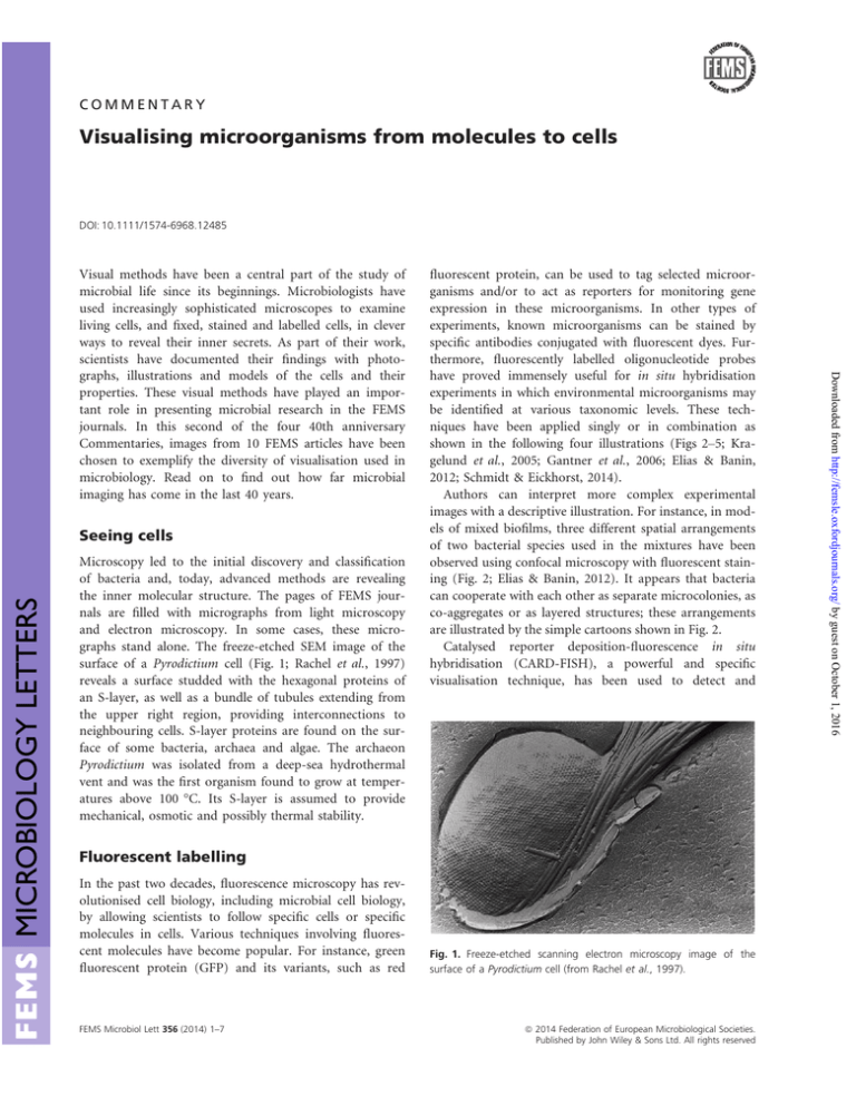
and electron microscopy. In some cases, these micrographs stand alone. The freeze-etched SEM image of the
surface of a Pyrodictium cell (Fig. 1; Rachel et al., 1997)
reveals a surface studded with the hexagonal proteins of
an S-layer, as well as a bundle of tubules extending from
the upper right region, providing interconnections to
neighbouring cells. S-layer proteins are found on the surface of some bacteria, archaea and algae. The archaeon
Pyrodictium was isolated from a deep-sea hydrothermal
vent and was the first organism found to grow at temperatures above 100 °C. Its S-layer is assumed to provide
mechanical, osmotic and possibly thermal stability.
fluorescent protein, can be used to tag selected microorganisms and/or to act as reporters for monitoring gene
expression in these microorganisms. In other types of
experiments, known microorganisms can be stained by
specific antibodies conjugated with fluorescent dyes. Furthermore, fluorescently labelled oligonucleotide probes
have proved immensely useful for in situ hybridisation
experiments in which environmental microorganisms may
be identified at various taxonomic levels. These techniques have been applied singly or in combination as
shown in the following four illustrations (Figs 2–5; Kragelund et al., 2005; Gantner et al., 2006; Elias & Banin,
2012; Schmidt & Eickhorst, 2014).
Authors can interpret more complex experimental
images with a descriptive illustration. For instance, in models of mixed biofilms, three different spatial arrangements
of two bacterial species used in the mixtures have been
observed using confocal microscopy with fluorescent staining (Fig. 2; Elias & Banin, 2012). It appears that bacteria
can cooperate with each other as separate microcolonies, as
co-aggregates or as layered structures; these arrangements
are illustrated by the simple cartoons shown in Fig. 2.
Catalysed reporter deposition-fluorescence in situ
hybridisation (CARD-FISH), a powerful and specific
visualisation technique, has been used to detect and
Fluorescent labelling
In the past two decades, fluorescence microscopy has revolutionised cell biology, including microbial cell biology,
by allowing scientists to follow specific cells or specific
molecules in cells. Various techniques involving fluorescent molecules have become popular. For instance, green
fluorescent protein (GFP) and its variants, such as red
FEMS Microbiol Lett 356 (2014) 1–7
Fig. 1. Freeze-etched scanning electron microscopy image of the
surface of a Pyrodictium cell (from Rachel et al., 1997).
ª 2014 Federation of European Microbiological Societies.
Published by John Wiley & Sons Ltd. All rights reserved
Downloaded from http://femsle.oxfordjournals.org/ by guest on October 1, 2016
Visual methods have been a central part of the study of
microbial life since its beginnings. Microbiologists have
used increasingly sophisticated microscopes to examine
living cells, and fixed, stained and labelled cells, in clever
ways to reveal their inner secrets. As part of their work,
scientists have documented their findings with photographs, illustrations and models of the cells and their
properties. These visual methods have played an important role in presenting microbial research in the FEMS
journals. In this second of the four 40th anniversary
Commentaries, images from 10 FEMS articles have been
chosen to exemplify the diversity of visualisation used in
microbiology. Read on to find out how far microbial
imaging has come in the last 40 years.
2
Commentary
uptake of radiolabelled acetate (black dots) in activated
sludge.
Seeing cell–cell communication
Fig. 3. Sequential hybridization reveals bacteria and archaea on a
rice root (from Schmidt & Eickhorst, 2014).
quantitate bacteria (green) and archaea (red) simultaneously on rice roots (Fig. 3; Schmidt & Eickhorst, 2014).
Betaproteobacteria dominated the bacterial populations on
root tips and elongation zones while methanogens were
particularly abundant in the rhizoplane.
Combined imaging techniques can show not only
which species are present in a natural environment, but
are also able to reveal their metabolic activities. In the
example given (Fig. 4; Kragelund et al., 2005), FISHMAR (fluorescence in situ hybridisation combined
with microautoradiography) simultaneously shows the
presence of filamentous eubacteria (green), the alphaproteobacterium Meganema perideroedes (red) and the
ª 2014 Federation of European Microbiological Societies.
Published by John Wiley & Sons Ltd. All rights reserved
Confocal scanning laser microscopy has been combined
with reporter gene technology to visualise bacterial signal
molecules (N-acyl-homoserine lactones) produced on
plant roots and to measure the ‘calling distance’, that is
the distance that such molecules can travel from a producer cell to a recipient cell. In the image shown (Fig. 5;
Gantner et al., 2006), the producer Pseudomonas putida
IsoF (red) emits signal molecules that, when sensed, paint
the recipient reporter Pseudomonas putida F117 green.
This frequently cited paper shows that cell–cell communication mostly occurs at distances of 5–10 lm on plant
roots, while a maximal distance of 78 lm has also been
measured.
Seeing organelles
In the past decade or so, researchers developed methods
of cellular tomography to add the third dimension to
electron microscopy. Several approaches may be taken. A
tomogram of yeast cells (Fig. 6; Perktold et al., 2007) was
created by imaging serial sections of a chemically fixed
cell. The sections were then interpreted manually or
FEMS Microbiol Lett 356 (2014) 1–7
Downloaded from http://femsle.oxfordjournals.org/ by guest on October 1, 2016
Fig. 2. Different spatial distribution of cells in mixed bacterial biofilms (from Elias & Banin, 2012; confocal images from Nielsen et al., 2000;
Rickard et al., 2006 and Hansen et al., 2007).
Commentary
3
Downloaded from http://femsle.oxfordjournals.org/ by guest on October 1, 2016
Fig. 4. Filaments of Meganema perideroedes
reveal metabolic activity (from Kragelund
et al., 2005).
Fig. 5. In situ production of Pseudomonas
putida signal molecules on tomato roots (from
Gantner et al., 2006).
semi-automatically and stacked up to create a threedimensional image showing the cell wall (CW), the endoplasmic reticulum (ER), a lipid particle (LP), mitochondria (M), the nucleus (N) and a vacuole (V).
FEMS Microbiol Lett 356 (2014) 1–7
Alternatively, a tomograph of magnetotactic bacteria
(Magnetospirillum) was obtained by cryoelectron microscopy (Fig. 7a) and by tilting the specimen and collecting
images from many angles (Fig. 7b) (Sch€
uler, 2008). These
ª 2014 Federation of European Microbiological Societies.
Published by John Wiley & Sons Ltd. All rights reserved
4
Commentary
(a)
(b)
€ler, 2008).
Fig. 7. The magnetosome chain in Magnetospirillum gryphiswaldense (from Schu
methods reveal the inner ultrastructure of cells, in particular the cytoskeletal magnetosome filament (Fig. 7a), but
the resolution is currently limited, allowing only the largest molecular complexes, such as ribosomes and the magnetosome filaments, to be seen.
Visualising molecules
Cell biology is increasingly becoming a molecular science
and microbiologists rely on a variety of visual tools to
ª 2014 Federation of European Microbiological Societies.
Published by John Wiley & Sons Ltd. All rights reserved
explore and present biomolecular structures. The image
from Joly et al. (2010) is an example of the rich visual
language for molecules (Fig. 8). It combines several representations to demonstrate different aspects of the
molecular structure of the bacterial phage shock protein
(Psp), including a cartoon ribbon to show the folding
of the chain, a surface to show the overall shape and
location of interaction sites, and a full atomic representation to show the binding of small molecules to the
protein. In addition, a schematic diagram of the amino
FEMS Microbiol Lett 356 (2014) 1–7
Downloaded from http://femsle.oxfordjournals.org/ by guest on October 1, 2016
Fig. 6. Transmission electron micrographs and
3D reconstructions of yeast cells (from
Perktold et al., 2007).
Commentary
5
Downloaded from http://femsle.oxfordjournals.org/ by guest on October 1, 2016
Fig. 8. Structure and motif organization of a phage shock protein (from Joly et al., 2010).
Fig. 9. Circular mitochondrial genome of Rhizoctonia solani (from Losada et al., 2014).
FEMS Microbiol Lett 356 (2014) 1–7
ª 2014 Federation of European Microbiological Societies.
Published by John Wiley & Sons Ltd. All rights reserved
6
Commentary
cellular ultrastructure, molecular concentrations and locations and, in the best cases, dynamics. The image from
Vendeville et al. (2011) shows a model from the LifeExplorer visualisation program, which is integrating these
types of information to create a model of bacterial cell
division (Fig. 10). Quite amazingly, experimental methods are currently narrowing this invisible gap, with continual improvement of micrographic resolution and
solution of larger and larger complexes by methods of
integrative structural biology.
David S. Goodsell
Department of Integrative Structural and Computational
Biology, The Scripps Research Institute, La Jolla, CA, USA
E-mail: goodsell@scripps.edu
Fig. 10. Model of the FtsZ ring by nucleoid occlusion and Min
systems in Escherichia coli (from Vendeville et al., 2011, reproduced
with permission from Thanbichler, 2010).
acid sequence is included to present the domain structure of the protein.
Graphical harnessing of data
A vast body of genome, proteome and interactome data
is currently available to researchers in microbiology, and
methods for visualising and harnessing these data are an
area of active research and development. Many of bacterial genomes have been sequenced, providing invaluable
insights into the structure and regulation of the cells’
genomic information. Most often, published reports
include summary illustrations, such as the image of the
circular mitochondrial genome from Rhizoctonia solani
(Fig. 9, Losada et al., 2014). These types of images are
perfect for obtaining an overall view; digital tools may
then be used to delve into the data in detail.
Modelling the mesoscale
Currently, there is a range of scale that is invisible to
experimental methods, between the nanoscale of atomic
structures and the microscale of microscopy. Computer
simulation is being used to create images of this ‘mesoscale’, by integrating information from microscopy and
molecular biology. This can be quite a treasure hunt to
find the necessary information on molecular structure,
ª 2014 Federation of European Microbiological Societies.
Published by John Wiley & Sons Ltd. All rights reserved
References
Elias S & Banin E (2012) Multi-species biofilms: living with
friendly neighbors. FEMS Microbiol Rev 36: 990–1004.
Gantner S, Schmid M, D€
urr C, Schuhegger R, Steidle A,
Hutzler P, Langebartels C, Eberl L, Hartmann A & Dazzo
FB (2006) In situ quantitation of the spatial scale of calling
distances and population density-independent
N-acylhomoserine lactone-mediated communication by
rhizobacteria colonized on plant roots. FEMS Microbiol Ecol
56: 188–194.
Hansen SK, Haagensen JAJ, Gjermansen M, Jorgensen TM,
Tolker-Nielsen T & Molin S (2007) Characterization of a
Pseudomonas putida rough variant evolved in a mixedspecies
biofilm with Acinetobacter sp. strain C6. J Bacteriol 189:
4932–4943.
Joly N, Engl C, Jovanovic G, Huvet M, Toni T, Sheng X,
Stumpf MPH & Buck M (2010) Managing membrane stress:
the phage shock protein (Psp) response, from molecular
mechanisms to physiology. FEMS Microbiol Rev 34: 797–827.
Kragelund C, Nielsen JL, Thomsen TR & Nielsen PH (2005)
Ecophysiology of the filamentous alphaproteobacterium
Meganema perideroedes in activated sludge. FEMS Microbiol
Ecol 54: 111–112.
Losada L, Pakala SB, Fedorova ND et al. (2014) Mobile
elements and mitochondrial genome expansion in the soil
fungus and potato pathogen Rhizoctonia solani AG-3. FEMS
Microbiol Lett 352: 165–173.
Nielsen AT, Tolker-Nielsen T, Barken KB & Molin S (2000)
Role of commensal relationships on the spatial structure of
a surface-attached microbial consortium. Environ Microbiol
2: 59–68.
Perktold A, Zechmann B, Daum G & Zellnig G (2007)
Organelle association visualized by three-dimensional
FEMS Microbiol Lett 356 (2014) 1–7
Downloaded from http://femsle.oxfordjournals.org/ by guest on October 1, 2016
Dieter Haas
Departement de Microbiologie Fondamentale, Universite de
Lausanne, Lausanne, Switzerland
Commentary
ultrastructural imaging of the yeast cell. FEMS Yeast Res 7:
629–638.
Rachel R, Pum D, Smarda J, Smajs D, Komrska J, Krzyzanek
V, Rieger G & Stetter KO (1997) II. Fine structure of
S-layers. FEMS Microbiol Rev 20: 13–23.
Rickard AH, Palmer RJ Jr, Blehert DS et al. (2006) Autoinducer
2: a concentration-dependent signal for mutualistic bacterial
biofilm growth. Mol Microbiol 60: 1446–1456.
Schmidt H & Eickhorst T (2014) Detection and quantification
of native microbial populations on soil-grown rice roots by
catalyzed reporter deposition-fluorescence in situ
hybridization. FEMS Microbiol Ecol 87: 390–402.
7
Sch€
uler D (2008) Genetics and cell biology of magnetosome
formation in magnetotactic bacteria. FEMS Microbiol Rev
32: 654–672.
Thanbichler M (2010) Synchronization of Chromosomedynamics
and Cell Division in Bacteria. Cold Spring Harbor Perspectives
in Biology. Cold Spring Harbor Press, Cold Spring Harbor,
NY.
Vendeville A, Lariviere D & Fourmentin E (2011) An
inventory of the bacterial macromolecular components
and their spatial organization. FEMS Microbiol Rev 35:
395–414.
Downloaded from http://femsle.oxfordjournals.org/ by guest on October 1, 2016
FEMS Microbiol Lett 356 (2014) 1–7
ª 2014 Federation of European Microbiological Societies.
Published by John Wiley & Sons Ltd. All rights reserved


