The stump capping procedure to prevent or treat terminal osseous
advertisement
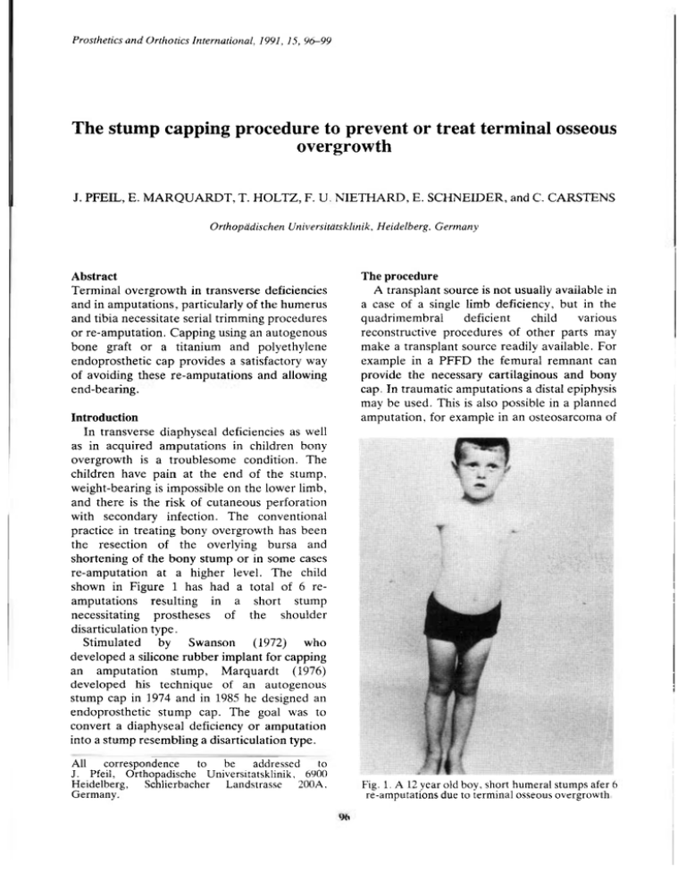
Prosthetics and Orthotics International, 1991, 15, 96-99 The stump capping procedure to prevent or treat terminal osseous overgrowth J. PFEIL, E. MARQUARDT, T. HOLTZ, F. U. NIETHARD, E. SCHNEIDER, and C. CARSTENS Orthopädischen Universitätsklinik, Heidelberg, Germany Abstract The procedure Terminal overgrowth in transverse deficiencies and in amputations, particularly of the humerus and tibia necessitate serial trimming procedures or re-amputation. Capping using an autogenous bone graft or a titanium and polyethylene endoprosthetic cap provides a satisfactory way of avoiding these re-amputations and allowing end-bearing. A transplant source is not usually available in a case of a single limb deficiency, but in the quadrimembral deficient child various reconstructive procedures of other parts may make a transplant source readily available. For example in a PFFD the femural remnant can provide the necessary cartilaginous and bony cap. In traumatic amputations a distal epiphysis may be used. This is also possible in a planned amputation, for example in an osteosarcoma of Introduction In transverse diaphyseal deficiencies as well as in acquired amputations in children bony overgrowth is a troublesome condition. The children have pain at the end of the stump, weight-bearing is impossible on the lower limb, and there is the risk of cutaneous perforation with secondary infection. The conventional practice in treating bony overgrowth has been the resection of the overlying bursa and shortening of the bony stump or in some cases re-amputation at a higher level. The child shown in Figure 1 has had a total of 6 reamputations resulting in a short stump necessitating prostheses of the shoulder disarticulation type. Stimulated by Swanson (1972) who developed a silicone rubber implant for capping an amputation stump, Marquardt (1976) developed his technique of an autogenous stump cap in 1974 and in 1985 he designed an endoprosthetic stump cap. The goal was to convert a diaphyseal deficiency or amputation into a stump resembling a disarticulation type. All correspondence to be addressed to J. Pfeil, Orthopädische Universitätsklinik, 6900 Heidelberg, Schlierbacher Landstrasse 200A. Germany. Fig. 1. A 12 year old boy, short humeral stumps afer 6 re-amputations due to terminal osseous overgrowth The stump capping procedure 97 Fig. 2. Osteosarcoma in the distal femur in a 10 year old boy. The distal epiphysis of the tibia is used as an autogenous stump cap at the time of amputation the distal femur (Fig. 2). Alternatively a graft may be taken from the dorsal part of the iliac crest. The surgical procedure consists of the resection of the bony spike, then the transplant is fixed with a screw or K-wires. The periosteum is reattached to the transplant, the remaining place is then filled with cancellous bone (Fig. 3). Fig. 3, The technique of the autogenous stump capping procedure. A medial longitudinal incision is made a few centimetres proximal to the end of the stump so as to avoid scarring the end-bearing area. The bony spike is resected and the cap fixed with Kwires or with a cancellous screw. To give you an idea of this procedure Figure 4 illustrates the follow-up X-rays of a 4 year old girl with very short epiphyseal stumps in bilateral PFFD. In her case the cartilaginous bony caps were preserved from the femur and a stump capping procedure was performed on both arms at the age of 4 years. A good end-bearing capacity was achieved on both sides. Eight years after the operation the patient was able to bring her arms together in front of the body. Following the stump capping procedure the skin at the end of the stump became tight but there was no risk of bony perforation. In this early case the incisions were made at the end of the stumps which is thought to be responsible for the narrowing of the soft tissues. When she was grown up a musculo-cutaneous flap was performed to give better soft tissue covering of the stumps. This procedure has, in our opinion, only cosmetic benefits, and does not bring any functional improvement. 98 J. Pfeil, E. Marquardt, T. Holtz, F. U. Niethard, E. Schneider, and C. Carstens Fig. 4. The use of osseo-cartilaginous transplant from the femur to the humerus bilateral, in a quadrimembral deficient child. Notice the growth of the humerus after this procedure in the follow up films. It is important to recognize that the success of this procedure depends not only on the surgical technique but also on a postoperative training programme. Approximately 3 months after the stump capping procedure a programme of endbearing training is started. Initially weights of 2 or 3 kg are used and gradually increased until the patient is able to take at least 50% of body weight directly over the stump end. Immediately after the end-bearing training the therapist teaches the patient the technique of skin stretching distally over the stump to prevent contractures or tightening of the skin over the reconstructed end. The prescription of and training in the use of the prostheses is also part of the postoperative care. The advantage of the autogenous stump capping is that no artificial material is needed, and it produces good long term results. There are some disadvantages in this procedure. In some cases a graft is not available. At lease two incisions are necessary. Fixation metal has to be removed at a later stage. Sometimes consolidation takes a long time, especially in cases where a purely cartilaginous transplant is used. In 1985 Marquardt developed in co-operation with Philipp Hannover, and the MECRON Company an endoprosthetic stump cap which is produced in different sizes. It consists of a titanium body which is fixed with at least three cortical screws to the bone. A polyethylene head covers the fixed titanium cap. Figure 5 shows the application of the endoprosthetic stump cap. First musculoperiostal flaps are raised and the spike resected, before the titanium cap is fixed with cortical screws. A polyethylene cap is fixed with a selflocking system. At the side of the polyethylene cap there are holes which allow the fixation of the musculoperiostal flaps. In this particular case it was possible to close the muscles over the polyethylene cap. Full end-bearing capacity at the femoral stump could be achieved. This method is also applied in short humeral deficiencies or in short tibial stumps. The advantages of the endoprosthetic stump cap are that no graft is needed and it is possible to achieve early weight-bearing. End-bearing can be started approximately 3 weeks postoperatively. A definitive prosthesis can already be used by the patient 6 weeks after this procedure. So far there is a lack of long term results. At present the number of cases treated in the University Clinic and a short postoperative follow-up prevents the provision of the complication rates of this procedure. The stump capping procedure 99 REFERENCES MARQUARDT E. (1976). Plastische Operationen bei drohender konchendurchspiessung am kindlichen Oberarmstumpf. Eine Vorlanfige Mittelung. Z. Orthop., 114, 711-714. SWANSON, A. B. (1972). Silicone-rubber implants to control the overgrowth phenomenon in the juvenile amputee. ICIB, 11 (9), 5-8. Fig. 5. Application of the endoprosthetic stump cap: (a). fixation of the titanium body: (b). fixation of the polyethylene cap with a selflocking system: (c). attachment of the muscles to the polyethylene cap.
