Diagnostic Imaging Pathways - Lower Urinary Tract Symptoms
Printed from Diagnostic Imaging Pathways
www.imagingpathways.health.wa.gov.au
© Government of Western Australia
Diagnostic Imaging Pathways - Lower Urinary Tract Symptoms
Population Covered By The Guidance
This pathway provides guidance on the imaging of adult patients with lower urinary tract symptoms.
Date reviewed: January 2012
Date of next review: January 2015
Published: April 2012
Quick User Guide
Move the mouse cursor over the PINK text boxes inside the flow chart to bring up a pop up box with salient
points.
Clicking on the PINK text box will bring up the full text.
The relative radiation level (RRL) of each imaging investigation is displayed in the pop up box.
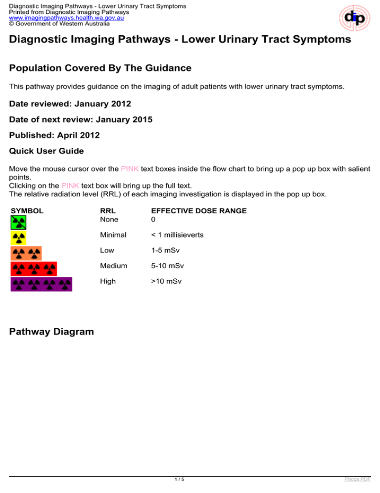
SYMBOL
RRL
None
EFFECTIVE DOSE RANGE
0
Minimal
< 1 millisieverts
Low
1-5 mSv
Medium
5-10 mSv
High
>10 mSv
Pathway Diagram
1/5
Phoca PDF
Diagnostic Imaging Pathways - Lower Urinary Tract Symptoms
Printed from Diagnostic Imaging Pathways
www.imagingpathways.health.wa.gov.au
© Government of Western Australia
Image Gallery
Note: These images open in a new page
1
Prostatitis
Image 1 (Ultrasound): Bulky prostate of approximately 49mL.
2
Neurogenic Bladder
Image 2 (Ultrasound): Thick-walled bladder consistent with neurogenic
bladder.
Teaching Points
2/5
Phoca PDF
Diagnostic Imaging Pathways - Lower Urinary Tract Symptoms
Printed from Diagnostic Imaging Pathways
www.imagingpathways.health.wa.gov.au
© Government of Western Australia
Lower Urinary Tract symptoms (LUTS) may be irritative and/or obstructive
Ultrasound is indicated in all patients to assess the upper tracts (for hydronephrosis) and the
bladder post-void residual volume
MSU is performed routinely
Cystoscopy may be required
If there is a suspicion of urinary calculi, non-contrast enhanced CT scan may be performed
Examinations
Lower Urinary Tract Symptoms (LUTS)
Ultrasound
Intravenous Pyelogram (IVP)
Intravenous Pyelogram (IVP)
Routine use is not indicated in every patient with symptoms of lower urinary tract symptoms 1,5,6
Indicated in patients with other associated findings such as 1,2
Stones on plain films
Haematuria
Atypical history
Imaging of upper urinary tract with IVP allows 1
Determination of the presence, degree and cause of upper urinary tract obstruction
(hydronephrosis)
Evaluation of the bladder
Detection of incidental upper tract (renal or ureteral) malignancies or stones
Limitations - less sensitive for evaluation of lower urinary tract
Disadvantages - requires the use of contrast agent and involves exposure to ionising radiation
Lower Urinary Tract Symptoms (LUTS)
Lower urinary tract symptoms (LUTS) is the complex of obstructive and irritative urinary symptoms
1
Symptoms include urinary hesitancy, poor "stream", straining, frequency, incomplete bladder
emptying, urgency, terminal urinary dribbling, and nocturia 1
LUTS may be caused by a variety of factors including changes in the bladder, prostate, urethra or
upper urinary tract. Common causes include urinary tract infection, benign prostatic hypertrophy,
urethral stricture, neurogenic bladder, bladder neck contracture, prostate and bladder cancer 1
Management of patients with LUTS is based on making a diagnosis and subjective measurements
of symptom severity and bother 1
Ultrasound
Transabdominal ultrasound is indicated in all patients with LUTS and renal insufficiency 1,2
Allows
Assessment of upper tract changes such as hydronephrosis
Determination of post-void residual 3
3/5
Phoca PDF
Diagnostic Imaging Pathways - Lower Urinary Tract Symptoms
Printed from Diagnostic Imaging Pathways
www.imagingpathways.health.wa.gov.au
© Government of Western Australia
Superior to IVP in detecting secondary changes of the bladder outlet obstruction such as bladder
wall thickening 4
Advantages - non-invasive, no ionising radiation and does not require the use of contrast agent
References
References are graded from Level I to V according to the Oxford Centre for Evidence-Based Medicine,
Levels of Evidence. Download the document
1. Grossfield GD, Coakley FV. Benign prostatic hyperplasia: clinical overview and value of
diagnostic imaging. Radiol Clin North Am. 2000;38(1):31-47. (Review article)
2. Scheckowitz EM, Resnick MI. Imaging of the prostate: benign prostatic hyperplasia. Urol Clin
North Am. 1995;22(2):321-32. (Review article)
3. Roehrborn CG, Chinn HK, Fulgham PF, et al. The role of transabdominal ultrasound in the
evaluation of patients with benign prostatic hypertrophy. J Urol. 1986;135(6):1190-3. (Level II
evidence). View the reference
4. Cascione CJ, Bartone FF, Hussain MB. Transabdominal ultrasound versus excretory
urography in preoperative evaluation of patients with prostatism. J Urol. 1987;137(5):883-5.
(Level II/III evidence)
5. Wasserman NF, Lapointe S, Eckmann DR, et al. Assessment of prostatism: role of intravenous
urography. Radiology. 1987;165(3):831-5. (Level II/III evidence)
6. De Lacey G, Johnson S, Mee D. Prostatism: how useful is routine imaging of the urinary
tract? Br Med J. 1988;296(6627):965-7. (Level II evidence). View the reference
Information for Consumers
Information from this website
Information from the Royal
Australian and New Zealand
College of Radiologists’ website
Consent to Procedure or Treatment
Computed Tomography (CT)
Radiation Risks of X-rays and Scans
Radiation Risk of Medical Imaging During
Pregnancy
Computed Tomography (CT)
Intravenous Pyelogram (IVP)
Radiation Risk of Medical Imaging for
Adults and Children
Ultrasound
Ultrasound
Ultrasound (Doppler)
4/5
Phoca PDF
Diagnostic Imaging Pathways - Lower Urinary Tract Symptoms
Printed from Diagnostic Imaging Pathways
www.imagingpathways.health.wa.gov.au
© Government of Western Australia
Copyright
© Copyright 2015, Department of Health Western Australia. All Rights Reserved. This web site and its
content has been prepared by The Department of Health, Western Australia. The information contained on
this web site is protected by copyright.
Legal Notice
Please remember that this leaflet is intended as general information only. It is not definitive and The
Department of Health, Western Australia can not accept any legal liability arising from its use. The
information is kept as up to date and accurate as possible, but please be warned that it is always subject
to change
.
File Formats
Some documents for download on this website are in a Portable Document Format (PDF). To read these
files you might need to download Adobe Acrobat Reader.
Legal Matters
5/5
Powered by TCPDF (www.tcpdf.org)
Phoca PDF


